Treating Dysfunctional Cardiac Tissue

a cardiac tissue and dysfunctional technology, applied in the field of improving medical devices, systems and methods, can solve the problems of affecting the normal affecting and affecting the function of the heart muscle, so as to reduce the distance between two points in tissue, less or minimally invasive, and reduce the overall size of the ventricle

Active Publication Date: 2009-04-09
BIOVENTRIX A CHF TECH
View PDF59 Cites 216 Cited by
  • Summary
  • Abstract
  • Description
  • Claims
  • Application Information

AI Technical Summary

Benefits of technology

[0011]Embodiments of the present invention provide improved medical devices, systems, and methods, in many cases for reducing the distance between two points in tissue, optionally in a less or minimally invasive manner. The present invention may find specific use in the treatment of a failing heart, particularly the alleviation of congestive heart failure and other progressive heart diseases by reconfiguring abnormal heart geometry that may be contributing to heart dysfunction. In many embodiments, an anchor is inserted through first and second walls of the heart. An arm of the anchor is deployed and rotationally positioned according to a desired contour. Application of tension to t

Problems solved by technology

However, in most cases congestive heart failure may be triggered by a heart attack or myocardial infarction.
Heart attacks can cause scar tissue that interferes with the heart muscle's healthy function, and that scar tissue can progressively replace more and more of the contractile heart tissue.
More specifically, the presence of the scar may lead to a compensatory neuro-hormonal response by the remaining, non-infarcted myocardium leading to progressive dysfunction and worsening failure.
People with heart failure may have difficulty exerting themselves, often becoming short of breath, tired, and the like.
The increased overall volume and higher intracardiac pressures result in congestion in the tissues.
Fluid may also collect in the lungs, interfering w

Method used


Image

  • Treating Dysfunctional Cardiac Tissue
  • Treating Dysfunctional Cardiac Tissue
  • Treating Dysfunctional Cardiac Tissue

Examples


Embodiment Construction

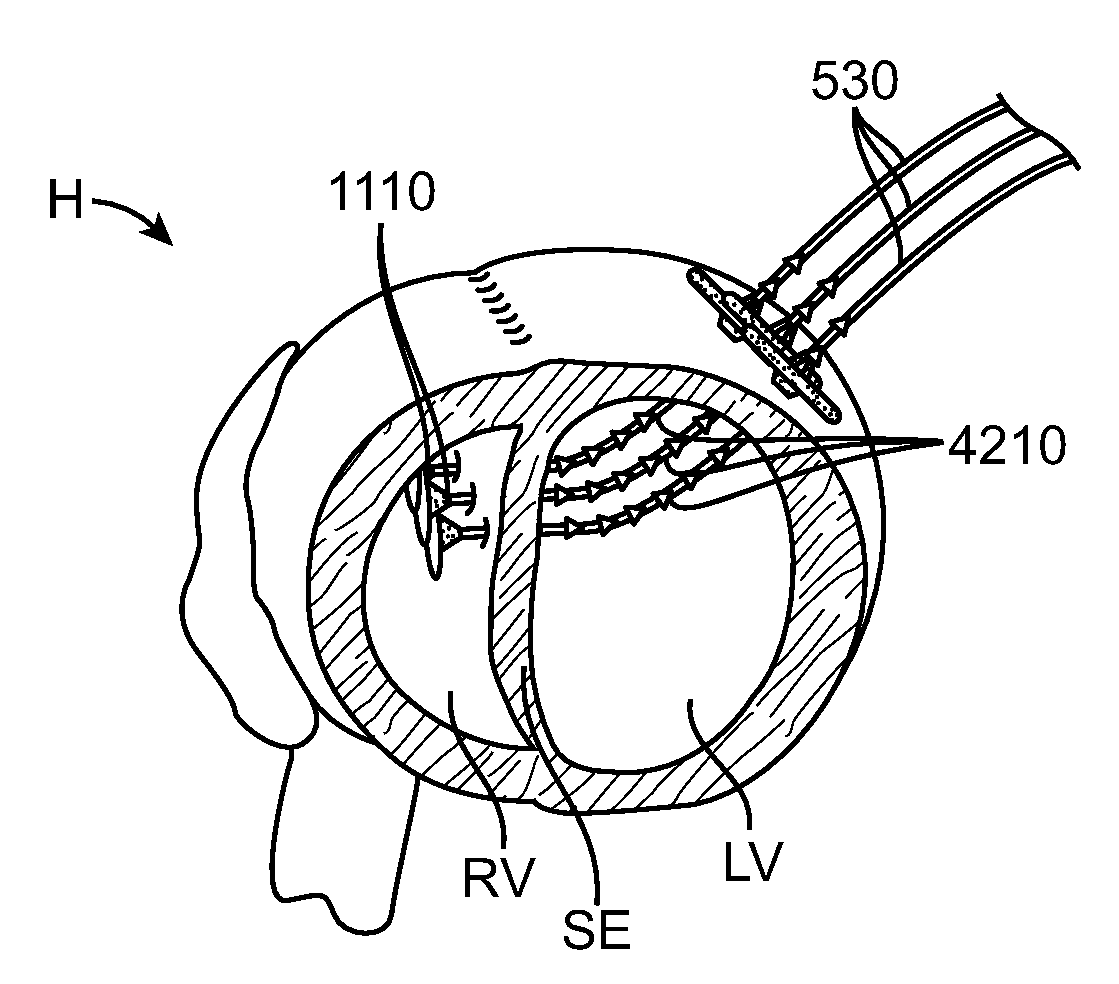
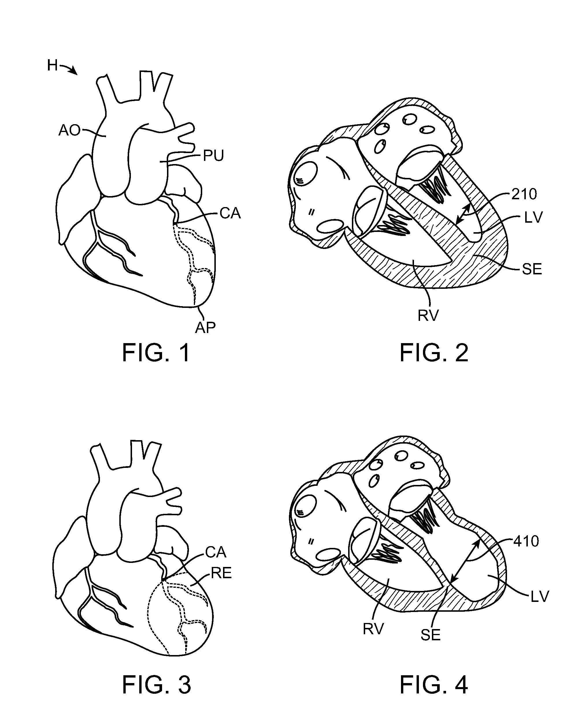
[0069]The present invention generally provides improved devices, systems, and methods for treatment of a heart. Embodiments of the invention may be particularly beneficial for treatment of congestive heart failure and other disease conditions of the heart. The invention may find uses as a prophylactic treatment, and / or may be included as at least a portion of a therapeutic intervention.

[0070]Embodiments of the invention may find use as a device applied to or implant placed in the heart of certain patients with congestive heart failure so as to reduce ventricular volume in a procedure called “Epicardial Catheter-based Ventricular Reconstruction,” or ECVR. The left ventricle of hearts affected with congestive heart failure may dilate or increase in size. This increase in size can result in a significant increase in wall tension and stress. With disease progression, the volume of the left ventricle gradually increases while forward blood flow gradually decreases. Scar tissue will often...


PUM


Abstract

Medical devices, systems, and methods reduce the distance between two points in tissue, often for treatment of congestive heart failure and often in a minimally invasive manner. An anchor is inserted along an insertion path through a first wall of the heart. An arm of the anchor is deployed and rotationally positioned according to a desired alignment. Application of tension to the anchor may draw the first and second walls of the heart into contact along a desired contour so as to effect a desired change in the geometry of the heart. Additional anchors may be inserted and aligned with the first anchor to close off a portion of a ventricle such that the ventricle is geometrically remodeled and disease progression is reversed, halted, and/or slowed.

Description

CROSS-REFERENCES TO RELATED APPLICATIONS[0001]This application claims the benefit of U.S. Provisional Patent Application No. 60 / 977,286 entitled “Method and Device for Treating Dysfunctional Cardiac Tissue,” filed on Oct. 3, 2007; and to U.S. Provisional Patent Application No. 61 / 082,438, entitled “Cardiac Anchor Structures, Methods, and Systems for Treatment of Congestive Heart Failure and Other Conditions,” filed on Jul. 21, 2008, the disclosures of which are hereby incorporated herein by reference.BACKGROUND OF THE INVENTION[0002]The present invention is related to improved medical devices, systems, and methods, with many embodiments being particularly useful for reducing the distance between two points in tissue in a minimally or less invasive manner. Specific reference is made to the treatment of a failing heart, particularly the alleviation of congestive heart failure and other progressive heart diseases. The provided devices, systems, and methods will often be used so as to r...

Claims


Application Information

Patent Timeline
IPC IPC(8): A61F2/24A61N1/362A61B17/03
CPCA61B17/00234A61B17/0401A61B17/0467A61B17/0487A61B19/026A61B2017/00243A61F2/2487A61B2017/0404A61B2017/048A61B2017/0488A61B2017/0496A61B2017/06052A61B2017/06176A61B2017/00469A61B2017/00247A61B2017/0237A61B2017/0408A61B2017/0417A61B2017/0464A61B50/30A61B17/0469A61M25/0138A61M25/0147A61B17/0057A61B17/06066A61B2017/00561A61B2017/00601A61B2017/00867A61B2017/0608A61B2017/061A61F2002/249A61M25/09A61M29/02
Inventor ANNEST, LON S.ARGENTO, CLAUDIOBUTLER, WILLIAMHEFLIN, ERNEST
Owner BIOVENTRIX A CHF TECH
Why Eureka
Try Eureka