The use of the lack of photons detected as the “
signal” from which an image is generated presents a number of inherent problems.
However,
high doses of radiation are typically undesirable due to the x-ray's potential to damage the body.
In particular, there is a direct relationship between the number of x-ray photons interacting with tissue and the
increased risk of radiation-induced
tissue damage and
cancer development.
In traditional x-ray imaging systems that rely upon attenuation-based x-ray imaging, a dilemma is created between the need for increased contrast achieved using elevated radiation doses and the potential for damage that such doses can yield.
However, these contrast agents also carry some risks.
Furthermore, enhancement of traditional x-ray imaging is limited to incremental reduction of
radiation exposure.
Unfortunately, these attempts for reducing
radiation exposure in traditional x-ray imaging systems have been only somewhat successful.
For example, modulating x-ray tube current as a function of
beam angle relative to the patient, while first proposed in the mid 1980s, still has not been found to be compelling.
A major problem here is that tuneable monochromatic x-ray sources are still not routinely available for clinical use.
While clearly being theoretically advantageous, velocity-based imaging systems have not been widely realized due to various formidable hurdles to actual implementation.
In particular, while previously demonstrated as operable by imaging small specimens, systems have not been successfully developed that are appropriate for humans or full body imaging.
These approaches are faced, however, with the problem of “unraveling” the many changes in
refraction (expressed as phase shifts) that occur as the
photon passes through 30 cm or more of tissue.
Hence, for a 30
centimeter (cm) human
abdomen, 6000 phase shifts would be expected, which is computationally cumbersome, if not currently impossible, to “unravel”.
However, to implement this approach, a perfect
silicon crystal is needed, which is difficult and costly to produce at the large scales necessary to image humans.
In this case, the optic
path length of the
reference beam should be stable to within 0.1 nanometer (nm), which presents another significant technological impediment to implementation.
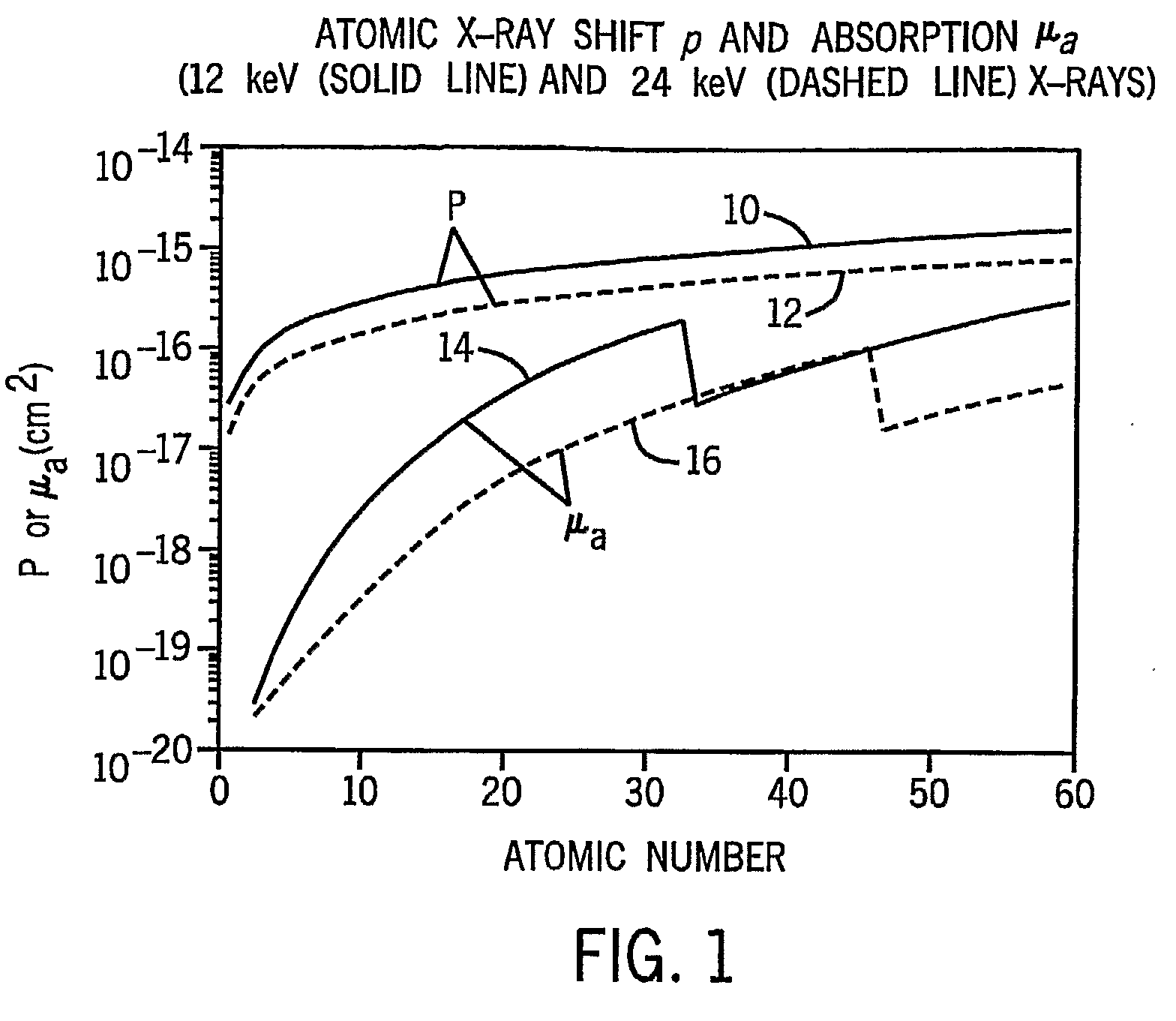
However, each of these methods, as well as the others described above, rely on rather low energy x-ray photons (approximately 10-30 keV) having relatively long wave lengths because the low energy x-ray photons yield more obvious interference patterns and greater
refraction deviation.
The lower energy x-ray photons are not suitable for imaging larger human subjects because the majority of the photons are stopped by the long tissue path lengths.
Thus, while these methods have been demonstrated as feasible on small specimens, they are not clinically viable for human patients.

