
Method and device to administer anesthetic and or vosactive agents according to non-invasively monitored cardiac and or neurological parameters

a vasoactive agent and cardiac and neurological parameter technology, applied in the direction of respiratory organ evaluation, diagnostic recording/measuring, respirator, etc., can solve the problems of affecting affecting the effect of patient consciousness, and affecting the patient's recovery, so as to improve the ability of patients to move freely and efficiently and safely, efficiently and safely assay equipotent doses, and efficient and safe teaching medical students

Inactive Publication Date: 2009-05-14
THE COOPER HEALTH SYST
View PDF13 Cites 111 Cited by
  • Summary
  • Abstract
  • Description
  • Claims
  • Application Information

AI Technical Summary

Benefits of technology

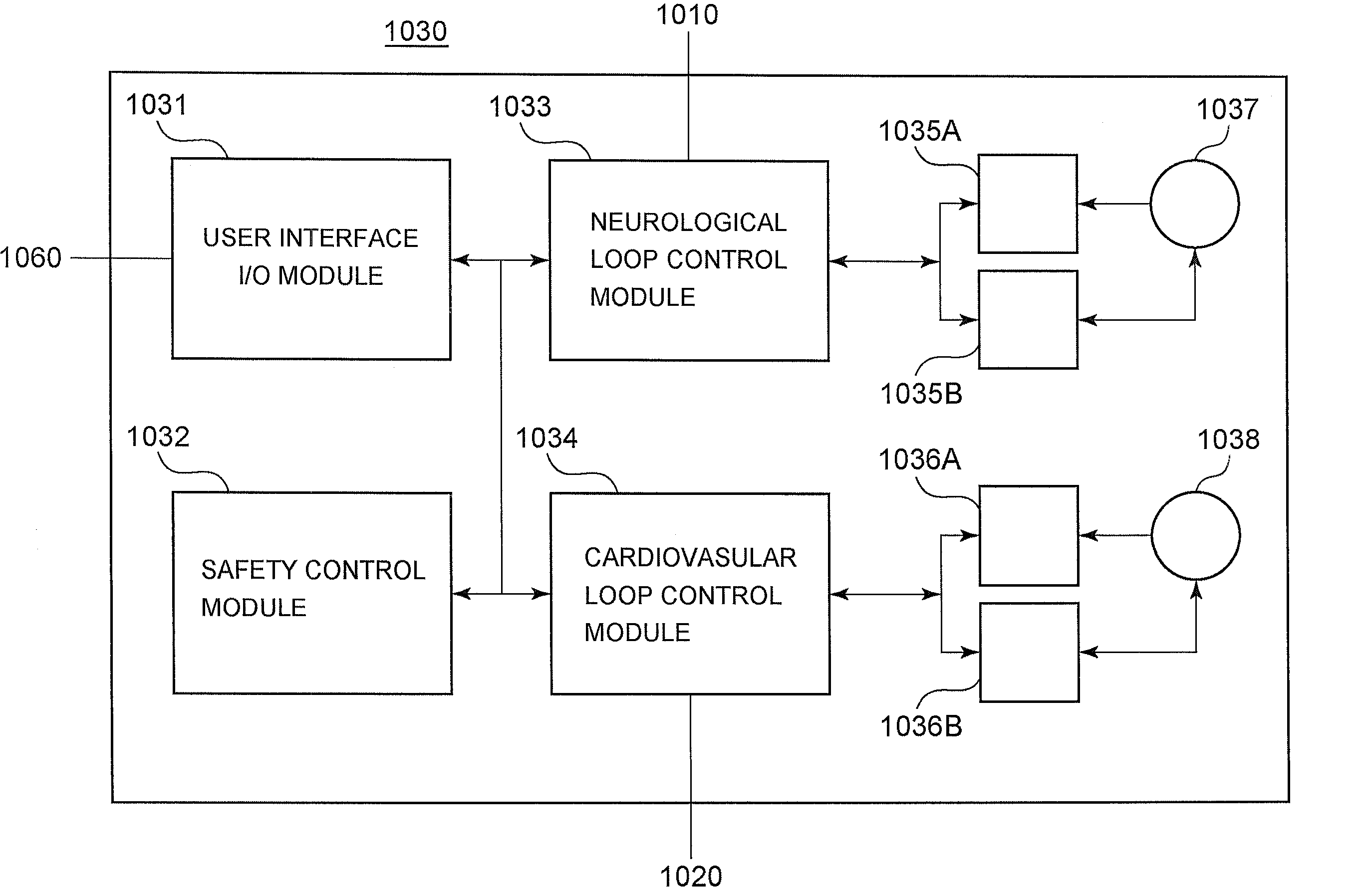
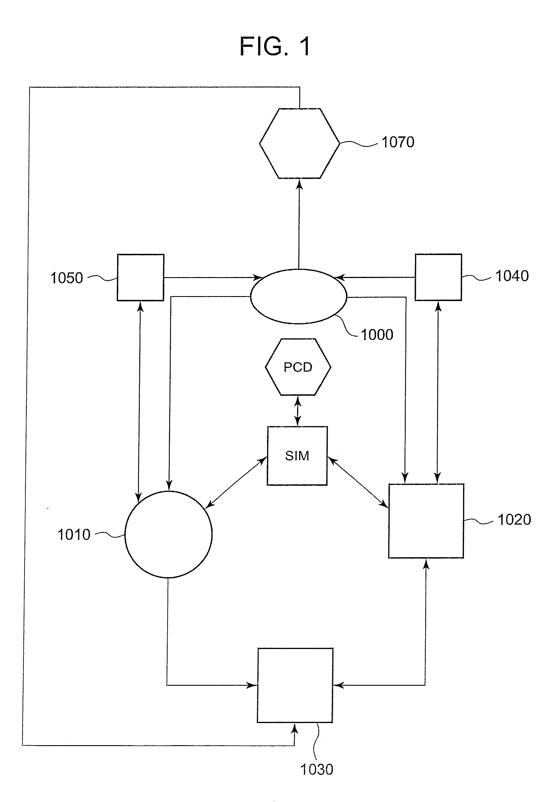
[0029]One object of the current invention is to permit the clinician to maintain a patient in a particular cardiovascular state and at a certain level of neurological depression with various vasoactive drugs with minimal or substantially no intervention. The cardiovascular homeostasis is automatically maintained with respect to cardiac output, preload, afterload, and contractility.

Problems solved by technology

A general anesthetic generally causes a progressive depression of the central nervous system and induces the patient to lose consciousness.
Firstly, the patient should be sufficiently anesthetized so that his / her movements are blocked.
If the patient's movements are not sufficiently blocked, the patient may begin to “twitch” (involuntary muscle reflexes) during the operation, which may move or disturb the operating field that is an area being operated.
Patients have sued for medical malpractice because they felt pain during the operation or were aware of the surgical procedure.
The muscle relaxant drugs, in the unconscious patient allow for a motionless surgical field without the profound central nervous system depression alluded to in to above, which exceeds what is necessary to preclude awareness, and is needed to prevent involuntary unconscious movement in the context of extremely painful stimuli, Using muscle relaxant drugs, which chemically and reversibly disconnect the effect of every voluntary nerve from every voluntary in the body, also increases the risk of intra-operative awareness.
Thirdly, the anesthesia should not be administered in an amount so as to lower blood pressure to the point where blood flow to the brain is reduced to a dangerous extent to cause cerebral ischemia and hypoxia.
For example, if the blood pressure is too low for over 10 minutes, the patient may not regain consciousness.
The depth is also called “plane of anesthesia.” However, there may be a number of problems with this approach.
Some operations may be prolonged for 10 to 15 hours, and the vigilance of the nurse-anesthetist or anesthesiologist may falter or fail.
Unfortunately, Preload, Afterload, and Contractility have been traditionally assessed by invasive methods.
This arrythmogenic procedure is usually reserved for the cardiac catheterization lab since it could be hazardous.
Swan-Ganz catheters are invasive, and their use can be the occasion of clinical mischief.
Although 2-D transesophageal echocardiography devices are prohibitively expensive and also require specialized image interpretation skills, they are still minimally invasive.
Because 2-D echo devices require the placement of a large probe in the esophagus, they cannot be used pre-operatively or post-operatively on spontaneously breathing and awake patients.
Likewise, the methods of Nuclear Medicine are expensive, requiring a cyclotron to produce specialized radiopharmaceuticals and specialized image interpretation skills.
Moreover, since Nuclear Ejection Fractions cannot be done continuously and in real time, they can be used to assess only baseline cardiac function and cannot be used to titrate fluid therapy and drug infusions from moment to moment.
Thus, it ignores about 30% of the total Cardiac Output and cannot measure Preload.
In addition, since the device sits in the thoracic esophagus, it cannot be used on people who are awake.
Although the non-invasively measured cardiac parameters accurately reflect the hemodynamic state of a patient, it is still difficult even for a seasoned medical professional to use vasoactive infusion pharmacotherapy to respond in a rational and timely manner to rapidly emerging changes and wide fluctuations in hemodynamic parameters encountered every day in the ordinary practice of anesthesiology.
Unfortunately, this current practice often results in large and frequent deviations from the norms of cardiovascular homeostasis.
Consequently, the patient may be unnecessarily brought into an even deeper state of neurological depression.
If a patient has too little fluid, the cardiac output becomes insufficient to perfuse vital organs like the brain, heart, and kidney, resulting in organ failure and death.
On the other hand, it a patient has too much fluid, the pumping capacity of the compromised left heart is overwhelmed, allowing fluid to back up into the lungs, causing a diffusion barrier to oxygenation.
Fluid welling up in the lungs effectively causes the patient to drown.

Method used


Image

  • Method and device to administer anesthetic and or vosactive agents according to non-invasively monitored cardiac and or neurological parameters
  • Method and device to administer anesthetic and or vosactive agents according to non-invasively monitored cardiac and or neurological parameters
  • Method and device to administer anesthetic and or vosactive agents according to non-invasively monitored cardiac and or neurological parameters

Examples


first embodiment

[0098]The cardiac data analysis unit 1022 transforms by multiplying the (EI, MAP, E-M) vector by a diagonal matrix as shown below. Let x be a vector in the non-invasive hemodynamic space M of the form (EI, MAP, E-M) Let A be the diagonal matrix shown below. If we represent x vertically as a column vector, we can multiply it by the matrix A such that Ax=b, where b is a vector of the form ((EI*MAP*E-M), (MAP*E-M), 1 / (E-M)), that is approximately equivalent to (LVEDP, SVR, dP / dtmax), and the first plurality of cardiac parameters responsive to external medicines as being demonstrated in the equation below.

(MAP*(E-M)000E-M0001 / (E-m)2)(EIMAP(E-M))=(EI*MAP*(E-M),MAP*(E-M),1 / (E-M))

[0099]The above operation of multiplying the vector by a matrix linearly transforms the vector x into the vector b. Vector b constitutes a new vector space N or a second Non-invasive Space whose axes are responsive to external medicines and fluid administration as being verified below. The three mutually perpendic...

second embodiment

[0104]Alternatively, the cardiac data analysis unit 1022 uses the diastolic filling interval (DI) to replace EI in eq. 8, which tracks Preload as LVEDP. The correlation is improved between (DI, EI, MAP, E-M), which is of the second plurality of non-invasively measured cardiac parameters and (LVEDP, SVR, dP / dtmax) or (P, A, C), which is the first plurality of invasive cardiac parameters in a In diastole, the left ventricular pressure is an exponential function of left ventricular volume, and this relation holds at any point during the diastolic filling interval including end-diastole. Therefore, LVEDP is an exponential function of Left Ventricular End Diastolic Volume (LVEDV). Physiologically, it makes intuitive sense that, other things being equal, the longer the length of time that the Left Ventricle fills in diastole, DI, the more volume of blood will fill the left ventricle at end-diastole with a higher resulting LVEDP, or Preload. To a reasonable approximation,

DI=T−EI  Eq. 17

wh...


PUM


Abstract

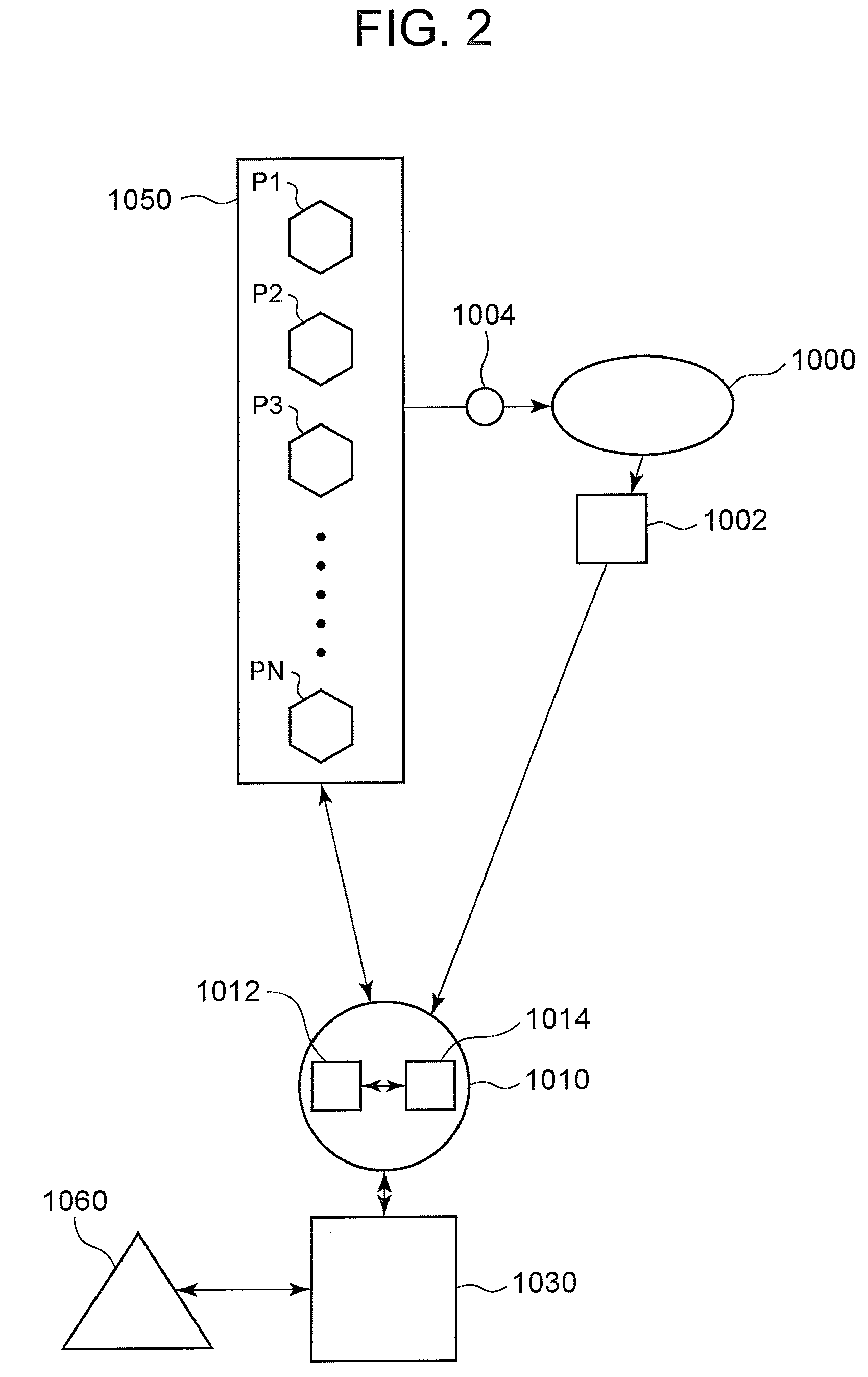
A method of and a device for non-invasively measuring the neurological depressed state and the hemodynamic state of a human patient and involving steps and units of non-invasively measuring EEG, cardiac cycle period, electrical-mechanical interval, mean arterial pressure, and ejection interval and converting the EEG into a neurological index as well as converting the measured electrical-mechanical interval, mean arterial pressure and ejection interval into the cardiac parameters such as Preload, Afterload and Contractility, which are the common cardiac parameters used by an anesthesiologist. A general anesthetic is administered based upon the converted neurological index. A vasoactive agent is independently administered based upon the converted cardiac parameters as necessary in order to restore cardiovascular homeostasis in the patient. The converted neurological and hemodynamic state of a patient are displayed on a screen as an index value and a three-dimensional vector with each of its three coordinates respectively representing Preload, Afterload and Contractility. Therefore, a medical practitioner looks at the screen and quickly obtains the important and necessary information.

Description

BACKGROUND OF THE INVENTION[0001]1. Field of the Invention[0002]The present invention relates to a method and device to administer anesthetic agents and vasoactive agents according to non-invasively monitored cardiac and or neurological parameters.[0003]2. Description of the Prior Art[0004]At the present time, since anesthetics or sedative-hypnotic drugs both induce loss of sensation, they are often used for surgical operations. A general anesthetic generally causes a progressive depression of the central nervous system and induces the patient to lose consciousness. In contrast, a local anesthetic affects sensation at the region where it is applied.[0005]Generally, prior to the operation, the patient is usually anesthetized by a specialized medical practitioner (“anesthesiologist”) who administers one or more volatile liquids or gases such as nitrous oxide, halothane, isoflurane, sevoflurane, desflurane, and etc. Alternatively, non-volatile sedative-hypnotic drugs such as pentothal,...

Claims


Application Information

Patent Timeline
IPC IPC(8): A61M21/00G16H20/17
CPCA61M5/142G06F19/3468A61M16/0051A61M16/01A61M16/18A61M2016/1025A61M2016/103A61M2202/0241A61M2202/0275A61M2202/048A61M2205/13A61M2205/17A61M2205/3569A61M2205/3592A61M2205/502A61M2205/52A61M2205/581A61M2230/04A61M2230/10A61M2230/202A61M2230/205A61M2230/65G06F19/3437A61M5/1723A61M16/024G16H50/50G16H20/17
Inventor HIRSH, ROBERT A.TORJMAN, MARC C.
Owner THE COOPER HEALTH SYST
Learn More