Compositions and methods for joining non-conjoined lumens

a technology of non-conjoined lumens and compositions, applied in the field of compositions, methods, and kits for joining together non-conjoined lumens, can solve the problems of long-term stenosis or fibrosis, calcified and diseased vessels providing mechanical challenges, and the geometry of distal portions is stabilized, so as to facilitate the joining

Active Publication Date: 2009-06-25
TAUTONA GROUP
View PDF77 Cites 18 Cited by
  • Summary
  • Abstract
  • Description
  • Claims
  • Application Information

AI Technical Summary

Benefits of technology

[0028]This invention is directed to the discovery that the stabilization of the geometry of the distal portions of tubular tissues or lumens facilitates joining such non-conjoined lumens.

Problems solved by technology

Because of the complexity and judgment required in suturing, automated techniques are not well accepted.
Calcified and diseased vessels provide mechanical challenges.
Sutures may, in some instances, cause a reaction resulting in long term stenosis or fibrosis.
Frequently, this is troublesome to the practitioner because the end of an tubular tissue—such as a clamped blood vessel devoid of blood—is not a perfectly round circle; rather it is in its unpressurized, deflated-looking state where a cross-sectional view of the circumference may be a circle, an oval or irregular, and, of course having no structural support from within, is unstably in any shape (unless the surrounding tissue possesses structural strength).
Microvascular anastomosis is often performed by hand under a microscope, and is tedious and painstaking work.
As a result, it can take many hours to complete just the microvascular anastomosis required to reconnect a severed body part or transplant an organ.
This system, however, is limited to use with two blood vessels having substantially the same diameter.
Further, manual eversion of a blood vessel having a diameter on the order of one millimeter is difficult and painstaking, particularly when the eversion is to be substantially even around the circumference of the ring.
This can slow or stop blood flow through the heart's blood vessels, leading to chest pain or a heart attack.
Multiple practitioners, additional complexity, and, as a practical matter, additional health care cost is involved over surgical procedures involving fewer practitioners and procedures.
Moreover, blood quality may be degraded as the heart-lung machine repetitively pumps the patient's blood through the systemic circulation.
The blood may embolize or clot in the distal circulation, or form clots which migrate to the distal vasculature, and cause a stroke.
Frequently, because the graft must be performed on arteries in locations directly affected by the beating heart, stabilizing mechanisms are not thoroughly effective, and the practitioner must suture the graft while the graft is moving in conjunction with the heart beat, at least to some extent.
Thus, the graft quality may be compromised.
After an hour, the risk of patient morbidity is thought to increase perhaps due to the heart-lung machine degrading the quality of the blood as it is circulated through the systemic circulation.
Bypass surgeries, however, often last three hours or longer.
Moreover, where the aorta is clamped and blood therefore cannot pass through, the blocked blood is thought to cause additional issues.
Anastomosis is time-consuming.
In “off pump” procedures where the heart remains beating, the difficulty of suturing an anastomosis graft on a moving surface of the heart may degrade the quality of such grafts completed on patients.
It can be appreciated that some of the sutures are easily made from on top of the conduit or blood vessel graft, while others are more difficult to complete as they are beneath the conduit.
It can be further appreciated that performing such complex suturing procedures on a moving platform, such as the beating heart, further increases the difficulty associated with such suturing procedures.
Improperly connecting blood vessel grafts to the patient may present substantial post-operative complications and / or increase operating room time spent correcting the improperly connected graft.

Method used


Image

  • Compositions and methods for joining non-conjoined lumens
  • Compositions and methods for joining non-conjoined lumens
  • Compositions and methods for joining non-conjoined lumens

Examples


example 1

Preparation and Property Determination of a Sol-Gel Composition of this Invention

[0252]Presented here is a protocol for preparing 100 mL (100 g) of a thermoreversible sol-gel composition having Formulation 1 listed in Table 1.

[0253]Materials:[0254]Phosphate buffered saline (“PBS”): 72 g, pH 7.4, with Ca2+ and Mg2+;[0255]Poloxamer 407 (BASF Pluronic® F 127): 17 g;[0256]Poloxamer 188 (BASF Pluronic® F 68): 11 g.

[0257]Procedure:

[0258]The above materials were mixed at 4° C. under high shear to facilitate dispersing the solid poloxamer in the solvent. The heterogenous mixture is then incubated at 4° C. for 12 hours to allow for complete dissolution of the poloxamer in the solvent. The mixture is then further stirred to ensure homogeneity.

[0259]Compositions having other formulations can be made similarly according to the total concentration of the poloxamers and the ratios of the two poloxamers.

[0260]Water or other aqueous solutions may replace PBS as the solvent. It is preferred that the...

example 2

An Anastomosis Operation Using a Sol-Gel Composition of this Invention

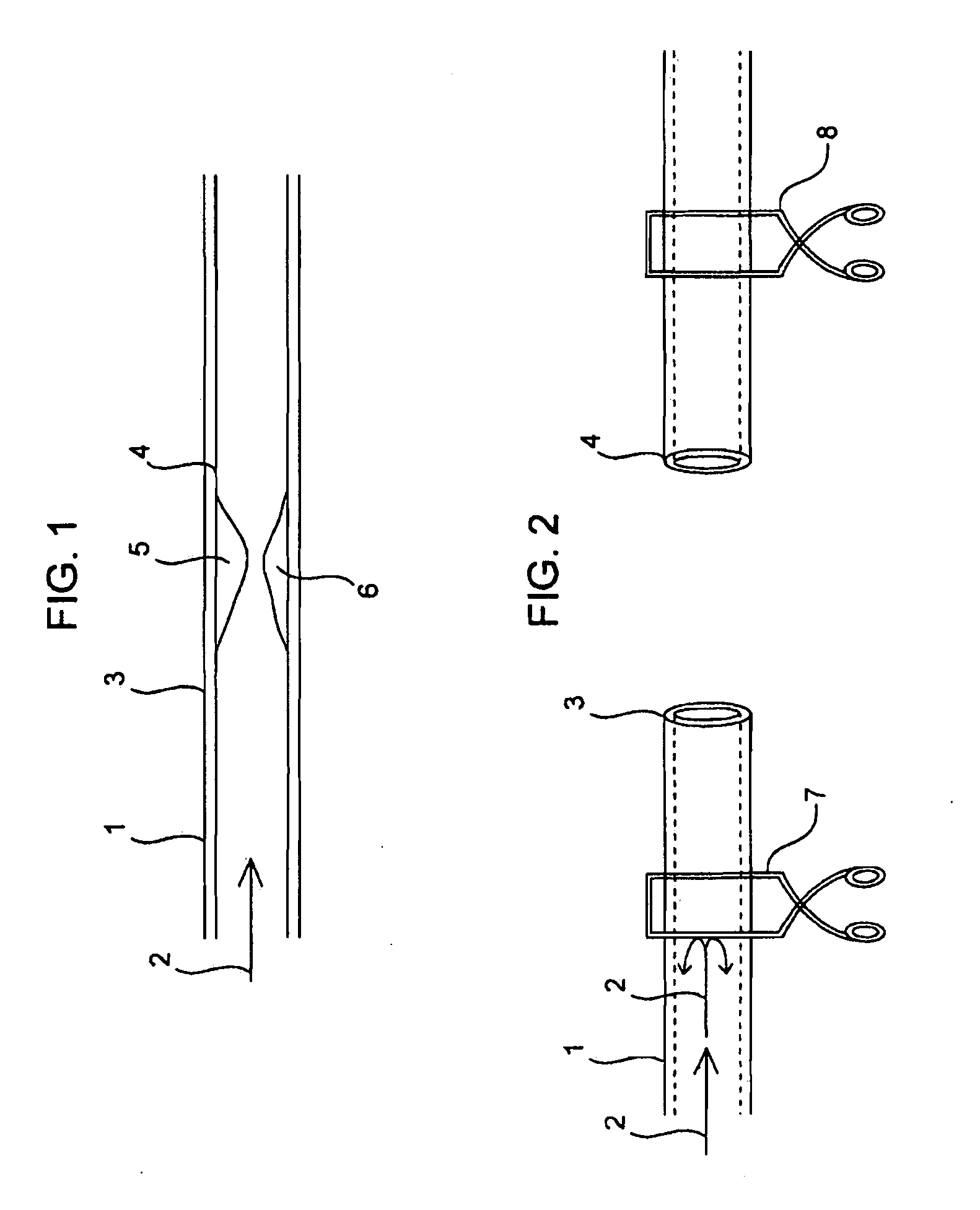
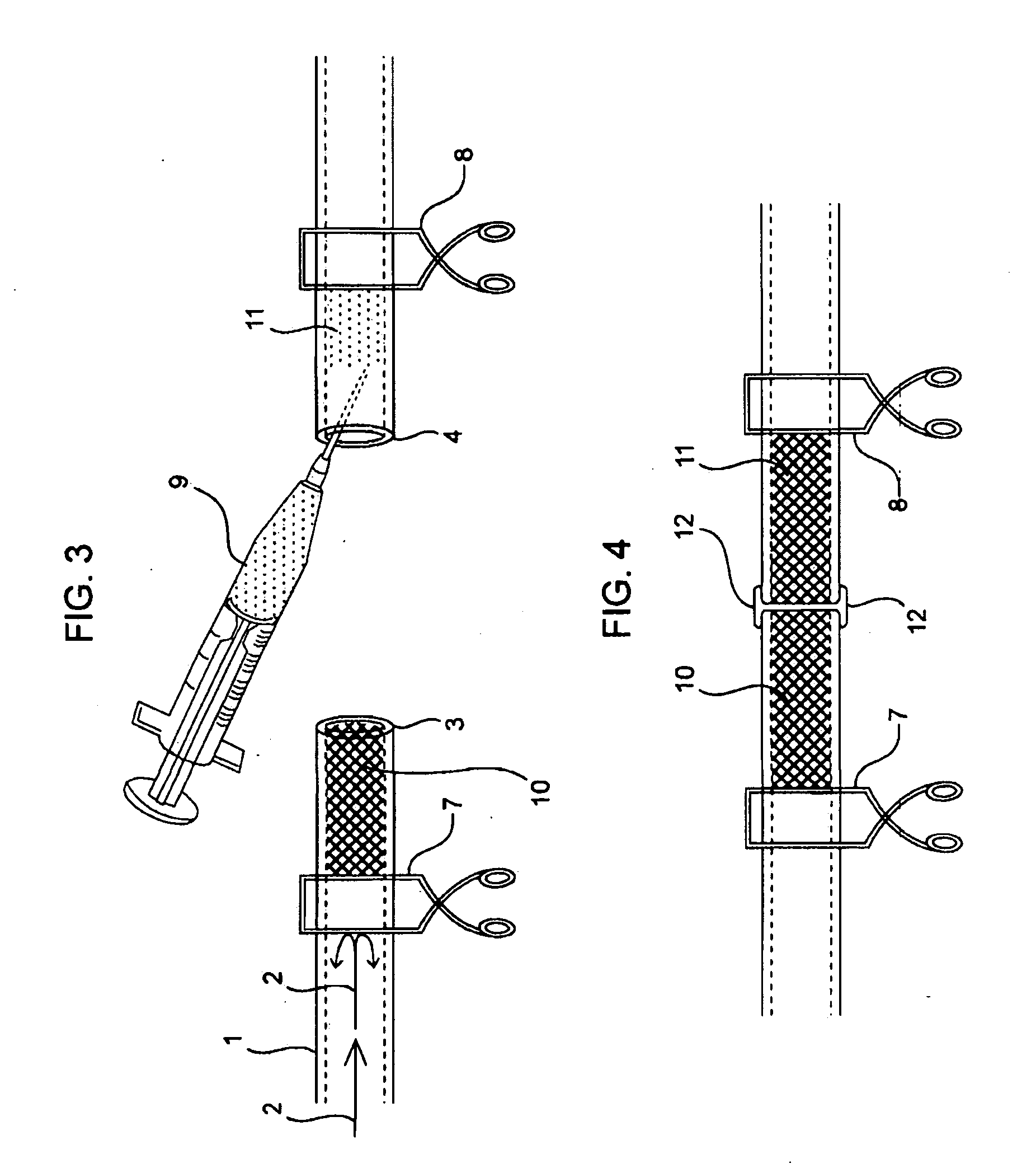
[0262]End to End Anastomosis Surgical Method Performed on Cardiac Tubular Tissues in Rats:

[0263]Rats, which are treated and cared for in accordance with all applicable laws and regulations, and in accordance with good laboratory practices, are anesthetized with isoflurane and prepped in sterile fashion with 70% ethanol. The aorta is exposed and isolated with blunt and sharp dissection through a midline laparotomy incision. The aorta is clamped proximally and distally, and subsequently divided and flushed with heparinized saline. In some cases the rat and a sol-gel composition of Formulation 1 are warmed to about 37° C. with sterile water, and then the sol-gel composition of Formulation 1 is injected into severed aortas in a semi-solid state. In other cases the sol-gel composition of Formulation 1 is injected as a liquid and then heated to about 37° C. with a convectional source to solidify it. After direct reappro...

example 3

Prophetic Example of Using a Sol-Gel Composition in a Vascular Grafting Procedure

[0266]In clinical practice, there are some instances where native vessels (i.e. blood vessels of the patient) are not available for bypass grafting or where, especially for older patients, native fistulas require a long maturation time or do not develop sufficiently. In these instances, physicians may choose to use artificial conduits such as grafts comprising polytetrafluoroethylene (PTFE), which can be formed in reinforced or nonreinforced configurations, for example expended PTFE (ePTFE) grafts, manufactured by Gore and others. Synthetic internal arteriouvenous (AV) fistulas or AV grafts, can be placed in many positions in the arms or legs or across the anterior chest wall.

[0267]The artificial grafts may be used instead of native vessels in all configurations: end-to-end inter-positional grafting, end-to-side for procedures such as artery to vein shunt for dialysis fistula access, end-to-side bypass ...


PUM

PropertyMeasurementUnit
elastic modulusaaaaaaaaaa
phase transition temperatureaaaaaaaaaa
transition temperatureaaaaaaaaaa

Abstract

Disclosed are compositions, methods, and kits for joining together non-conjoined lumens in a patient's body including vascular lumens. More particularly, in various aspects, this invention provides compositions, methods, and kits for joining such non-conjoined lumens, including small lumens typically requiring microsurgical technique

Description

CROSS-REFERENCE TO RELATED APPLICATIONS[0001]This application claims the benefit under 35 U.S.C. § 119(e) of provisional Patent Application Ser. No. 61 / 015,660, filed on Dec. 20, 2007, which is incorporated herein by reference in its entirety.FIELD OF THE INVENTION[0002]This invention relates generally to compositions, methods, and kits for joining together non-conjoined lumens in a patient's body including vascular lumens. More particularly, in various aspects, this invention provides compositions, methods, and kits for joining such non-conjoined lumens, including small lumens typically requiring microsurgical techniques.BACKGROUND OF THE INVENTION[0003]Non-conjoined lumens arise in a variety of settings including surgical settings where the non-conjoined lumens are intentionally created or arise from lacerations or puncture wounds. Intentionally created non-conjoined lumens include those arising during surgical repair of e.g., treatment of a blockage in a lumen by bypass procedure...

Claims


Application Information

Patent Timeline
Patent Type & Authority Applications(United States)
IPC IPC(8): A61K9/10A61B17/11A61K31/727
CPCA61L27/14
Inventor FULLER, GERALD G.RAPPLEYE, C. TRAVISMANDRUSOV, EVGENIA
Owner TAUTONA GROUP
Why Eureka
Try Eureka