
Implantable cartilaginous tissue repair device

a cartilaginous tissue and implantable technology, applied in the field of surgical devices, can solve the problems of pain and reduced mobility in old people, injury to the fibrocartilage of the meniscus, and small volume for discharging energy, and achieve the effects of convenient fixation, high modulus, and strong and tough attachment to the bon

Active Publication Date: 2009-07-02
ORTHOX LTD
View PDF13 Cites 75 Cited by
  • Summary
  • Abstract
  • Description
  • Claims
  • Application Information

AI Technical Summary

Benefits of technology

[0037]The present invention also provides a substantially bioresorbable implantable cartilaginous tissue repair device which allows and encourages the gradual infiltration and replacement of at least some parts of the device with autologous collagen and proteoglycans (i.e. collagen and proteoglycans produced by the patient's own body) more effectively than prior art devices. The porosity of the hydrogel allows, in particular, for infiltration by mesenchyme or stem cells.
[0038]The present invention provides a device of composite construction which enables the mechanical properties of the prosthesis to be tuned locally to provide appropriate stress / strain environments throughout the prosthesis to avoid the problem of stress shielding thereby encouraging the de novo formation of the correct connective tissues appropriate to the different locations of a complex tissue.
[0039]The three-dimensional fibre lay and constrained porous hydrogel impart cartilage-like properties to the implantable device enabling it to distribute stresses during compressive loading and, in the case of menisci and intervertebral discs, to act as a shock absorber.
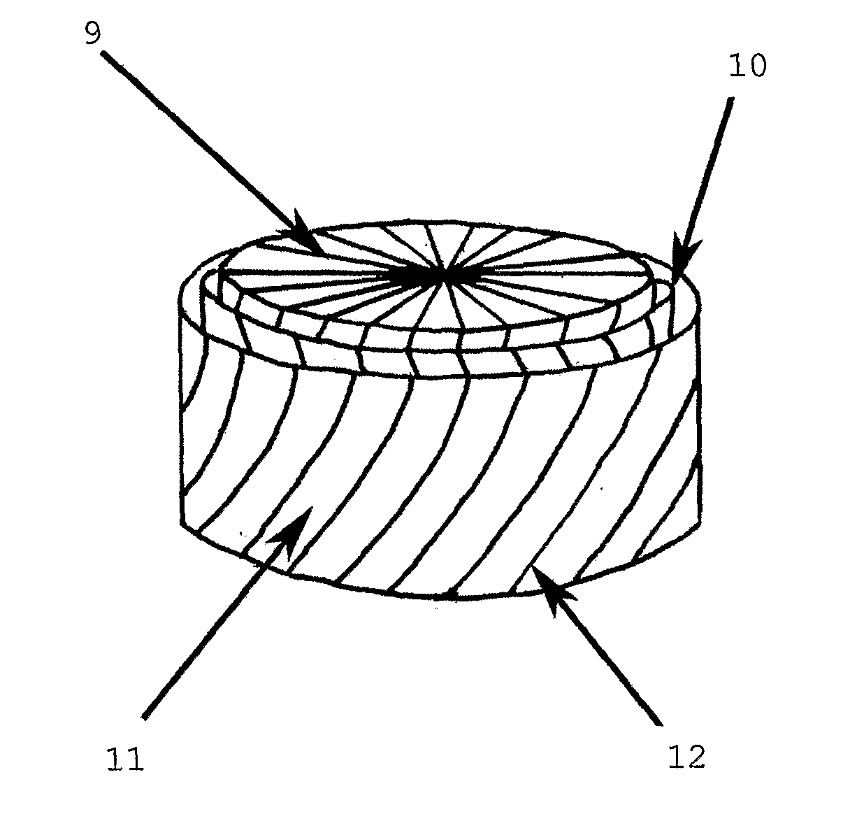
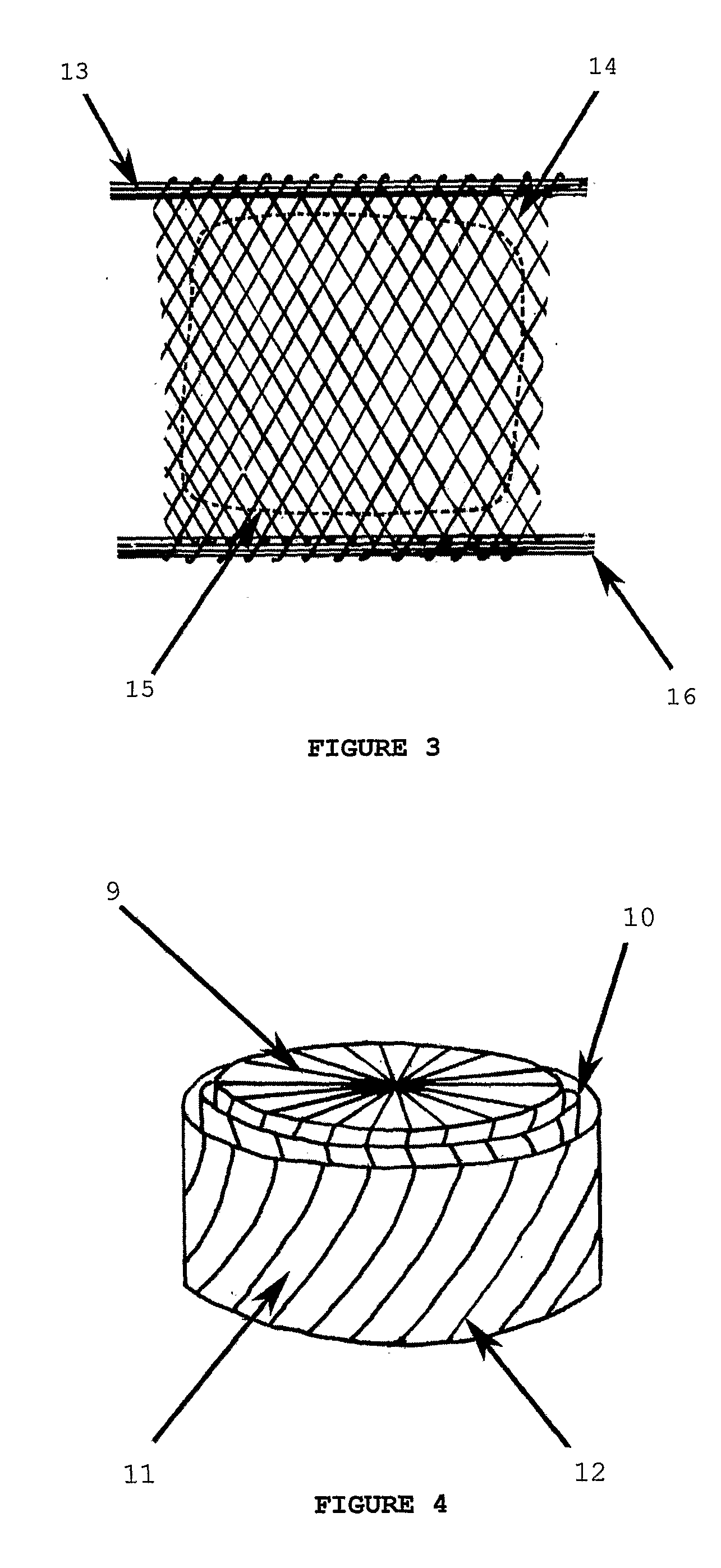
[0042]In some embodiments, an integral means of attaching or anchoring the implantable device firmly to the bone of a patient helps to overcome a persistent weakness of many orthopaedic devices—mechanical failure at the bone / prosthesis interface. The mineralisation of such an integral means of firmly attaching or anchoring the device, as used in some embodiments of the invention, provides a tough, compression-resistant material which would also facilitate fixation with a polylactic acid screw (‘poly screw’) or equivalent device in a drilled tunnel in the bone, thereby providing a strong and tough attachment to the bone.
[0044]In some embodiments of the invention, the resorption rates of silk fibres in the three-dimensional fibre lay and / or of silk protein hydrogel can be regionally tuned by controlled covalent cross-linking, controlled formation of β-sheet crystallinty and / or by tuning the hydrophobicity of the protein by alkylating with hydrophobic side chains. This enables the planning of an orderly ‘hand over’ of stress to the newly forming connective tissues during resorption without stress shielding.
[0045]In some embodiments, regionally specific growth factors are incorporated into some or all of the implantable device which encourage appropriate cell recruitment, differentiation and secretion which thus further facilitates the conversion over time of the device to regionally appropriate connective tissue.

Problems solved by technology

Osteoarthrosis is caused by the progressive damage and breakdown of articular cartilage most commonly in the hip and knee and is an important cause of pain and reduced mobility in old people.
Injury to the fibrocartilage of the meniscus is a common sports injury and is also seen as a result of road traffic accidents.
This is because its volume for dissipating energy is very small compared to that of bone and because it actually increases in stiffness with increasing strain rate making it an inappropriate material for use as a shock absorber.
No wholly satisfactory procedure exists for replacing damaged articular cartilage in osteoarthrosis and instead artificial prostheses are most commonly used to replace the entire hip and knee joints.
While these increase mobility and reduce pain they suffer from progressive wear, mechanical failure, adverse tissue reactions and loosening at their interphase with the bone.
For this reason damage to these fibres increases the risk of secondary osteoarthrotic damage to the condylar cartilages as the normal load distribution and shock-absorbing functions are impaired.
However, even with partial mensicectomy, secondary osteoarthrosis is still a long-term consequence.
However there is no proof that replacement of the meniscus with an allograft can re-establish some of the important meniscal functions, and thereby prevent or reduce the development of osteoarthrosis secondary to meniscectomy (Messner, K. and Gao, J.
The major problems are the lack of remodelling of the graft resulting in inferior structural, biochemical and mechanical properties and insufficient fixation to bone.
Further disadvantages include the shortage of suitable donors, difficulties with preservation techniques, the possible transfer of diseases, difficulty in shaping the implant to fit the donor and possible immunological reactions to the implant (Stone, K. R. Clin. Sports Med. 1996, 15, 557-571).
Total knee replacement cannot be considered as treatment for uncomplicated meniscal injury.
Partial or total meniscal replacements made from collagen, Teflon fibre, carbon fibre, reinforced polyester, or polyurethane-coated Dacron showed high failure rates resulting from poor fixation, mechanical failure or severe inflammatory response.
However, the mechanical properties of the polyurethanes tested did not match native meniscus very well and this may help to explain why only poorly orientated collagen was found in the regenerating fibro-cartilage in the implanted devices in place of the well-orientated collagen in normal meniscus.
A further potential problem was that the polyurethane materials produced a Stage I inflammatory response (giant cells and some macrophages).
This material has low biocompatibility and would be likely to trigger a severe synovial reaction.
The suggested materials have low biocompatibility and would be likely to trigger a severe synovial reaction.
This structure improves on the mechanical properties of the constructs of WO 89 / 00413, but still struggles to reach those of cartilage.

Method used


Image

  • Implantable cartilaginous tissue repair device
  • Implantable cartilaginous tissue repair device
  • Implantable cartilaginous tissue repair device

Examples


Embodiment Construction

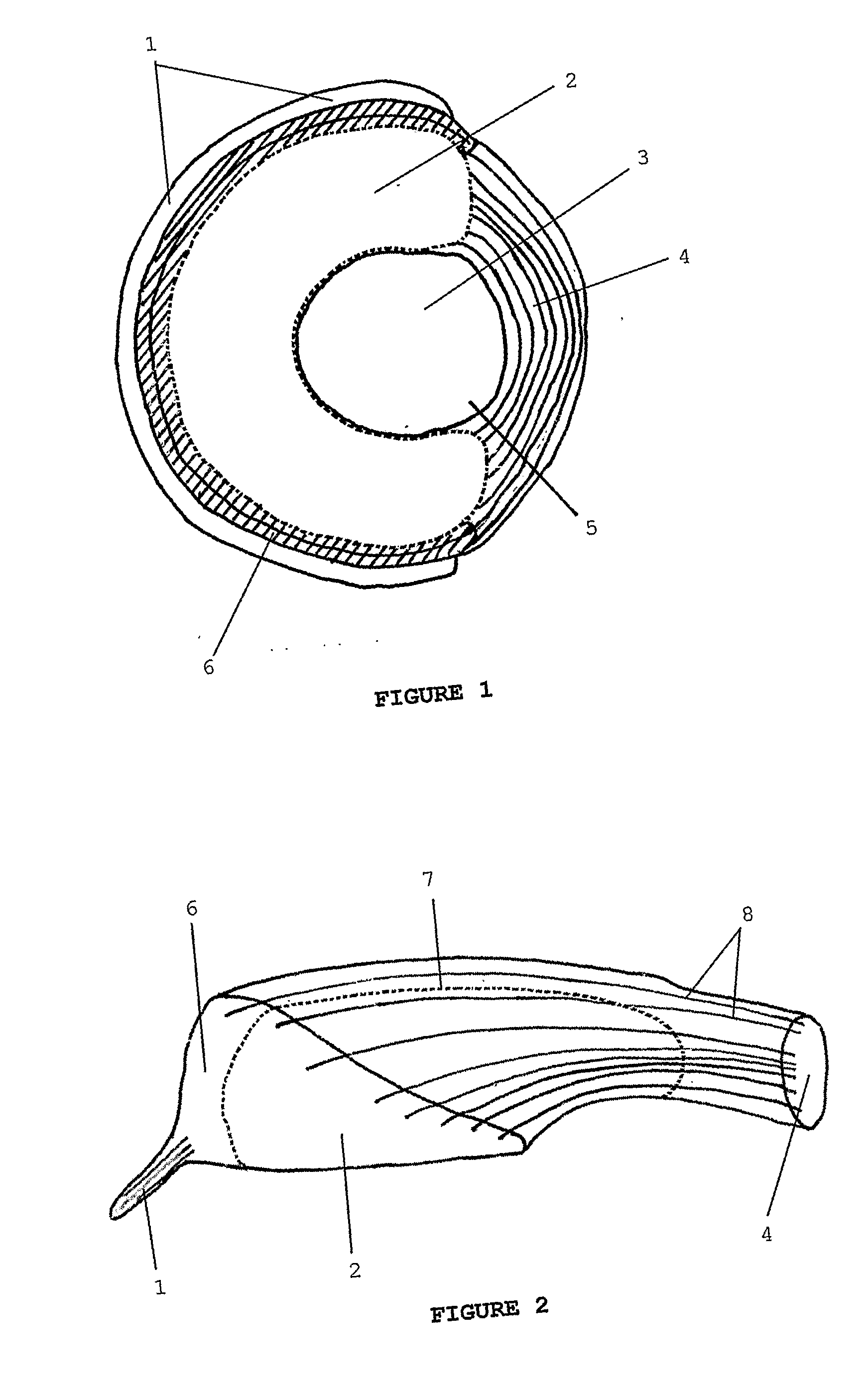
[0057]FIGS. 1 and 2 are respectively a schematic plan view and a schematic cross-sectional perspective view of an embodiment of a meniscal repair device according to the invention.

[0058]A three-dimensional fibre lay of silk fibres—both circumferential (8) and radial (not shown)—has been impregnated partially with a porous regenerated silk fibroin hydrogel and partially with a non-porous regenerated silk fibroin hydrogel to form the device.

[0059]The device of FIGS. 1 and 2 consists of a coronary ligament analogue (1) attached to a porous peripheral band (6) which is in turn attached to both a central, non-porous region (2) and a meniscofemoral ligament analogue (4). The attachment of the porous peripheral band (6) to the non-porous region occurs along a limit of the porous peripheral band (7). A central former (3) has been used to facilitate manufacture of the meniscal repair device. This central former (3) is excised prior to use along a line of cut (5).

[0060]Thus the anatomically-s...


PUM

PropertyMeasurementUnit
pore diameteraaaaaaaaaa
pHaaaaaaaaaa

Abstract

A cartilaginous tissue repair device with a biocompatible, bioresorbable three-dimensional silk or other fibre lay and a biocompatible, bioresorbable substantially porous silk-based or other hydrogel partially or substantially filling the interstices of the fibre lay; with or without an integral means of firmly anchoring the device to a patient's bone.

Description

FIELD OF THE INVENTION[0001]The present invention relates generally to surgical devices for approximating, repairing or regenerating damaged or diseased cartilage, and to manufacturing methods for such devices. In particular, the present invention relates to a device for the replacement, partial replacement or augmentation of damaged cartilage applicable to the repair of articular cartilage, the meniscus of the knee, the tempero-mandibular joint or an intervertebral disc.BACKGROUND OF THE INVENTION[0002]Cartilage in the adult mammalian body occurs in three principal forms: hyaline cartilage; white fibrocartilage; and yellow elastic cartilage. Hyaline cartilage is chiefly present as articular cartilage in the synovial diarthroidal joints of the hip and shoulder and between the long bones where it forms the stiff and smooth articulating surfaces. White fibrocartilage is present in the menisci of the knee and temporomandibular joint of the jaw and in the intervertebral discs. Yellow el...

Claims


Application Information

Patent Timeline
Patent Type & Authority Applications(United States)
IPC IPC(8): A61F2/28A61K9/00A61F2/00A61F2/02A61F2/30A61F2/38A61F2/44A61L27/22A61L27/48A61L27/52A61L27/56
CPCA61F2/30756A61F2/3872A61F2/442A61F2002/30062A61F2002/30075A61F2002/30766A61L27/56A61F2002/4495A61F2210/0004A61F2210/0061A61L27/227A61L27/48A61L27/52A61F2002/3092A61L2430/06B29C70/30A61L27/46A61L27/58B29C44/1276B29C44/3419B29C70/222B29K2105/041B29K2105/0809B29K2311/10
Inventor MANN, STEPHENKNIGHT, DAVID PHILIPSKAER, NICHOLAS JAMES VAVASOURBERTRAM, CAROLINEOREFFO, RICHARD O.C.ROACH, HELMTRUD I.COLLINS, ANDREW MICHAEL
Owner ORTHOX LTD
Learn More