

Endothelial predecessor cell seeded wound healing scaffold
a scaffold and endothelial technology, applied in the direction of prosthesis, peptide/protein ingredients, drug compositions, etc., can solve the problems of significant inhibition of wound healing in the patient, fewer cells available for migration or differentiation, and various approaches to promote wound healing
- Summary
- Abstract
- Description
- Claims
- Application Information
AI Technical Summary
Benefits of technology
Problems solved by technology
Method used
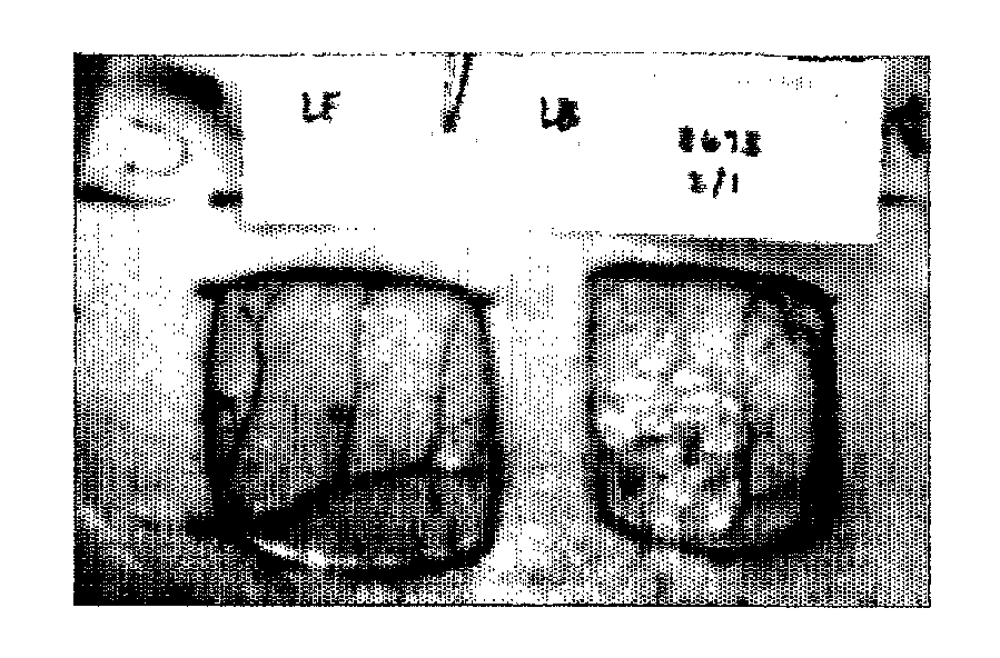
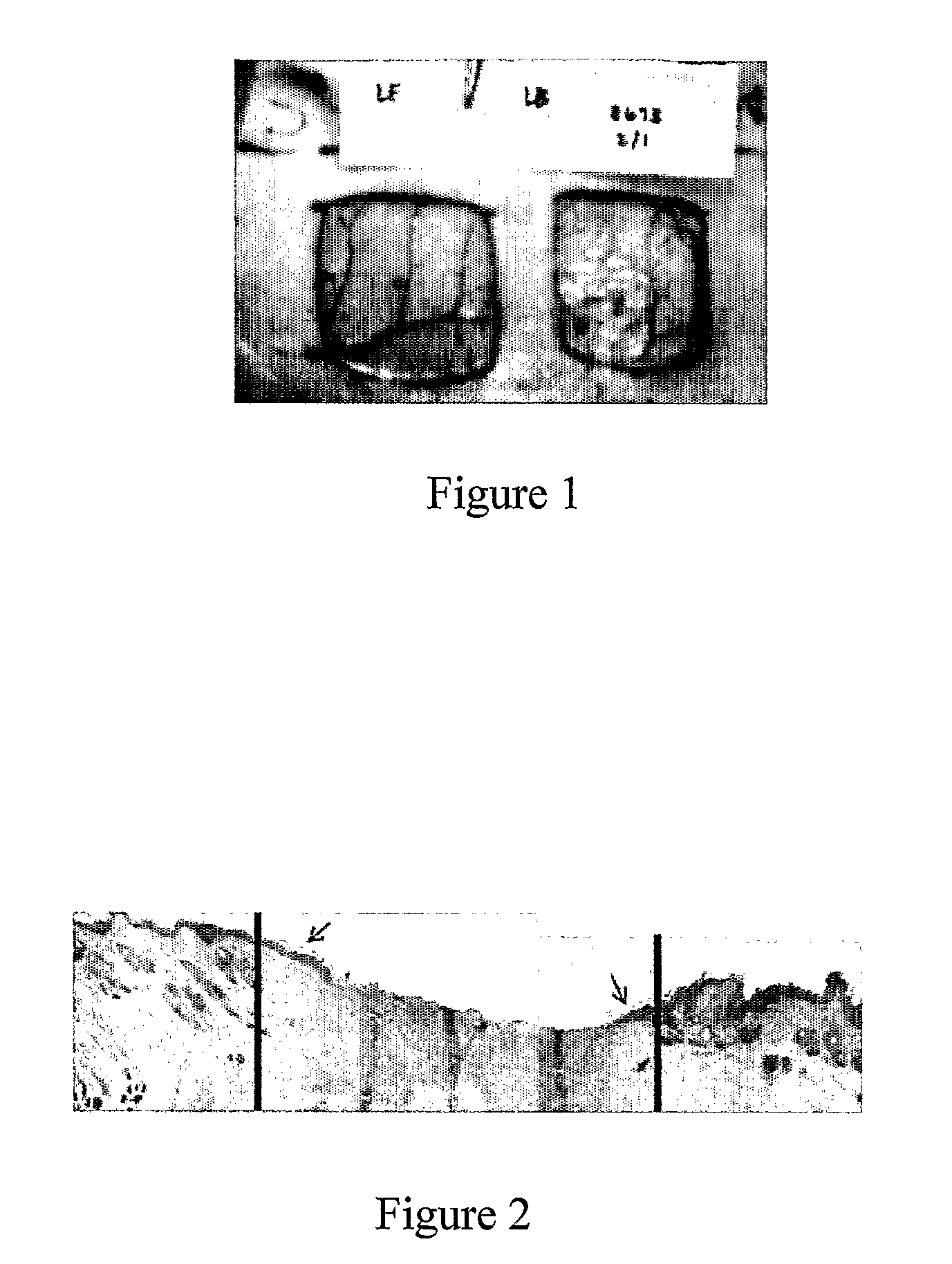
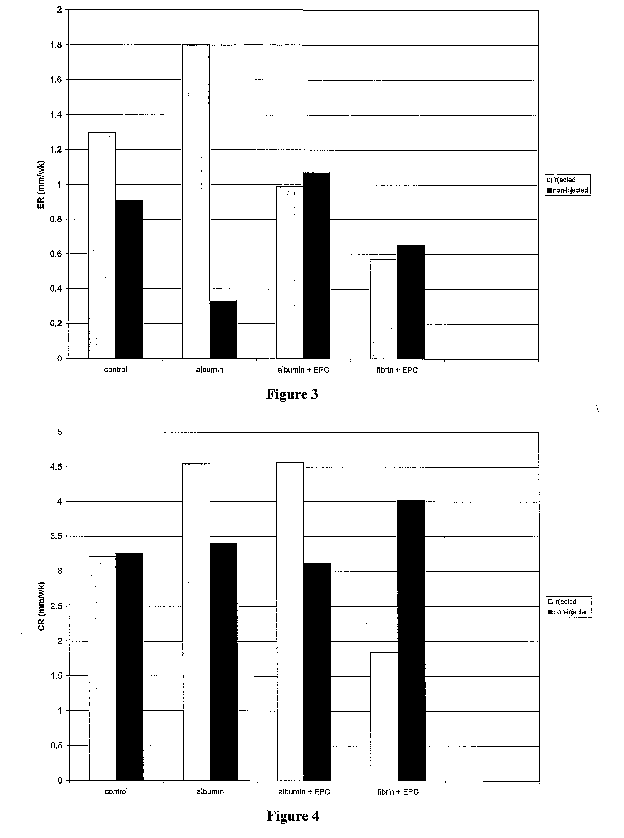
Image

Examples
example 1
Separation of Peripheral Blood Mononuclear Cells (PBMC)
[0067]Peripheral blood is collected from the donor using 50 ml syringes coated with 1 ml heparin. Between 100-125 ml of blood is usually collected during each draw. 15 ml of phosphate buffered saline (PBS) without calcium or magnesium is added to 50 ml centrifuge tubes. One tube is used for every 20 ml of blood drawn. 20 ml of collected peripheral blood is mixed with PBS in the tubes. 14 ml Histopaque solution (1.077 g / ml, Sigma) is added to the bottom of the tube to separate the blood cells based on a density gradient. The tubes are then spun in a centrifuge at 1800 rpm (450 g), 20° C., and no brake for 24 minutes. The blood separates to form a layer of red blood cells in the bottom of the tube, a layer of clear Histopaque solution, a cloudy band of PBMC, and the remaining yellowish portion of the plasma. The plasma layer is aspirated, then the PBMC band is harvested from the tube using a 10 ml pipette. The harvested PBMC cells...
example 2
Separation of CD34+Predecessor Cells
[0069]The PBMC pellet of Example 1 is resuspended in PBS for a final volume of 300 microliters per 108 cells. 100 microliters per 108 cells of Human IgG (Miltenyi Biotech, Inc.) is added to the cell suspension. The blocking reagent inhibits unspecific binding, or Fc-receptor mediated binding, of CD34 Microbeads to non-target cells. CD34+ cells are labeled by adding 100 microliters per 108 cells of the CD34 Microbeads (Miltenyi Biotech Inc.) and mixing well. Alternatively, cells are first incubated with a primary anti-CD34 antibody (QBEND / 10, Miltenyi Biotech Inc.), which is modified with a hapten, then the CD34+ cells are magnetically labeled with Anti-Hapten MicroBeads (Miltenyi Biotech Inc.). Cells are incubated for 30 minutes in the refrigerator. Incubating for longer may lead to unspecific cell labeling. The cells are then washed with 50 ml MACS® buffer solution (Miltenyi Biotech Inc.) and spun at 1500 rpm for 8 minutes. The MACS® buffer solut...
example 3
[0071]The separation of CD34+ predecessor cells from bone marrow is accomplished by the method described in Example 2.
PUM
| Property | Measurement | Unit |
|---|---|---|
| Fraction | aaaaa | aaaaa |
| Fraction | aaaaa | aaaaa |
| Fraction | aaaaa | aaaaa |
Abstract
Description
Claims
Application Information