Hand-held electric field imager for measuring the electric field in mammalian skin and other epithelial structures

an electric field and imager technology, applied in the field of hand-held devices for measuring the electric field of skin or epithelial structures, can solve the problems of inconvenient use, limited usefulness of conventional methods for determining this information, and no consistent methodology for such treatment, so as to minimize the risk and discomfort of the subject.

Inactive Publication Date: 2009-11-12
NEWBEM
View PDF19 Cites 4 Cited by
  • Summary
  • Abstract
  • Description
  • Claims
  • Application Information

AI Technical Summary

Benefits of technology

[0012]The present invention provides a hand-held, noninvasive diagnostic device for measuring the electric fields in mammalian skin and other epithelial structures. An embodiment of the invention provides a device that measures the electric fields in the skin of a subject at any angle whi...

Problems solved by technology

Manipulation of electric fields near the wound could have direct application in enhancing wound healing, but because there is a lack of reliable information regarding the electric fields associated with normally healing wounds, in humans, no consistent methodology has been established for the use of electric fields in such treatment.
Thus far, the conventional techniques for determining this information have been limited in their usefulness.
This method has several disadvantages including risk to the subject from broken or damaged electrodes, difficulty positioning the electrodes, and interference from movement of the subject.
In addition, to reduce noise, the measurement setup must be placed in an electromagnetically shielded cage, which severely hampers the portability and ultimate patient utility of the measurement system.
A variation on this direct method uses skin surface electrodes, however this approach is problematic because the electrodes are placed on the highly resistive stratum corneum while the signal that they must detect is beneath that layer at the stratum granulosum.
The resistance of the stratum corneum varies from day to day and in different b...

Method used


Image

  • Hand-held electric field imager for measuring the electric field in mammalian skin and other epithelial structures
  • Hand-held electric field imager for measuring the electric field in mammalian skin and other epithelial structures
  • Hand-held electric field imager for measuring the electric field in mammalian skin and other epithelial structures

Examples


Embodiment Construction

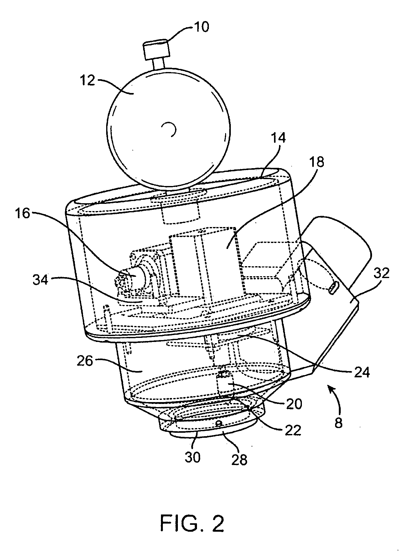
[0032]The present invention provides a device for measuring the electric fields in epithelial tissue that is hand-held, noninvasive and suitable for use on human subjects, particularly for use in outpatient or other clinical settings. FIG. 2 shows a partially transparent perspective view of the hand-held device 8 according to an embodiment of the invention. As can be seen in the figure, there is a push-button actuator 10 at the top of the device attached to, and extending through, positioning handle 12. Positioning handle 12 is designed to be gripped by one hand of an operator in order to position and orient the device, thus it is preferably a spherical shape, but may be any other suitable shape for gripping. The arrangement of the positioning handle 12 with push-button actuator 10 allows an operator to activate and position the device with a single hand by wrapping a finger or fingers around the positioning handle 12 and engaging the push-button actuator 10 with a single finger. At...


PUM


Abstract

The present invention provides a hand-held, noninvasive diagnostic device for measuring the electric fields in mammalian skin and other epithelial structures. The device includes an outer housing that contacts the skin, providing stability and allowing the device to move along with minor movement of the skin. Recessed within the outer housing is a probe that acts as a sensor to measure the electric field in the skin through an aperture in the bottom surface of the outer housing. By applying a series of known voltages while the probe is vibrating, the skin's local surface potential can be measured and the lateral electric field can be calculated from the spatial distribution of surface potential measurements. Active feedback is used to maintain a constant distance between the probe and the skin surface.

Description

FIELD OF THE INVENTION[0001]This application is directed to a device for acquiring information from skin and other epithelia, and particularly to a hand-held device for measuring the electric field of skin or epithelial structures in a clinical setting.BACKGROUND OF THE INVENTION[0002]Electric fields exist in many forms within the bodies of mammals, such as the electrocardiogram of the heart and the electroencephalogram of the brain. In addition to these more well-known fields, electric fields are also present in the epithelium, which is a tissue composed of layers of cells that line the cavities and surfaces of organs throughout the body, as well as forming part of the skin itself. The epidermis of the skin is composed of stratified squamous epithelial cells and normally generates a voltage across itself called the transepithelial potential. This voltage difference is generated by pumping positive ions from the apical side to the basal side of the epidermis.[0003]FIG. 1A shows a ty...

Claims


Application Information

Patent Timeline
IPC IPC(8): A61B5/05
CPCA61B5/442A61B5/444A61B5/6844A61B2562/0214A61B5/04A61B2560/04A61B5/0051A61B5/24
Inventor NUCCITELLI, RICHARDNUCCITELLI, PAMELALIAU, SHR-KAIPLIQUETT, UWE F.LI, CHANGYINARSING, SUMANATHOS, BRIANSHEIKH, SALEH
Owner NEWBEM
Why Eureka
Try Eureka