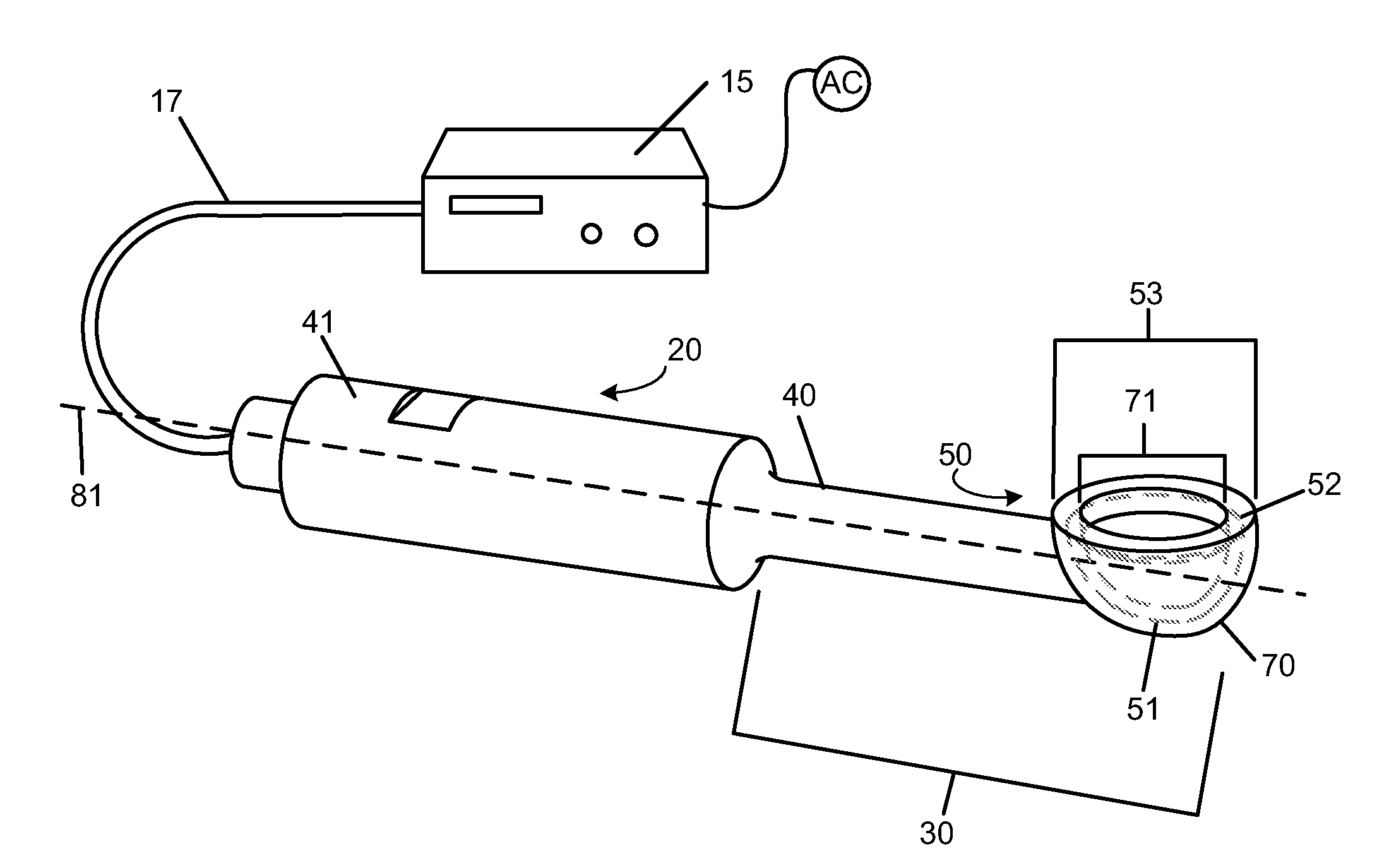
[0008]With the present invention, the apparatus comprises an ultrasound generator driving an ultrasound
transducer. An ultrasound horn is mechanically coupled to the ultrasound
transducer. The ultrasound horn consists of a shaft and an ultrasound tip. The ultrasound horn receives the ultrasound
waves from the ultrasound generator and transmits the ultrasound
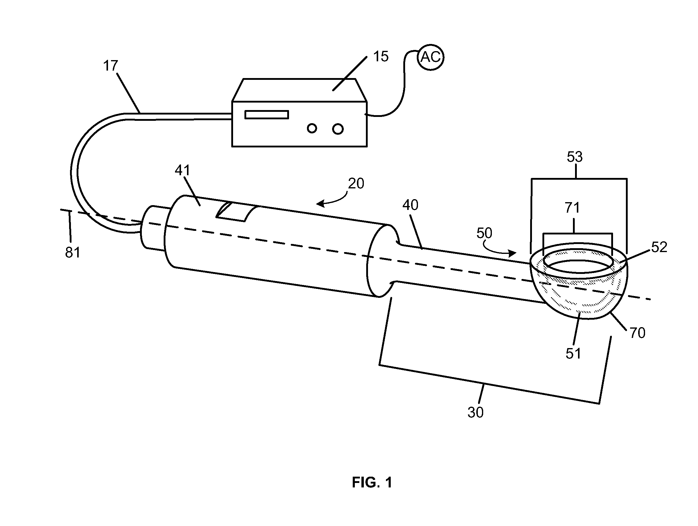
waves to the distal end of the ultrasound tip. The shaft and the ultrasound tip may be integral parts or may be mechanically coupled. The ultrasound tip comprises at least one
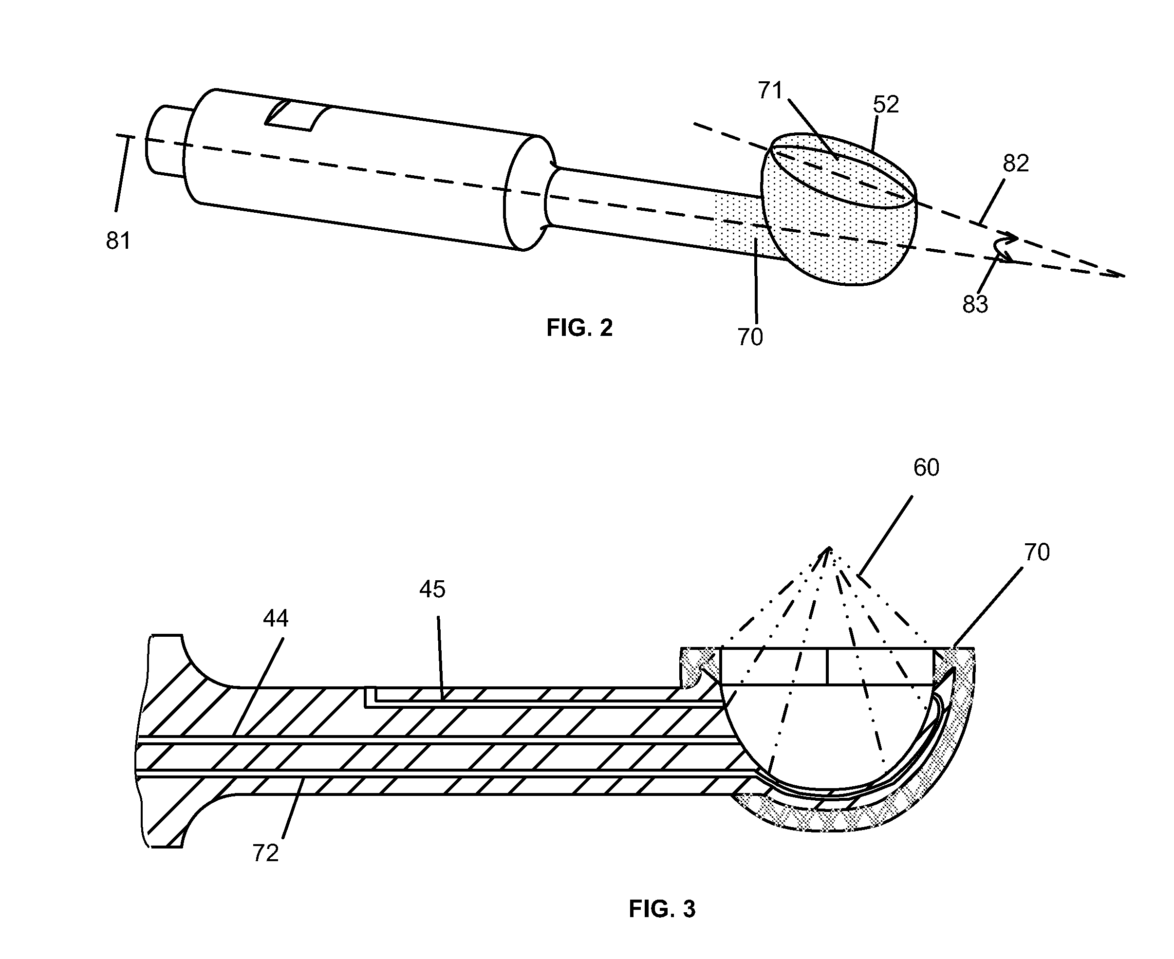
radial surface, a cavity or some other form of a hollowed out area within at least one of the radial surfaces, and a radial edge circumventing the opening of the cavity. In a preferred embodiment, a tapered edge or point is provided to concentrate vibrations passing through the tip. A
coupling fluid is used to enhance transmission of ultrasound
waves from the cavity.
[0010]The ultrasound tip adjoins a non-metallic sheath preventing concentrating elements on the tip from contacting the patient's skin. The sheath is preferably made of a flexible removable material such as rubber, plastic,
fluoropolymer or other
polymer. The material is chosen so that it is sufficiently elastic so that; 1) it may be installed over the wide portions of the ultrasound tip, 2) once installed it will attach to the ultrasound tip so it will not be dislodged during use, and 3) it may be easily replaced after each use. The sheath may be constructed of a segmented design to facilitate installation and removal of the shield. An example of this would be having the segments substantially independent with one or more points of attachment for the segments, such as the petals of a flower. A
lubricant or gel such as
silicone based materials may be used to displace air between the shield and the ultrasound tip to modify the ultrasound transmission characteristics to the patient's skin.
[0011]Alternatively, the sheath may be a reusable permanent
polymer coating such as fluropolymer,
epoxy or a
plastic polymer integrally deposited over the ultrasound tip that is cleaned after each use. In either embodiment, the sheath prevents the
metal surfaces and edges of the tip from contacting the patent's skin and potentially breaking the skin layer. This allows the device and method to be used by physical therapists and other medical personnel that would not be certified to use a device capable of breaking the skin layer during use.
[0012]
Ultrasound may be applied to the skin and underlying tissues by at least two mechanisms. The sheath may be contacted to the skin allowing
ultrasound energy to be transferred directly from the device, such as from a
radial surface or radial edge. In addition, the
ultrasound energy may be focused by the cavity of the radial surface to the skin. In this case, the
coupling fluid serves as a transfer medium to allow transfer of the ultrasound waves to the
skin surface to greatly enhance the ultrasound
transfer efficiency across the
air interface. Typically, the focal point of the cavity is at or below the skin layer so that the
therapeutic effect of the ultrasound is directed to treating the
vascular tissue causing spider veins which are generally located just below the
skin surface.
[0013]
Ultrasound energy may be optimized to achieve the desired effects by effectively utilizing its various properties including;
thermal treatment,
cavitation, microstreaming and
harmonic resonance. At higher intensities, the use of the
thermal energy produced from the ultrasound waves and the focused
cavitation and microstreaming effects are particularly effective at disrupting or destroying unwanted tissue such as the
vascular tissue of spider veins. Focusing the ultrasound allows precise placement and control of the levels of energy released from
cavitation and microstreaming. Specifically, the concentrated ultrasound waves from the concentrating elements on the edge circumventing the cavity may collapse the
vein which is then ablated / disrupted by the
focused ultrasound energy emanating from the walls of the cavity. Before, after and / or during the treatment of spider veins the operating parameters may be altered to achieve additional therapeutic effects. For example, the intensity of ultrasound may be increased and / or the focus shifted away from the blood vessels being treated as to induce cavitations and / or microstreaming capable of inducing the
ablation of tissue at a
secondary site. In combination or in the alternative, the intensity of the
ultrasound energy could be lowered as to aid in the facilitation of
angiogenesis. In combination with or in the alternative to adjusting the intensity of the ultrasound delivered, the frequency and / or amplitude of the ultrasound may be adjusted as to correspond with the
harmonic resonance of different tissues as to optimize the interaction with the tissue.

