Valvuloplasty Catheter And Methods

a valvuloplasty and catheter technology, applied in the field of percutaneous transcatheter and transapical cardiac valve implantation, can solve the problems of poor valve function, undersized estimation of the diameter of the annulus,

Active Publication Date: 2010-04-15
INTERVALVE MEDICAL INC
View PDF99 Cites 230 Cited by
  • Summary
  • Abstract
  • Description
  • Claims
  • Application Information

AI Technical Summary

Benefits of technology

[0014]One embodiment according to the present invention is directed to a dog-bone-shaped balloon catheter for performing valvuloplasty on a stenotic aortic or pulmonary valve or for opening up any stenotic constriction within a tubular member of the body. The tubular member could be, for example, any blood vessel of the body including a coronary artery, peripheral artery, veins of the body, esophagus, trachea, intestinal vessels, bile ducts, ureter, and the like. This embodiment has additional utility for use in predilatation of the aortic valve leaflets prior to placing a percutaneous aortic valve or other prosthetic device used for aortic valve repair, replacement, or implant. This embodiment may also be formed with a larger or smaller diameter balloon and used in arteries, veins, body orifices, or other hollow organs of the human body where dilatation along with a diameter measurement are needed. It provides advantages over the standard cylindrically-shaped valvuloplasty balloon due to the dog-bone shape for the balloon as well as the construction of the balloon.
[0015]Generally, the dog-bone shape allows the bulbous portions of the balloon to self-center on each side of the aortic annulus and position the narrower diameter waist adjacent to the annulus. The larger bulbous proximal end region of the balloon is positioned into contact with the aortic valve leaflets such that inflation of the balloon pushes the leaflets outward against the aortic sinus. The bulbous proximal portion of the balloon allows the aortic valve leaflets to be cracked or broken at or near their base and hyperextended outwards toward the sinus in a manner that provides greater benefit than that provided by a standard cylindrical balloon without the concern for dissecting the annulus. The narrow waist of the dog-bone balloon is formed such that the smaller diameter waist will not dissect the narrower annulus region. The distal bulbous region, which is located in the left ventricular outflow tract (LVOT), helps to prevent the balloon from migrating downstream during inflation due to blood pressure generated from the beating heart.
[0016]The dog-bone-shaped balloon of the present invention is preferably formed with a semi-compliant material in the smaller diameter waist region and with a non-compliant material for the proximal and distal bulbous end regions. The waist region functions to more accurately measure the diameter of the annulus than what can be attained using standard echocardiographic measurements. The waist also serves to measure the compliance characteristics of the annulus and thereby helps the physician to perform the valve dilatation procedure with a greater degree of safety to the patient against possible annular dissection. Inaccuracies with the standard echo measurements exist due in part to the anatomically oval shape of the annulus which results in typically undersized estimates for the diameter of the annulus. Such undersizing often can lead to incorrect sizing of the percutaneous valve and resultant poor valve function. The semi-compliant waist of the present invention is able to firmly contact the oval waist, readjust its shape, and provide a more accurate measurement of its true diameter while ensuring that the annulus is not exposed to dilating forces that could cause annular dissection.

Problems solved by technology

Inaccuracies with the standard echo measurements exist due in part to the anatomically oval shape of the annulus which results in typically undersized estimates for the diameter of the annulus.
Such undersizing often can lead to incorrect sizing of the percutaneous valve and resultant poor valve function.

Method used


Image

  • Valvuloplasty Catheter And Methods
  • Valvuloplasty Catheter And Methods
  • Valvuloplasty Catheter And Methods

Examples


Embodiment Construction

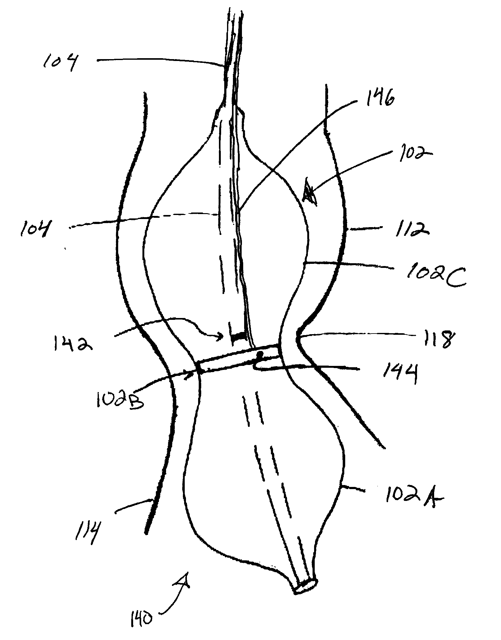
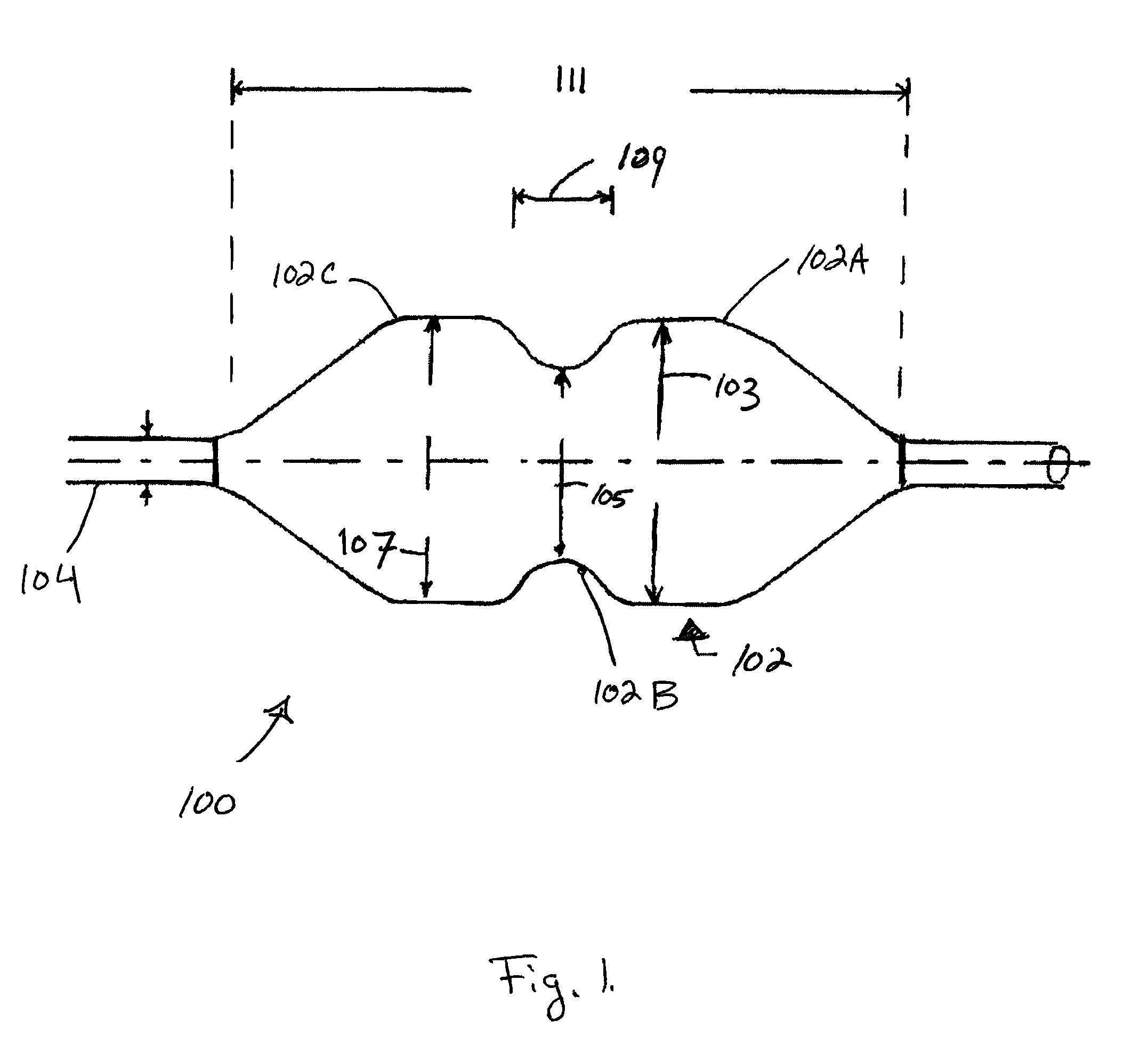
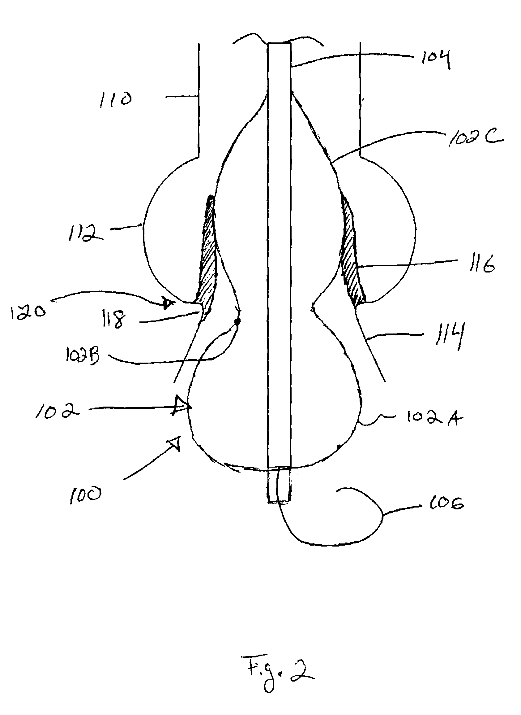
[0056]Specific embodiments of the invention will now be described with reference to the accompanying drawings. This invention may, however, be embodied in many different forms and should not be construed as limited to the embodiments set forth herein; rather, these embodiments are provided so that this disclosure will be thorough and complete, and will fully convey the scope of the invention to those skilled in the art. The terminology used in the detailed description of the embodiments illustrated in the accompanying drawings is not intended to be limiting of the invention. In the drawings, like numbers refer to like elements.

[0057]Unless otherwise defined, all terms (including technical and scientific terms) used herein have the same meaning as commonly understood by one of ordinary skill in the art to which this invention belongs. It will be further understood that terms, such as those defined in commonly used dictionaries, should be interpreted as having a meaning that is consis...


PUM


Abstract

A valvuloplasty catheter has a dog-bone shaped balloon with semi-compliant smaller diameter waist and non-compliant larger diameter bulbous end regions. The balloon centers across the valve with the waist adjacent to the annulus. One bulbous region serves to hyperextend the valve leaflets and the other assists in stabilizing the balloon position to reduce migration. The semi-compliant waist increases in diameter as fluid enters the balloon until it comes into contact with the valve annulus. The pressure within the balloon per unit of volume delivery has a greater slope after contact with the annulus than before resulting in a change in slope for the pressure versus volume curve. The diameter of the balloon and annulus are determined at this inflection point when the balloon contacts the annulus.

Description

RELATED APPLICATIONS[0001]This application claims priority to U.S. Provisional Application Ser. No. 61 / 104,636 filed Oct. 10, 2008 entitled Valvuloplasty Catheter And Methods, U.S. Provisional Application Ser. No. 61 / 112,566 filed Nov. 7, 2008 entitled Valvuloplasty Catheter And Methods, and U.S. Provisional Application Ser. No. 61 / 145,705 filed Jan. 19, 2009 entitled Valvuloplasty Catheter And Methods, all of which are hereby incorporated herein by reference.FIELD OF THE INVENTION[0002]This invention relates to percutaneous transcatheter and transapical cardiac valve implantation. More specifically, this invention relates to a device to better dilate the aortic valve leaflets than prior art and assess aortic valve annulus.BACKGROUND OF THE INVENTION[0003]Calcific aortic stenosis is a common cause of acquired valvular heart disease with substantial morbidity and mortality. Its incidence increases exponentially in older patient populations. Fibrosis, degeneration and subsequent calci...

Claims


Application Information

Patent Timeline
Patent Type & Authority Applications(United States)
IPC IPC(8): A61M31/00A61B5/00
CPCA61M25/1002A61M25/1011A61M25/1018A61M25/1029A61M29/02A61M2025/0002A61B2017/22098A61M2025/1047A61M2025/1075A61M2025/1086A61M2025/1088A61B17/22012A61M2025/1031A61M2025/1059A61M25/10186A61M25/10188
Inventor DRASLER, WILLIAMPEDERSEN, WESUNGS, MARK
Owner INTERVALVE MEDICAL INC
Why Eureka
Try Eureka