Use of a reference source with adaptive optics in biological microscopy

a reference source and optics technology, applied in the field of microscopic imaging, can solve problems such as general accuracy, and achieve the effect of improving the strehl ratio of microscopy

Active Publication Date: 2010-04-29
RGT UNIV OF CALIFORNIA
View PDF14 Cites 19 Cited by
  • Summary
  • Abstract
  • Description
  • Claims
  • Application Information

AI Technical Summary

Benefits of technology

[0005]Optical aberrations are introduced during optical imaging through biological tissue. The invention encompasses methods of microscopy using adaptive optics technology to improve the Strehl ratio of microscopical imaging when imaging biological tissue.

Problems solved by technology

For example the Hartman method could be used, although this is generally less accurate.

Method used


Image

  • Use of a reference source with adaptive optics in biological microscopy
  • Use of a reference source with adaptive optics in biological microscopy
  • Use of a reference source with adaptive optics in biological microscopy

Examples


Embodiment Construction

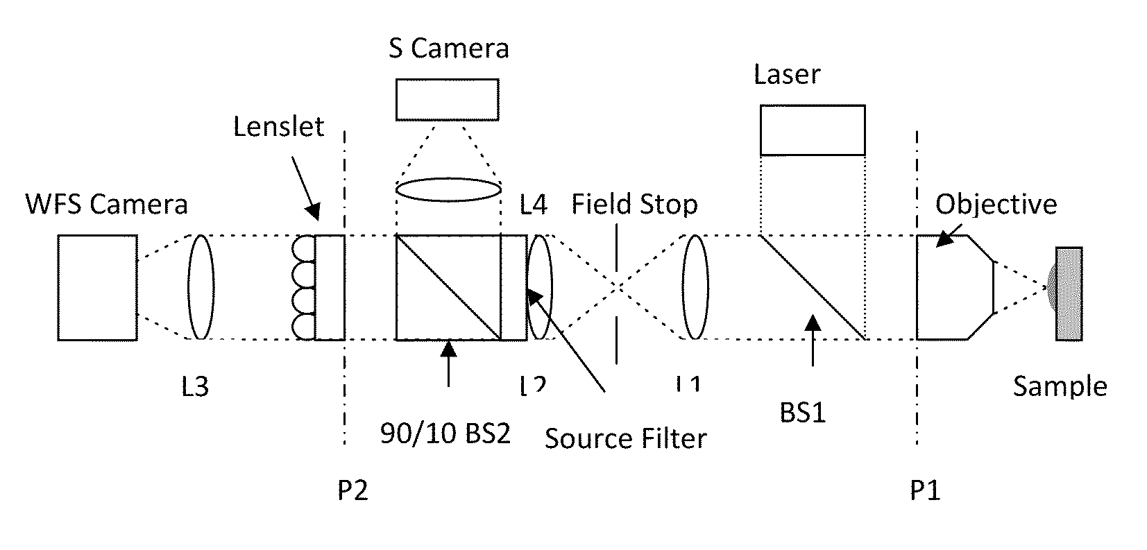
[0021]Described are methods of microscopic imaging of biological tissue using adaptive optics technology to improve the image focus and sharpness. Wavefront measurements are taken by using a novel method of seeding biological tissue by using a fluorescent microsphere as a “guide star” as a natural point-source reference. The current methods are capable of improving the Strehl ratio of modern biological microscopes as much as 15 times.

[0022]The invention encompasses methods for measuring aberrations introduced during optical imaging through biological tissue and using adaptive optics technology to improve the Strehl ratio of microscopes when imaging biological tissue. The methods comprise seeding a medium with a reference source, applying a biological tissue to the seeded medium, subjecting the biological tissue on the seeded medium to adaptive optics microscopy, and determining the aberrations in light wavelength caused by imaging through the biological tissue. In one aspect of the ...


PUM

PropertyMeasurementUnit
wavelengthsaaaaaaaaaa
diameteraaaaaaaaaa
half-widthaaaaaaaaaa

Abstract

Methods of microscopic imaging of biological tissue using adaptive optics technology to improve the image focus and sharpness. Wavefront measurements are taken by using a novel method of seeding biological tissue by using a fluorescent microsphere as a “guide star” as a natural point-source reference. The current methods are capable of improving the Strehl ratio of modern biological microscopes as much as 15 times.

Description

RELATIONSHIP TO OTHER APPLICATIONS AND CLAIM OF PRIORITY[0001]This application claims priority to and the benefit of U.S. provisional patent application No. 61 / 197,291 filed 24 Oct. 2009 entitled “Use of a reference source with adaptive optics in biological microscopy”.GOVERNMENT SUPPORT CLAUSE[0002]This invention was made with government support under National Science Foundation Agreement Nos. 8976783 and PHY 0120999. The government has certain rights in the invention.FIELD OF THE INVENTION[0003]The invention relates to methods of microscopic imaging using adaptive optics technology to improve the Strehl ratio (and therefore image focus and sharpness) of microscopic imaging of biological tissue. Wavefront measurements are made by using a novel method of seeding biological tissue by using a fluorescent microsphere as a “guide star” as a natural point-source reference.BACKGROUND[0004]Changes in the index of refraction due to tissue composition limit the resolving power of biological ...

Claims


Application Information

Patent Timeline
Patent Type & Authority Applications(United States)
IPC IPC(8): C12Q1/00
CPCG01N21/41G01N21/6456G02B26/06G02B21/16G01N2496/00
Inventor AZUCENA, OSCARKUBBY, JOELCAO, JIANSULLIVAN, WILLIAM
Owner RGT UNIV OF CALIFORNIA
Why Eureka
Try Eureka