Method and apparatus for early diagnosis of Alzheimer's using non-invasive eye tomography by Teraherts

a technology of terahertz imaging and tomography, applied in the field of non-invasive detection of can solve the problems of only promising cures and/or successful treatment of alzheimer's, unable to detect early development of alzheimer's, and unable to achieve the effects of ad diagnosis, no ambiguity, and high confiden

Inactive Publication Date: 2010-05-06
MOTAMEDI MANOUCHEHR +1
View PDF8 Cites 7 Cited by
  • Summary
  • Abstract
  • Description
  • Claims
  • Application Information

AI Technical Summary

Benefits of technology

[0019]The disclosed method and apparatus use terahertz waves instead of laser for scanning the eye of the AD patient to precisely image the amyloid layer non-invasively in real time to determine the deposited plaque of amyloid with very high resolution by dividing the amyloid layer thickness to a large number of very thin microlayers. Each microlayer independently is scanned by terahertz waves and all microlayers' images are integrated to generate a well defined signature of the AD patient eyes. The accuracy and high resolution of this method can easily distinguish the abnormality of an AD patient with the signature of a normal person and the signature of a person with common age-related cataracts.
[0020]Sensing with terahertz segment of spectrum has several significant advantages over sensing at other sides of spectrum. Terahertz radiation is completely unionized with photon energies more than six orders of magnitude less than soft x-rays. Most terahertz applications require less than one microwatt of power makes terahertz radiation completely safe for use by humans or on human subjects.
[0024]Besides detecting the presence of AD, the present method and apparatus is also valuable to determine how advance is the concentration of Amyloid plaques as well as information regarding whether the progression has stopped or has slowed down. A map showing, the difference between AD plaques and age-related cataracts, can aid in the accurate diagnosis of AD with no ambiguity. This is in contrast to the poor resolution of the prior art using laser techniques. When using terahertz systems such as disclosed herein, the combination of the spatial resolution and the high spectral resolution of a terahertz imaging system can be utilized to detect Alzheimer's progression with high level of confidence.
[0025]The present method and apparatus gathers data in three spatial dimensions. Initially, a spot is selected on the outer ring of the lens of the eye which is the location of the AD plaques deposition. The spot travels in depth where a line image is constructed resulting optical arrangement and synchronizing, using methods of optical coherence topography (OCT) and other similar methods. The depth of the RNFL is binned according to the desired spatial resolution, signal-to-noise ratio, and the characteristic of the rapid optical delay line. Then, the incident beam will be scanned in two directions collecting three dimensional data of a rectangular area alongside of the deposited ring. To improve the reliability of this method several rectangular areas of the outer ring of the patient's eye are selected for similar experiment. A tomography will then be constructed from the gathered overall data.

Problems solved by technology

Alzheimer's generally appears in older persons, continuing steadily to disorder brain and gradually destroys a person's memory and ability to make judgments, communicate and carry out daily activities.
There is not a cure and / or successful treatment for Alzheimer's.
This techniques and other similar methods are only promising if Alzheimer's has been detected at its early development.
Presently, there are no simple methods like blood or urine test that can detect Alzheimer's.
1. Testing the patient's memory by asking questions and studying the patient's family history to determine if Alzheimer's is present. This is not very reliable technique and it mostly is done by a family doctor as initial warning for Alzheimer's.
2. Brain scanning by such techniques as CT (computed tomography) and MRI (magnetic resonance imaging) could be used for AD diagnosis. The problem of these techniques is that they are only effective at the late stages of Alzheimer's which the damage is already done.
3. Researchers at New York University Medical Center have reported EEG (electroencephalogram) testing that measures electrical activity in the brain to pinpoint the early signs of Alzheimer's as warning signal. In this method, the measured brain activity of patient is compared to a standard activity of a patient with no Alzheimer's disease. This method still is at initial study stage and according to the scientists at Alzheimer's Association, there is concern that EGG study is too limited and the findings are too preliminary.
4. Researchers in the Netherlands reported that they can predict the early detection of Alzheimer's by measuring the size of two parts of the brain, the hippocampus, and amygdala. For those patients who have smaller sizes of these two parts against some standard have several time more likely to suffer from the Alzheimer's decease. The technique is still at study stage and mostly is looking for any type of dementia; they are not focusing on Alzheimer's. Another drawback of this technique is direct scanning of the brain which is likely harmful to patient.
5. There is a new technique recently reported by several institutes including Harvard Medical School and Brigham and Women's Hospital in Boston. The technique is detecting early phases of Alzheimer's disease by measuring the level of Beta-amyloid protein plaques deposited in the lens of the patient's eye using a non-invasive laser scanning and imaging. This new discovery could revolutionize early detections of Alzheimer's disease including most of the dementia disorders. All the previous measuring techniques have to use part of the brain for detection which is not advisable. The fact that the plaque (abnormal beta-amyloid protein) which deposits in the brain of Alzheimer's patient also will deposit simultaneously in the lens of the patient's eye similar to formation of cataracts, opens a new avenue for early diagnosis and continuous monitoring of Alzheimer's.
In this patent the laser power is not known and its safety is questionable.
Even the inventors are not sure that the method is practical and there is no proof that the RNFL is affected by AD.
The first one is high intensity laser scanning which is harmful to the patient's eyes and sometimes extremely dangerous if it has to penetrate inside the layers of the deposited plague.
The second problem of the existing disclosed methods is the requirement of large, heavy, and expensive apparatus which makes testing inconvenient to be done in Doctors' office and the patient has to go to hospital.
The third drawback is the lack of a very sensitive and highly efficient testing process which is required for early detection of the AD symptoms.

Method used


Image

  • Method and apparatus for early diagnosis of Alzheimer's using non-invasive eye tomography by Teraherts
  • Method and apparatus for early diagnosis of Alzheimer's using non-invasive eye tomography by Teraherts
  • Method and apparatus for early diagnosis of Alzheimer's using non-invasive eye tomography by Teraherts

Examples


Embodiment Construction

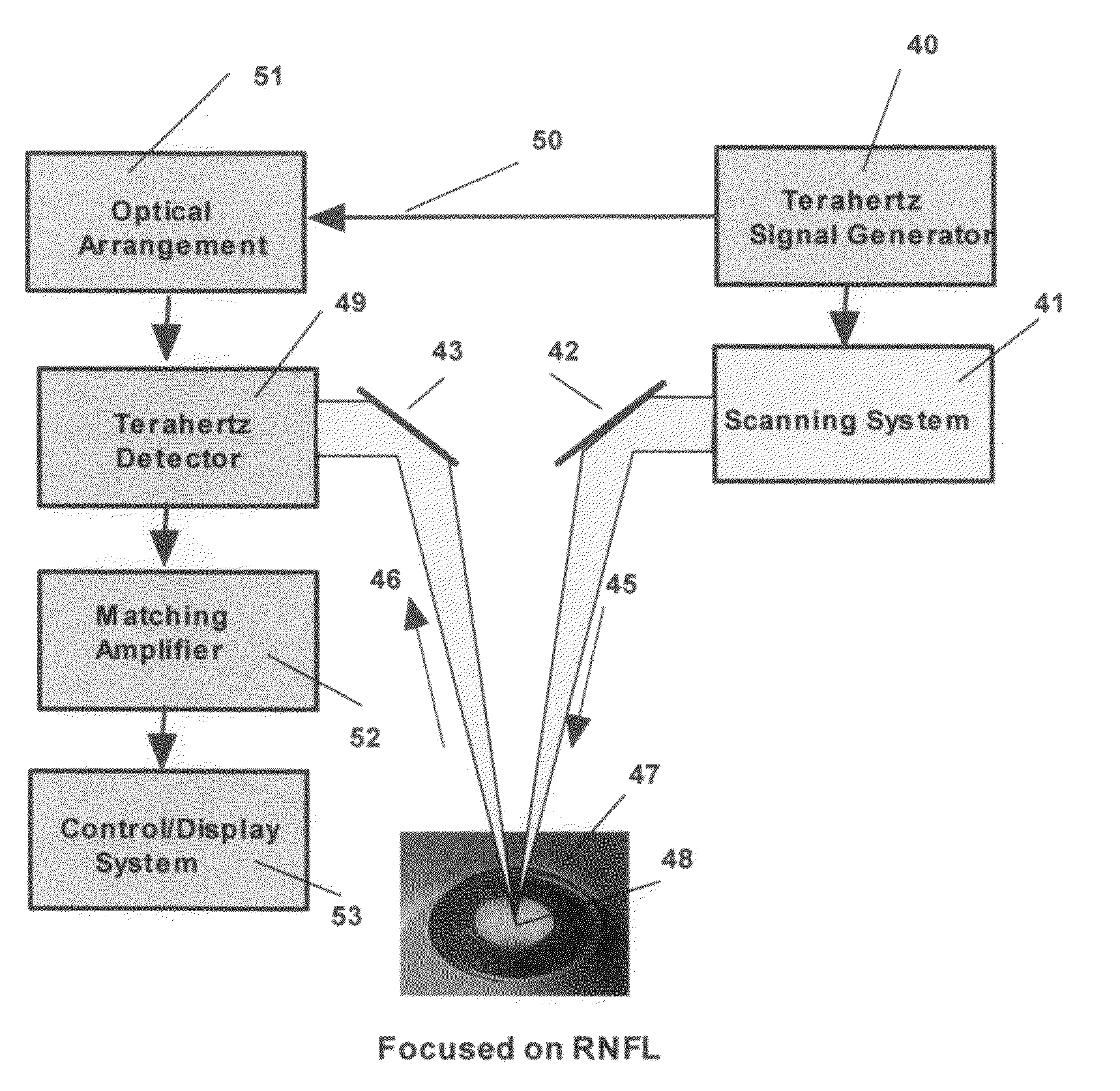
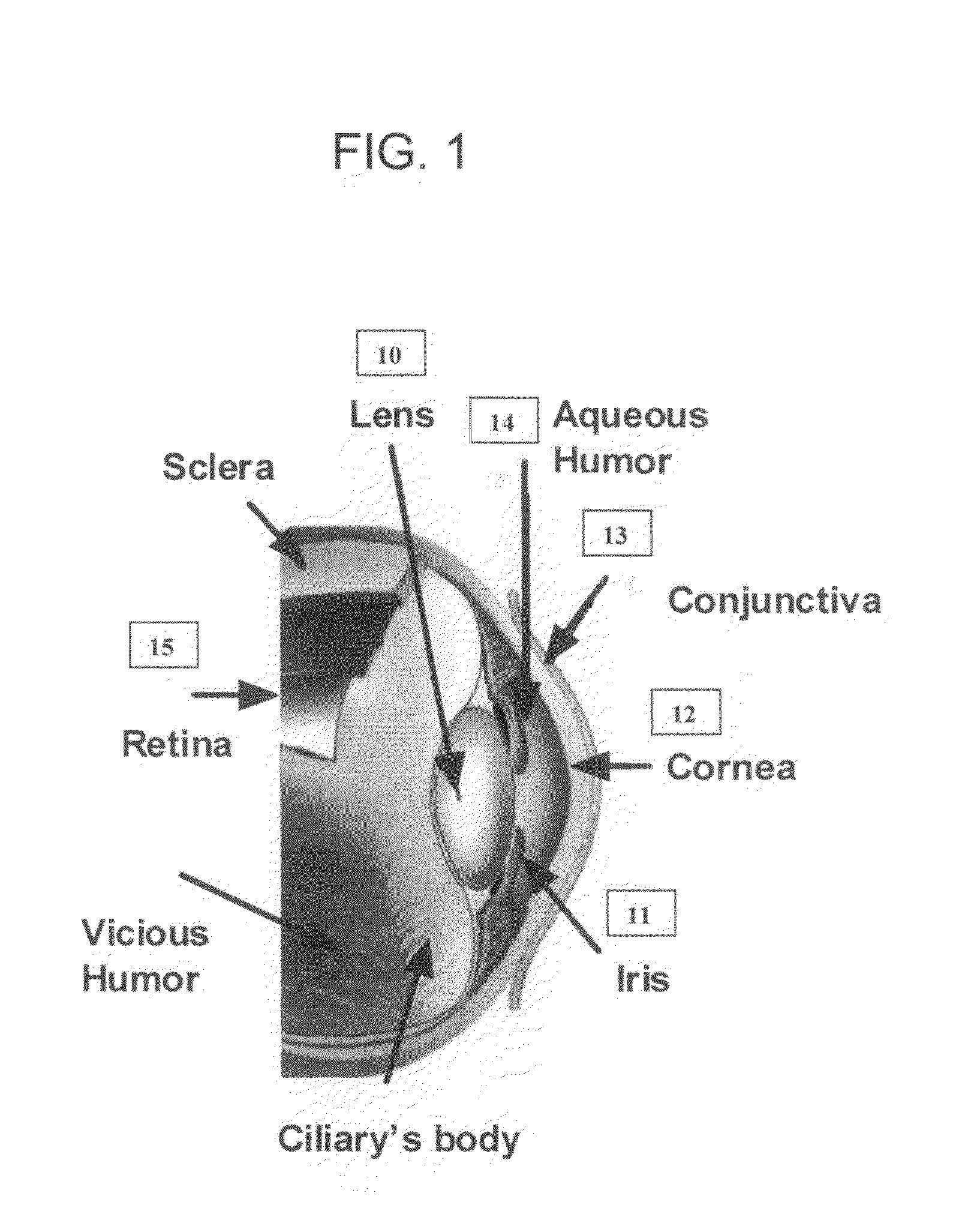
[0031]FIG. 1 is a diagram of the human eye's structure which shows the lens 10 dimension relative to other elements of the eye. The lens focuses light rays onto the retina. An iris 11, the color part of the eye changes the amount of the light requires for optical processing. The eye acts as a perfect high resolution camera. It uses its variable index of reflection to dynamically focus a subject. Cornea 12 is bounded between conjunctiva 13 and aqueous Humor 14. Retina 15 works as an image processor to convert incoming rays into nerves signals where the brain analyzes the signals and creates the image.

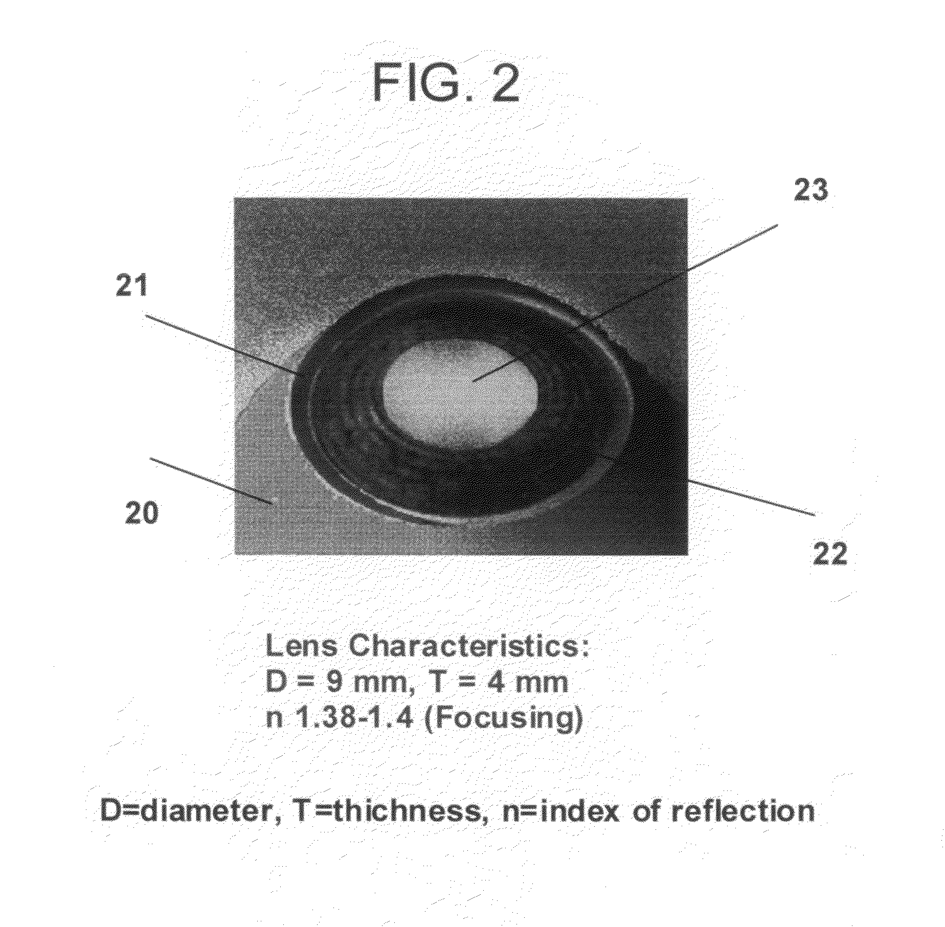
[0032]FIG. 2 is a close up picture of the eye 20 where the lens 21 is partially exposed by iris 22 acts as a diaphragm and partially exposed the lens in the opening pupil 23. The lens normal characteristics have shown in FIG. 2. FIG. 3 shows a comparison between Alzheimer's plaques deposited in the lens of the patient's eye and the cataracts plaques deposited similarly in the lens of the...


PUM


Abstract

The primary objective of the present method and apparatus is to provide a transportable diagnosis system for a reliable method to examine the conditions of a human eye lens. The method provides precise and safe technique for the early diagnosis of Alzheimer's Disease (AD) which is not involved brain surgery or biopsy. The detection of deposited amyloid plaque in the eyes of the patient has been discovered to be a possible diagnosis solution. The disclosed method uses Terahertz waves in both time and frequency domains instead of laser for scanning the eye of the AD patient to precisely image the amyloid layer non-evasively in real time and to map the deposited plaque with high resolution which can easily distinguish the abnormality of an AD patient with the comparison of the patient eye lens test to the available signature of a normal person and the signature of a person with common age-related cataracts.

Description

[0001]Continuous-in-part of application Ser. No. 11 / 880,911FIELD OF THE DISCLOSED METHOD AND APPARATUS[0002]This method and apparatus is related to non-invasively detecting early development of Alzheimer by scanning patient eye and, in particular, relates to a method and apparatus for diagnosis and tomography using Terahertz imaging.BACKGROUND OF PRIOR ARTS[0003]Alzheimer's disease (AD) in a medical term is classified as a form of dementia, a group of conditions that gradually destroy brain cells. Alzheimer's generally appears in older persons, continuing steadily to disorder brain and gradually destroys a person's memory and ability to make judgments, communicate and carry out daily activities. Scientists now believe that the cause of AD is most likely due to the concentration of Beta-amyloid deposited in some part of the brain. As Alzheimer's progresses, patients may also experience changes in behavior during their personal lives, such as anger, agitation and sometimes even delusi...

Claims


Application Information

Patent Timeline
Patent Type & Authority Applications(United States)
IPC IPC(8): A61B3/10
CPCA61B3/102
Inventor MOTAMEDI, MANOUCHEHRDABIRI, ALI
Owner MOTAMEDI MANOUCHEHR
Why Eureka
Try Eureka