
System for differentiating the lengths of nucleic acids of interest in a sample

Active Publication Date: 2010-05-27
CAPLIPER LIFE SCI INC
View PDF34 Cites 21 Cited by
  • Summary
  • Abstract
  • Description
  • Claims
  • Application Information

AI Technical Summary

Benefits of technology

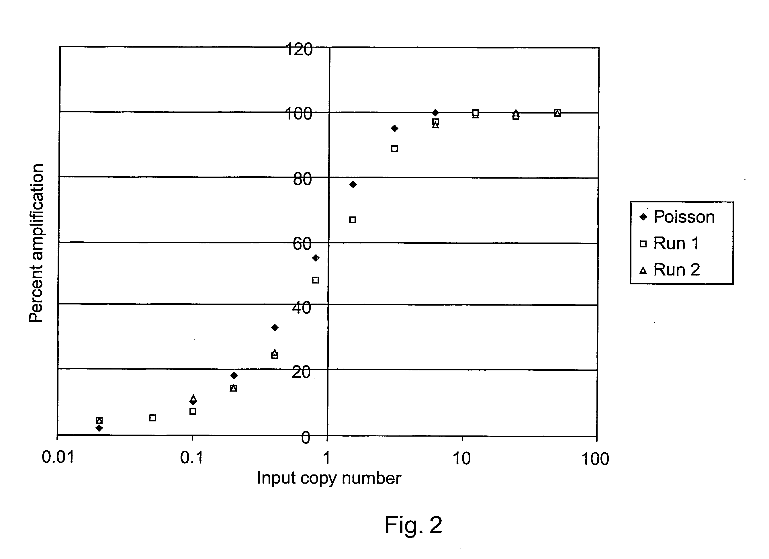
[0024]The concentration of the nucleic acids of interest and / or any additional nucleic acid is optionally low in the methods of the invention, e.g., about 1 molecule per aliquot. For example, the sample can be diluted to a concentration of about 1 molecule of interest per nanoliter or less. Optionally, diluted aliquots are each diluted to the same degree; however, diluted aliquots can also be differentially diluted (e.g., to form a dilution series). The volume of the aliquots can be quite low to keep reagent costs low, e.g., in microfluidic applications. For example, the aliquots can be less than about 100 nl in volume, e.g., less than about 10 nl in volume, or, e.g., about 1 nl in volume or less.
[0031]Optionally, the components of the system can be treated with one or more reagents between operational runs to reduce cross contamination between operations. For example, the amplification channel can have acid or base flowed into the channel between amplification reactions to reduce unwanted contamination from one or more previous amplification products.
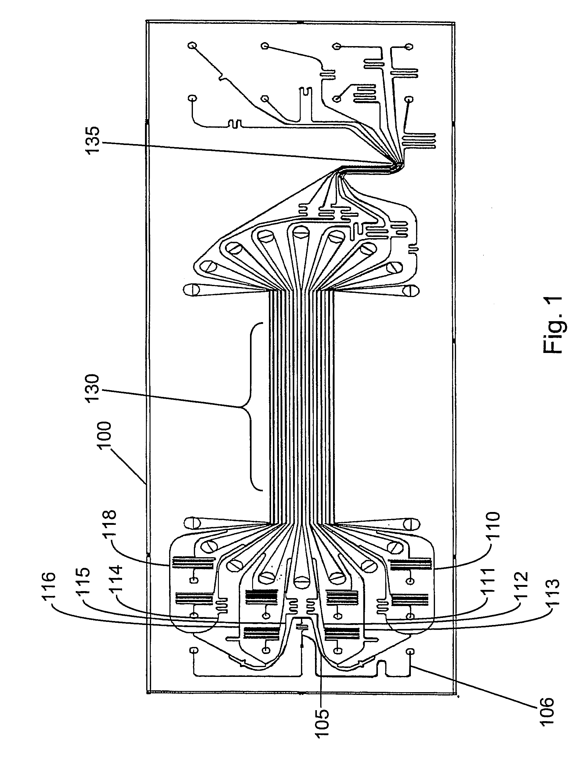
[0044]In a preferred embodiment of differentiating lengths of nucleic acids of interest in a sample, an amplification reaction can be used to enhance the sensitivity of the assay. The nucleic acid of interest is contacted with a first primer pair and a second primer pair having at least one primer that is outside of the sequence defined by the first primer pair, the nucleic acid of interest is amplified in a reaction mixture in a microchannel or microchamber with polymerase extensions from the primers to produce first amplicons defined by the first primer pair or second amplicons defined by the second primer pair. First and second probes complementary to the first and second amplicon and having detectable markers are introduced into the reaction mixture to hybridize with available complementary sequences, and one or more signals are detected from the probes. Detection of a signal from only one of the probes indicates a fragmented nucleic acid of interest and detecting signals from both probes indicates a nucleic acid that is not fragmented. In preferred embodiments of this sensitive assay, the reaction mixture detected contains only a single copy of the nucleic acid of interest. Reaction mixtures detected in the methods can be homogenous mixtures, e.g., not requiring separation of labeled constituents before detection of signals.
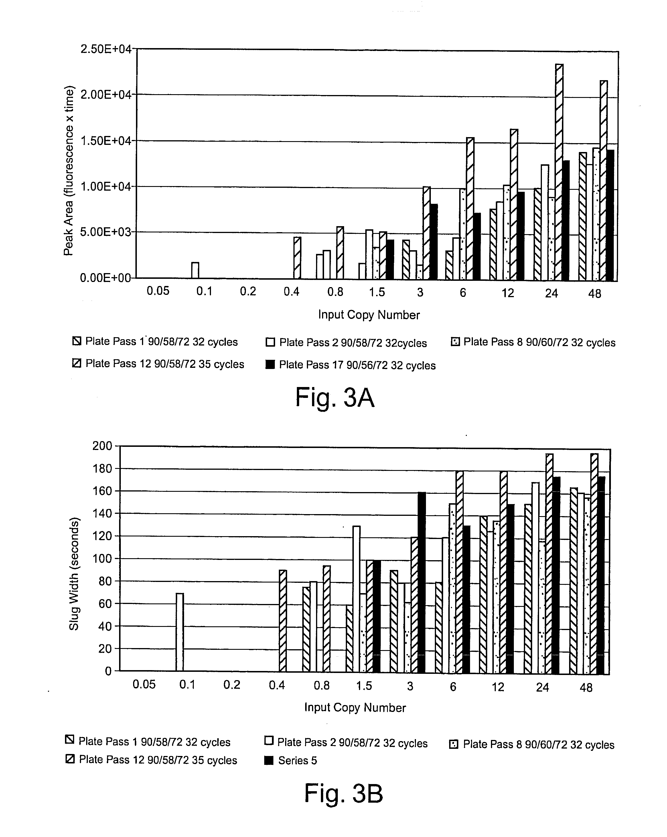
[0051]Samples containing unknown amounts of a nucleic acid of interest can be quantified by comparing to their signal peaks to sets of standard signal peaks. For example, a nucleic acid of interest in a sample can be quantified by amplifying a dilution series of standard materials containing known amounts of the nucleic acid of interest through a certain number of amplification cycles, detecting signals associated with standard amplicons produced from the standard materials, amplifying the sample nucleic acid of interest the number of amplification cycles, detecting a signal associated with sample amplicons produced from the sample nucleic acid of interest, and comparing one or more standard amplicon signals to the sample amplicon signal to determine a concentration value for the nucleic acid of interest in the sample. Sample and standard signal parameters for comparison can include, e.g., the shape of their signal peaks, points of inflection on the signal peaks, slopes of the signal peaks, signal peak amplitudes, signal peak areas, signal peak widths at half height, and / or the like. The reliability of results can be enhanced through various schemes of repeated testing. For example, the amplifying, detecting, and comparing steps can be repeated one or more times, with different numbers of amplification cycles, to determine additional concentration values for the sample nucleic acid of interest for statistical evaluation providing more precise or more accurate concentration value results for the nucleic acid of interest in the sample.
[0052]Improved assay results can be obtained by gathering signal data after amplifications through two or more different numbers of cycles. A major benefit of running the quantitative assay at different amplifications is to broaden the usable range of the assay. Typically, statistical evaluation of the additional data provided by analysis at multiple amplification levels can enhance other assay parameters, such as precision, accuracy, and sensitivity. Quantifying a nucleic acid of interest in a sample based on detection of multiple amplifications can include: amplifying the nucleic acid of interest through more than one number of amplification cycles, detecting signals associated with amplicons produced from two or more of the amplification cycle numbers, preparing a sample curve of a signal parameter versus number of amplification cycles, and comparing one or more identifiable points from the sample curve to a standard curve of the identifiable points versus concentration to quantify the nucleic acid of interest. Exemplary identifiable points from signal curves include points of inflection, points having a certain slope, points having a certain signal amplitude, points having a certain fraction of a maximum signal amplitude, and / or the like.

Problems solved by technology

Despite the wide-spread use of amplification technologies and the adaptation of these technologies to truly high throughput systems, certain technical difficulties persist in amplifying and detecting nucleic acids, particularly rare copy nucleic acids.
For example, if a set of primers hybridizes to a high copy nucleic acid, as well as to a low copy nucleic acid in a given sample, the geometric amplification of the high copy nucleic acid proportionately dominates the amplification reaction and it is difficult or impossible to identify the low copy nucleic acid in any resulting population of amplified nucleic acids.
Thus, low copy number alleles of a gene can be very difficult to detect, e.g., where a primer set cannot easily be identified that only amplifies the rare nucleic acid (and the practitioner will realize that perfect reagent specificity is rare or non-existent in practice).
It is worth noting that these problems simply have not been addressed by the prior art.
(2001) Anal. Chem. 73:565-570), none of these prior approaches are suitable for detection of rare copy nucleic acids in samples.
That is, none of these approaches are suitable to high throughput automation and the devices in the prior art cannot be adapted to practicably detect rare copy nucleic acids.
This cumbersome process results in few amplification reactions being made and analyzed in any useful time period and required almost continuous user intervention to make the system operate.
In addition, none of the prior methods unambiguously determine the length of a nucleic acid of interest from a homogenous mixture, e.g., without additional steps of size selective chromatography.
Another difficulty with amplification methods that is completely unaddressed in the prior art is that it can be quite difficult to perform quantitative analysis on rare nucleic acids.
The problems noted above for detection apply to quantitative analysis as well, with the additional problem that quantification is impacted by the presence of high copy number nucleic acids in the sample, even if the rare nucleic acid can be amplified.
Thus, it is not generally possible to assess accurately the concentration of rare nucleic acids in a sample, particularly where the components of the system have not previously been characterized or purified (it is, of course, somewhat simpler to assess amplification products quantitatively if the materials selected for amplification are already characterized).
While amplification of materials that have already been fully characterized is of academic interest, this approach is of little practical value if it cannot be adapted to characterization of unknown materials.
However, this method can be nonspecific, slow and labor intensive, fail to confirm separated amplicons were amplified from the same nucleic acid strands (i.e., ambiguous), fail to distinguish between long nucleic acids with marginally different lengths, fail to determine length directly from a homogenous mixture, and fail to detect nucleic acids of interest against a background of other nucleic acids in many complex clinical samples.

Method used


Image

  • System for differentiating the lengths of nucleic acids of interest in a sample
  • System for differentiating the lengths of nucleic acids of interest in a sample
  • System for differentiating the lengths of nucleic acids of interest in a sample

Examples


examples

[0172]The following examples are offered to illustrate, but not to limit the claimed invention. It is understood that the examples and embodiments described herein are for illustrative purposes only and that various modifications or changes in light thereof will be suggested to persons skilled in the art and are to be included within the spirit and purview of this application and scope of the appended claims.

Single Molecule Amplification and Detection of DNA in a Microfluidic Format

Introduction

[0173]The amplification of a desired region of DNA by polymerase chain reaction (PCR) has revolutionized the field of molecular biology. In conventional formats of PCR, which use many microliters of fluids during amplification, the starting DNA copy number is typically at least hundreds to tens of thousands of molecules. Recent advances in microfluidics have demonstrated that it is feasible to miniaturize PCR down by a thousand fold to a nanoliter-reaction volume range. When the sample concent...


PUM

PropertyMeasurementUnit
Fractionaaaaaaaaaa
Fractionaaaaaaaaaa
Fractionaaaaaaaaaa

Abstract

Systems for differentiating the lengths of nucleic acids of interest in a sample are provided. The system includes a microfluidic device, a detector, and a software system. The microfluidic device includes an amplification microchannel or microchamber containing a reaction mixture under conditions that provide one or more amplicons of the nucleic acid of interest. The detector is integral with or proximal to the microfluidic device and is configured to detect the amplicons as one or more signals from a homogenous mixture. The software system interprets one or more coincidentally detected signals to indicate lengths of one or more individual nucleic acid molecules of interest, thereby differentiating the lengths of the nucleic acids of interest.

Description

CROSS REFERENCE TO RELATED APPLICATIONS[0001]This application is a divisional of patent application Ser. No. 10 / 845,996, “SINGLE MOLECULE AMPLIFICATION AND DETECTION OF DNA LENGTH,” filed May 14, 2004, now abandoned, which is a continuation-in-part of patent application Ser. No. 10 / 741,162, “SINGLE MOLECULE AMPLIFICATION AND DETECTION OF DNA IN A MICROFLUIDIC FORMAT,” filed Dec. 19, 2003, now abandoned, which is a regular utility application corresponding to Provisional Patent Application U.S. Ser. No. 60 / 518,431 “SINGLE MOLECULE AMPLIFICATION AND DETECTION OF DNA IN A MICROFLUIDIC FORMAT” filed Nov. 6, 2003, Provisional Patent Application U.S. Ser. No. 60 / 462,384 “SINGLE MOLECULE AMPLIFICATION AND DETECTION OF DNA IN A MICROFLUIDIC FORMAT,” filed Apr. 11, 2003, and Provisional Patent Application U.S. Ser. No. 60 / 436,098 “SINGLE MOLECULE AMPLIFICATION AND DETECTION OF DNA IN A MICROFLUIDIC FORMAT,” filed Dec. 20, 2002. The subject application claims priority to and benefit of each o...

Claims


Application Information

Patent Timeline
IPC IPC(8): C12M1/00B01L3/00B01L7/00C12P19/34C12Q1/68
CPCB01L3/5027B01L7/52B01L2200/10B01L2200/16B01L2300/1827B01L2400/0406C12Q2531/113C12Q1/6851C12Q1/686C12Q1/6837C12Q2565/629C12Q2527/143C12Q2547/107C12Q2537/157
Inventor KNAPP, MICHAEL R.BAKER, JILL M.CHOW, ANDREA W.KOPF-SILL, ANNE R.SPAID, MICHAEL
Owner CAPLIPER LIFE SCI INC
Learn More