Image contrast enhancement for in vivo oxygenation measurements during surgery

a tissue oxygenation and contrast enhancement technology, applied in the field of real-time monitoring, can solve the problems of inability the standard method used in the assessment of organ viability during surgery is limited to visual cues, and there is no in vivo methodology to monitor the oxygenation of the renal parenchymal organ during laparoscopic surgery. , to achieve the effect of best contrast and enhanced contrast between oxygenated and deoxy

Inactive Publication Date: 2010-07-22
ELSTER ERIC A +4
View PDF1 Cites 14 Cited by
  • Summary
  • Abstract
  • Description
  • Claims
  • Application Information

AI Technical Summary

Benefits of technology

[0013]Furthermore, the system and method of present invention utilize standard laparoscopic equipments to provide real-time or near real-time visualization of tissue oxygenation without needing additional equipments, extra medical preparation of patients, or extensive training of the surgeon.
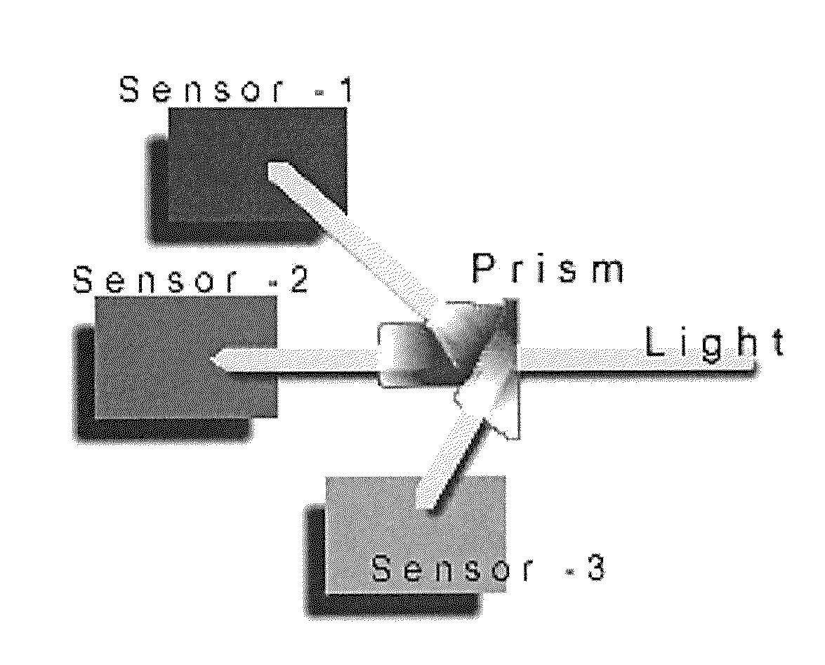
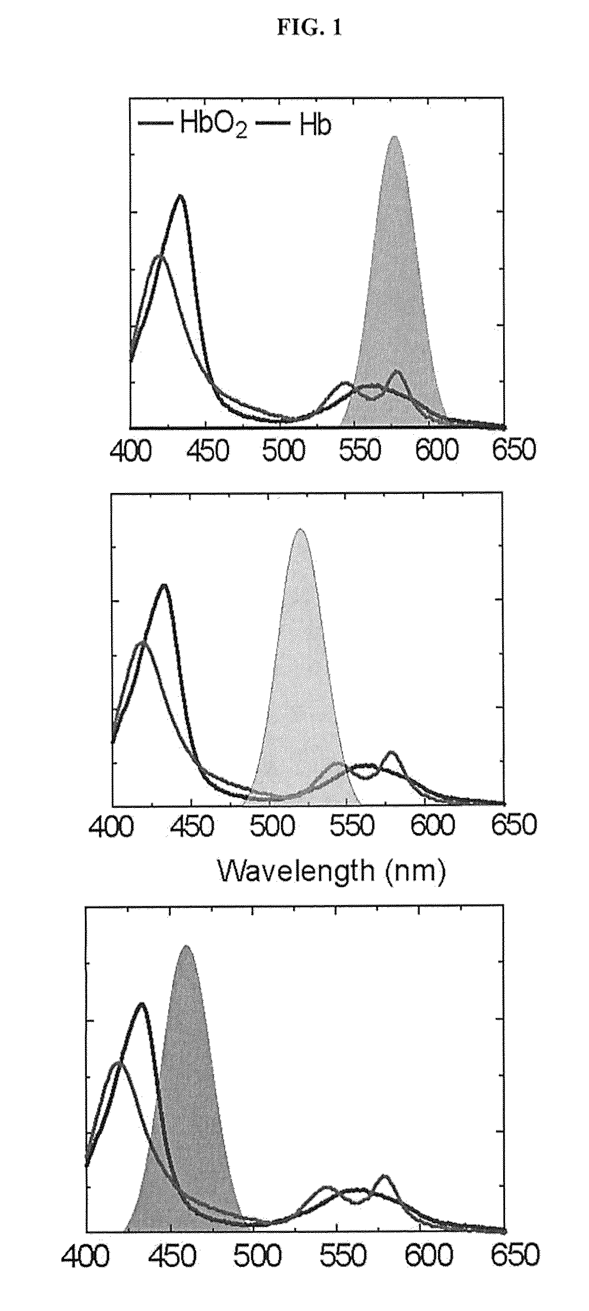
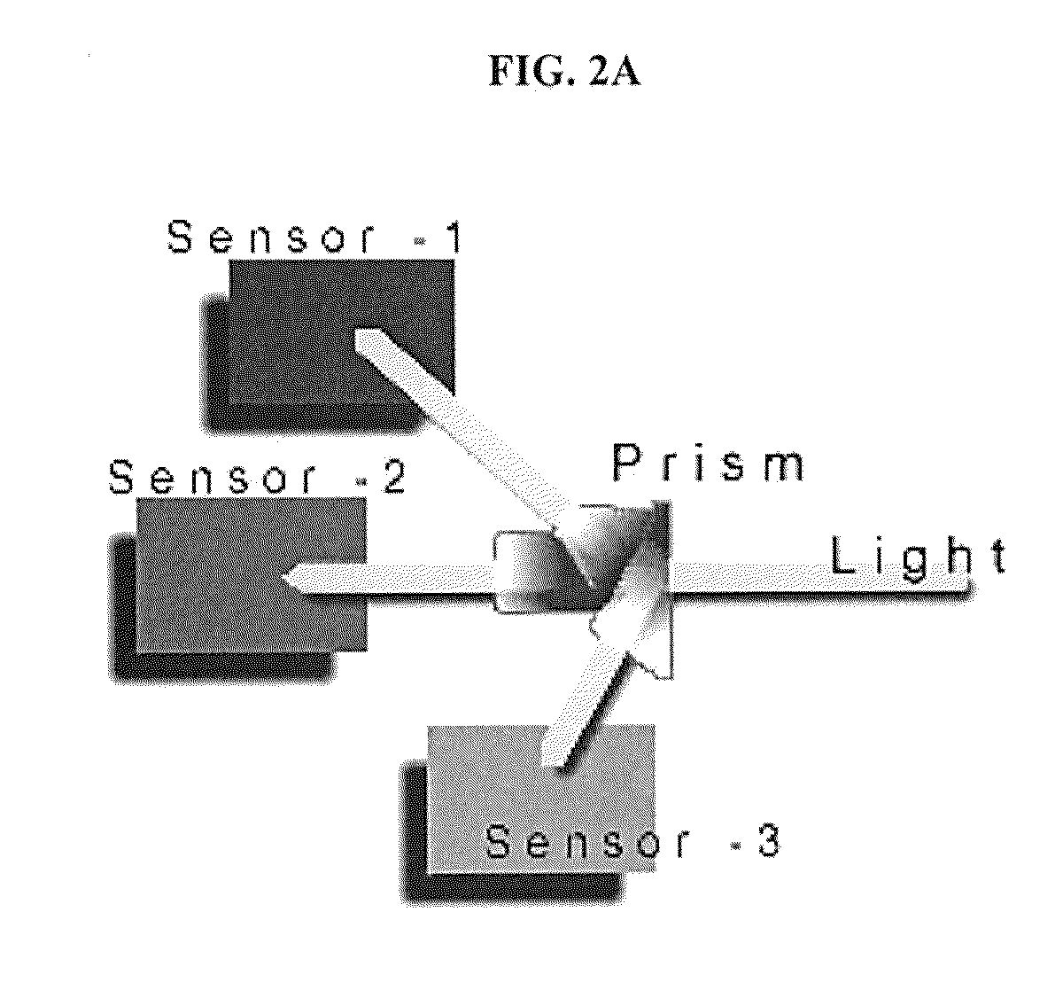
[0014]In one embodiment, a 3-CCD camera is used to acquire continuous video images of a target area of tissue or organ. The video images are stored. Depending on the need of the surgeon, the images may be directly displayed on a monitor or contrast enhanced to provide a visualization of the ischemic condition of the target tissue. To begin image enhancement, a selected group of image frames are extracted from the video footage and the contrast between oxygenated and deoxygenated tissue are enhanced using an image processor. More specifically, the blue CCD response is subtracted from the red CCD response, and the resultant image is plotted using a modified colormap optimized to provide the best contrast. The contrast image is then overlaid onto the original image frame under a predetermined transparency range of 40-50%, and displayed on a monitor. The surgeon thus is able to identify oxygenated tissue and organ in one color and deoxygenated tissues in a different color.

Problems solved by technology

The current standard method used in assessment of organ viability during surgery is limited to visual cues and tactile feedback.
However, during laparoscopic surgery, these assessment techniques are greatly impaired.
Currently, there is no in vivo methodology to monitor renal parenchymal oxygenation during laparoscopic surgery.
However, disadvantages of laparoscopic surgery include slightly longer warm ischemic times, and increased incidences of delayed graft function [1] [2].
Ischemia of tissue occurs when oxygen delivery to the tissue is inadequate to meet the metabolic demands.
Although, systemic hypoxia is readily diagnosed and treated, ischemic insults to individual tissue can be difficult to diagnose clinically, particularly when tissue cyanosis cannot be visually appreciated.
Indolent organ injury can have a delayed impact on function.
These issues, while minor in most donors, are increasingly problematic in situations utilizing older donors, or organs intended for use in very small children [3] [4].
This is of particular concern during partial nephrectomies since organ damage results in acute renal failure in 50% of such cases [15].
These methodologies are limited by their inability to assess the organ directly.
Unfortunately, to date there has not been a method to evaluate tissue oxygenation laparoscopically in a time frame that is clinically relevant.
For example, during the course of the operation, blood supply to the organ becomes impaired by the technical manoeuvres done during dissection (i.e., approaching the vessels from the posterior aspect).
However, this method does not provide regional information.
Ultrasound Doppler technique is another common clinical tool used to measure blood flow in large vessels, but is rather insensitive to blood flow in smaller vessels, and do not readily permit continuous measurements.
Laser Doppler techniques have also been used recently to measure tissue oxygenation, but are typically limited to tissue surface.
Magnetic resonance imaging (MRI) has high temporal and spatial resolution, and has become a gold standard technique in noninvasive measurement of blood flow and metabolic response, but its clinical use is limited by high cost and poor mobility.
However, to date most (but not all) applications of DCS have been in small animal studies wherein source-detector separations were comparatively small.
In addition, pulse oximetry and fluorescein have a high risk of failure for detecting tissue necrosis.
One disadvantage of a technique like NCLBF is that the measurement is made via a pencil probe, appropriate for open surgery but not laparoscopic surgery.
While the technique shows promising results and allows for real time in vivo imaging, it requires special instrumentation and is not easily converted to a format for use with a laparoscopic tower.
However, the open lens probe of the endoscope samples and evaluates only a small portion of the tissue during a single measurement.
Evaluation of the kidney as a whole would require a large number of sampling points, proving inefficient in a time limited scenario.
While the OXYLITE® probe is very effective for sampling tissue oxygenation within the tissue itself [13, 14], it suffers from the same limitations as the erythrocyte velocity measurement.

Method used


Image

  • Image contrast enhancement for in vivo oxygenation measurements during surgery
  • Image contrast enhancement for in vivo oxygenation measurements during surgery
  • Image contrast enhancement for in vivo oxygenation measurements during surgery

Examples


##ic example 1

Prophetic Example 1

Real-Time Validation Using Porcine Model

[0056]The 3-CCD (Stryker) camera and the tower light were mounted to the overhead operating room light such that both kidneys were evenly illuminated and in the field of view. The renal vein and artery were partially and sequentially clamped with Satinsky clamps until the vessels were completely occluded (i.e. immediately following 1 click, then 5 minutes post-click, immediately following a total of 2 clicks, then 5 minutes post-click, immediately following a total of 3 clicks, then 2 and 5 minutes post-click, immediately following 4 clicks, then 1, 2 and 5 minutes post-click).

[0057]At each time point of interest, an image was captured directly by the camera and processed immediately and presented as a 3-CCD contrast enhanced image. The surgeon defined the areas for ROIs and the algorithm performed and displayed a real-time calculation of the mean intensity value and its corresponding sO2. In addition, at each time point, re...


PUM


Abstract

A system and method for real-time or near real-time monitoring of tissue / organ oxygenation through visual assessment of contrast enhanced images of the target area of tissue or organ. Video of a target tissue / organ was acquired during surgery, selected image frames were extracted. Each extracted image is separated into red, green and blue CCD responses. A modified contrast image was created by subtracting blue CCD responses from red CCD responses, and plotting the resultant image using a modified colormap. Overlaying said modified contrast image onto the original extracted image frame under a selected transparency range, and display it for review.

Description

CROSS-REFERENCE OF RELATED APPLICATION[0001]This application claims priority to U.S. Provisional Application No. 61 / 120,971 filed Dec. 9, 2008.FIELD OF INVENTION[0002]The present invention relates generally to real-time monitoring of the oxygen levels of a target tissue area or a target organ. More specifically, the invention relates to a system and method for real-time assessment of tissue and organ oxygenation during surgery.BACKGROUND OF THE INVENTION[0003]The current standard method used in assessment of organ viability during surgery is limited to visual cues and tactile feedback. However, during laparoscopic surgery, these assessment techniques are greatly impaired. A concern in laparoscopic surgery is the loss of three-dimensional assessment of organs and tissue perfusion. This is of particular relevance during laparoscopic renal donation, where the condition of the kidney must be optimized despite considerable manipulation. Currently, there is no in vivo methodology to monit...

Claims


Application Information

Patent Timeline
Patent Type & Authority Applications(United States)
IPC IPC(8): A61B6/00H04N7/18
CPCA61B90/361
Inventor ELSTER, ERIC A.TADAKI, DOUG K.CRANE, NICOLE J.HUFFMAN, SCOTT W.LEVIN, IRA W.
Owner ELSTER ERIC A
Why Eureka
Try Eureka