
Protein G-Oligonucleotide Conjugate

a technology of oligonucleotide and conjugate, which is applied in the field of protein g conjugate, can solve the problems of macromolecules losing orientation and activity to bind to antigens, less reproducibility, and asymmetric antibodies

Inactive Publication Date: 2010-08-12
KOREA RES INST OF BIOSCI & BIOTECH
View PDF6 Cites 2 Cited by
  • Summary
  • Abstract
  • Description
  • Claims
  • Application Information

AI Technical Summary

Benefits of technology

[0007]It is an object of the present invention to solve the problem that antibodies lose their orientation upon binding to an immunosensor, and to provide techniques for easily immobilizing antibodies on a variety of solid supports in a consistent orientation using well-defined DNA surfaces.Technical Solution
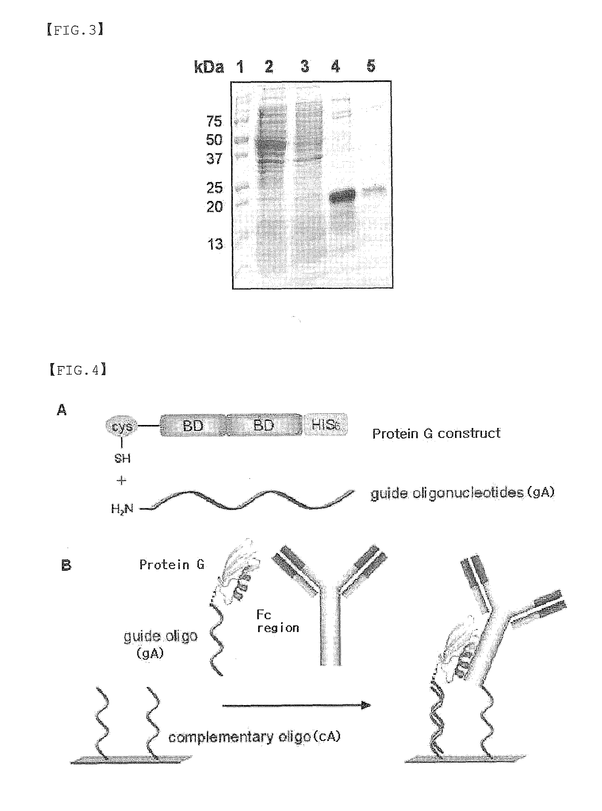
[0008]Previously, the present inventors have prepared an N-terminal cysteine-tagged protein G variant (Korean Patent Application No. 10-2007-0052560), and confirmed its usefulness through experiments, in order to solve the problem that antibodies lose their orientation upon binding to an immunosensor. Also, based on the invention, the present inventors have prepared a protein G conjugate (gA-G) by chemically linking an oligonucleotide (gA) having an amine group with the cysteine-tagged protein G variant using a linker capable of selectively reacting with both amine and thiol groups. They found that antibodies can be easily immobilized on a variety of solid supports in a consistent orientation and on intended areas of the surfaces by using the protein G conjugate, thereby completing the present invention.

Problems solved by technology

The physical immobilization techniques (Trends Anal. Chem. 2000 19, 530-540) have been minimally used because they cause denaturation of the protein, and the results are less reproducible.
However, when immobilization of antibodies is performed using a chemical immobilization technique, the antibodies, being asymmetric macromolecules, often lose their orientation and activity to bind to antigens (Analyst 121, 29R-32R).
However, there is a problem that this protein G itself also loses orientation and its ability to bind to an antibody when bound to the support.
However, this method is directed to thiolating the amino groups of amino acids having an amino group (Arg, Asn, Gln, Lys), instead of thiolating any specific site, and thus the method results in low specificity and requires additional purification processes after chemical treatments (Biosensors and Bioelectronics, 2005, 21, 103-110).
However, both methods have a drawback in that a small molecule or DNA has to be linked to the antibody, so as to cause loss of its orientation or chemical modification of antigen-binding site.

Method used


Image

  • Protein G-Oligonucleotide Conjugate
  • Protein G-Oligonucleotide Conjugate
  • Protein G-Oligonucleotide Conjugate

Examples


example 1

Protein Expression Analysis of Cysteine-Tagged Streptococcal Protein G Variant

[0055] Gene Preparation of Cysteine-Tagged Streptococcal Protein G Variant

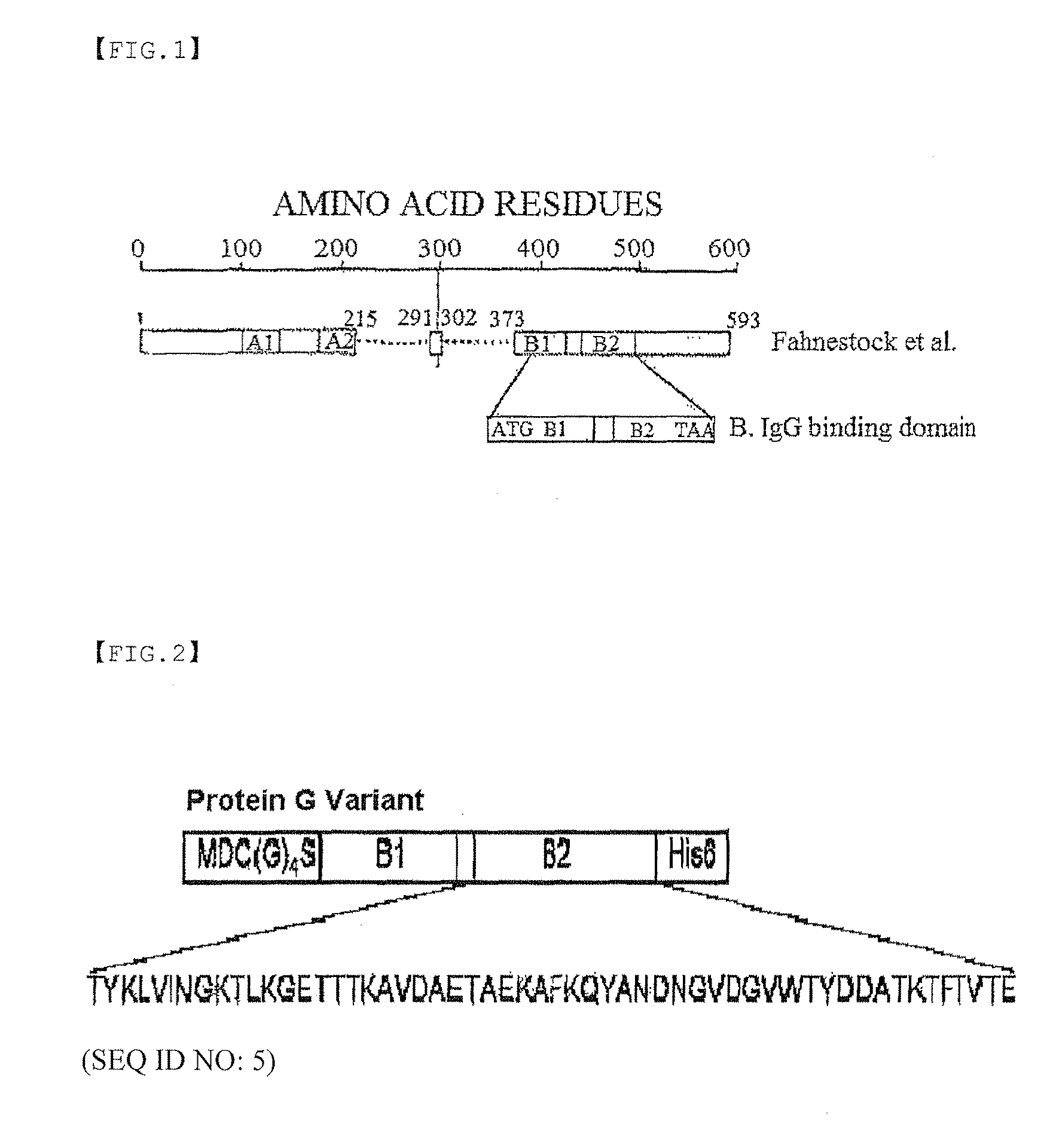
[0056]Two primers were prepared in order to tag with cysteine at the N-terminus. In the base sequence of the 5′-primer, an initiation codon (ATG) was followed by GAT (Asp codon) and TGC (cysteine codon), and in order to provide a link to protein G, GGC GGC GGC GGC AGC (four Gly codons and one Ser codon) were included. Furthermore, in order to insert the gene into an expression vector pET21a (Novagen), the NdeI restriction site was introduced into the N-terminal primer and the XhoI restriction site was introduced into the C-terminal primer. The Streptococcal genomic gene was obtained, and a polymerase chain reaction (PCR) was performed with the primers. Thus, only the amino acid regions (B1 [a form having 10 initial amino acid residues cleaved], B2), which are known as domains to which an antibody binds, were obtained. The obtained fr...

example 2

Preparation of Protein G Conjugate (gA-G)

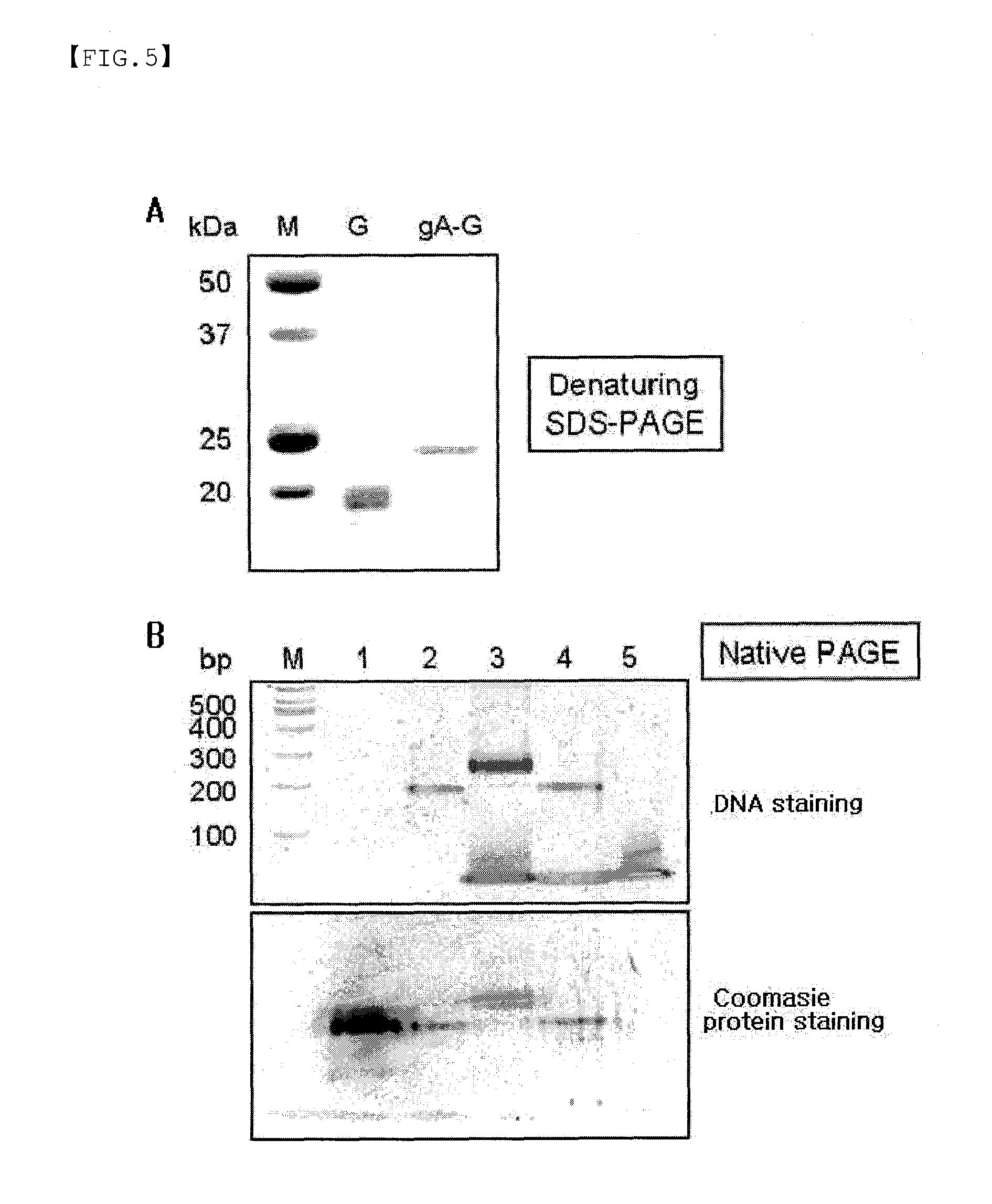
[0066]Using Sulfo-SMCC (Sulfosuccinimidyl 4-(N-maleimidomethyl)cyclohexane-1 carboxylate), an oligonucleotide (gA) modified with an amine group and a Streptococcal protein G variant tagged with one cysteine were chemically linked to each other to prepare a protein G conjugate (gA-G).

[0067]In particular, 60 nmol of the oligo DNA (gA) modified with an amine group at 5′-end was dissolved in 400 μl of 0.25 M phosphate buffer, and then reacted with 1.5 mg of Sulfo-SMCC (3400 nmol) dissolved in 75 μl of DMF solution. The mixture was reacted at normal temperature for 1 hr, and then the activated oligo DNA (gA) was separated from the excess unreacted Sulfo-SMCC using a binding buffer (20 mM Tris, 50 mM NaCl, 1 mM EDTA pH7.0) by Sephadex G25 gel filtration. While performing the activation of oligo DNA, the cysteine tagged-protein G variant was reacted with 20 mM DTT for complete reduction, followed by gel filtration to remove DTT. Consequently, the ob...

example 3

Fabrication of Protein G Conjugate (gA-G)-Immobilized Biosensor and Biochip

[0071]The oligo DNA (gA) was chemically linked to the one cysteine-tagged Streptococcal protein G variant, and then reacted with the surface of gold thin film, on which the oligo DNA (cA) complementary to oligo DNA (gA) was linked, to fabricate a protein G conjugate (gA-G)-immobilized biosensor and biochip.

[0072]In particular, the oligo DNA (cA) was reacted with the surface of gold thin film, and then changes in the surface plasmon resonance signal were measured by means of a surface plasmon resonance (SPR)-based biosensor to detect the immobilization reaction of the complementary oligo DNA (gA), protein G conjugate (gA-G), and noncomplementary control oligo DNA (gB) in real-time.

[0073]As a result, when the noncomplementary control oligo DNA (gB) was injected, there was little change in the surface plasmon resonance signal. When the complementary oligo DNA (gA, 7.5 kDa) was injected, the surface plasmon reson...


PUM

PropertyMeasurementUnit
pHaaaaaaaaaa
pHaaaaaaaaaa
lengthaaaaaaaaaa

Abstract

The present invention relates to a protein G conjugate, which is prepared by linking an N-terminal cysteine-tagged protein G variant with an oligonucleotide via a linker. The conjugate binds in a directional manner on the surface of a biochip and biosensor, thereby providing a biochip and biosensor having improved antibody immobilization ability.

Description

TECHNICAL FIELD[0001]The present invention relates to a protein G conjugate (gA-G) which is prepared by linking an N-terminal cysteine-tagged protein G variant with an oligonucleotide using a linker, a method for preparing the same, and a biochip and a biosensor fabricated by using the conjugate.BACKGROUND ART[0002]Antibodies have been widely used in medical studies concerning diagnosis and treatment of diseases as well as in biological analyses, because of their property of specifically binding to an antigen (Curr. Opin. Biotechnol. 12 (2001) 65-69, Curr. Opin. Chem. Biol. 5 (2001) 40-45). Recently, as an immunoassay, immunosensors have been developed, which require the immobilization of an antibody on a solid support to measure changes in current, resistance and mass, optical properties or the like (Affinity Biosensors. vol. 7: Techniques and protocols). Among them, a surface plasmon resonance-based immunosensor making use of optical properties has been commercialized. The surface...

Claims


Application Information

Patent Timeline
Patent Type & Authority Applications(United States)
IPC IPC(8): G01N33/543C07K14/00
CPCC07K14/315G01N33/54393C07K19/00
Inventor CHUNG, BONG HYUNJUNG, YONG WONLEE, JEONG MIN
Owner KOREA RES INST OF BIOSCI & BIOTECH
Learn More