Articular cartilage, device and method for repairing cartilage defects

a technology of cartilage and cartilage, applied in the field of articular cartilage for repairing cartilage defects, can solve the problems of insufficient treatment of medium or greater surfaces, insufficient simple biological reparation methods available for the time being, and insufficient long-term examination results, so as to achieve optimal structure and improve the efficiency of their use

Inactive Publication Date: 2010-08-19
PECSI TUDOMANYEGYETEM
View PDF15 Cites 57 Cited by
  • Summary
  • Abstract
  • Description
  • Claims
  • Application Information

AI Technical Summary

Benefits of technology

[0019]The invention is based on the recognition that thin cartilage allografts of great surfaces are optimal for the replacement of defected joint cartilages, and the efficiency of their use may be improved if the side intended to face the bone is provided with incisions, and preferably with cartilage cells as well. It is also reco

Problems solved by technology

Joint cartilage defects or deceases can result in progressive impairment of life quality.
However, the simple biological reparation methods available for the time being (abrasion, drilling, debridement, shaving or microfracture) proved in long term examinations to be insufficient, as the produced fibrous cartilage is mechanically weak.
Such osteochondral (bone based cartilage graft) replacements may be sufficient for reparing smaller (2) defects, but medium or great

Method used


Image

  • Articular cartilage, device and method for repairing cartilage defects
  • Articular cartilage, device and method for repairing cartilage defects
  • Articular cartilage, device and method for repairing cartilage defects

Examples


example 1

[0038]Several hundred milligrams of hyaline cartilage was collected with arthroscopy for repairing the cartilage damage of a young sportsman. The collected cartilage was delivered to a cell culturing laboratory.

[0039]After having obtained the required number of cells, they were suspended, poured onto the side of the matrix provided with incision, and left for properly sedimenting.

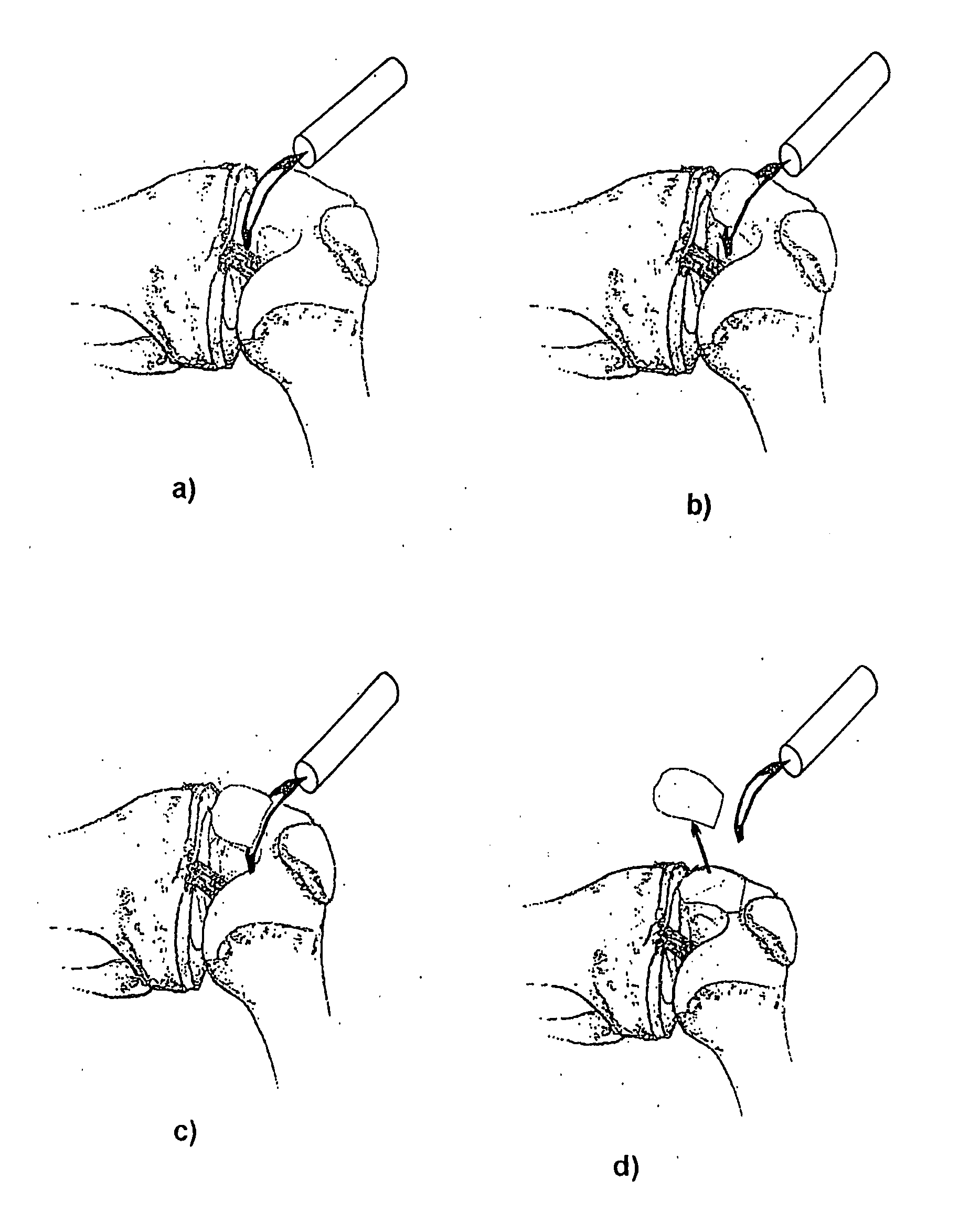
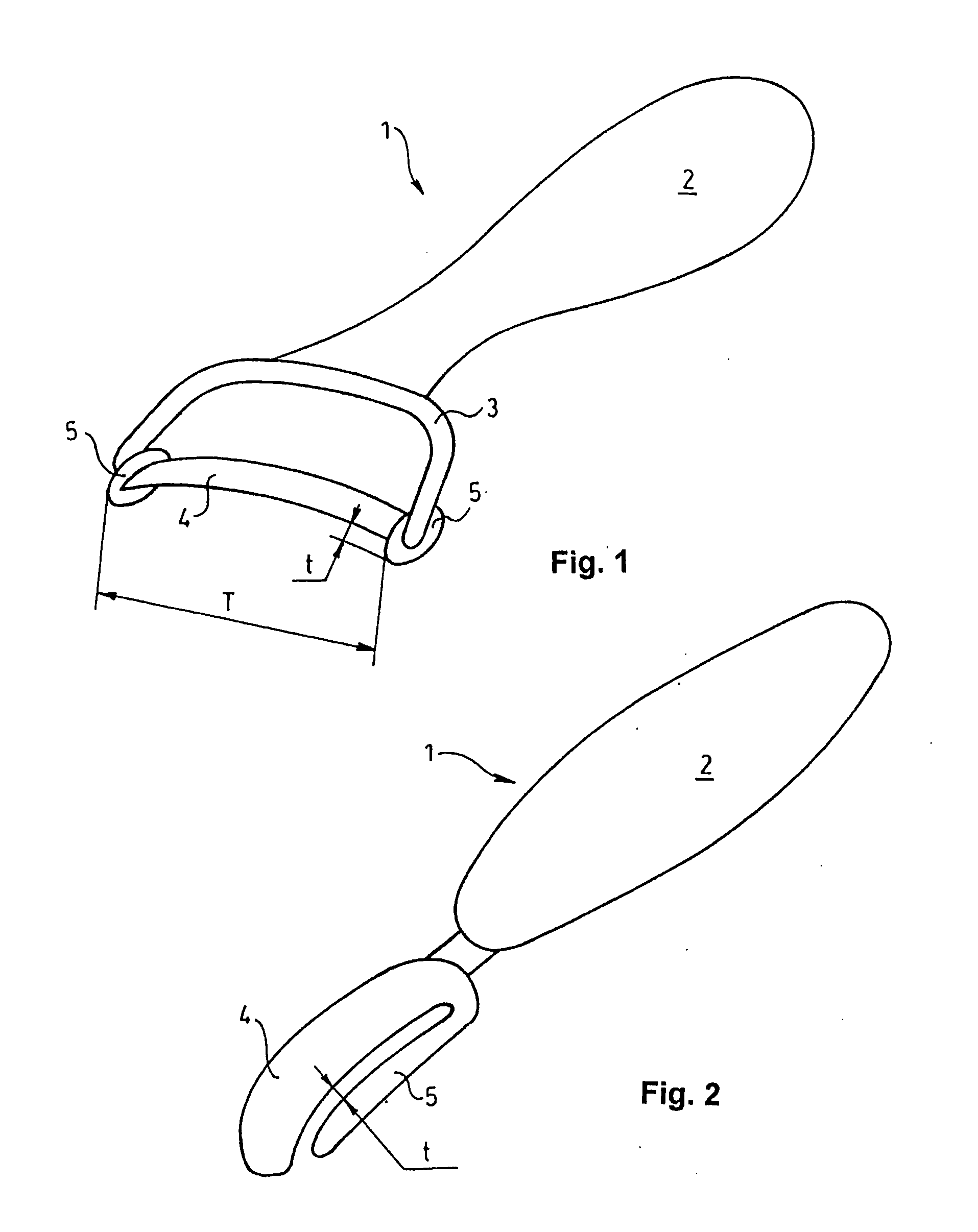
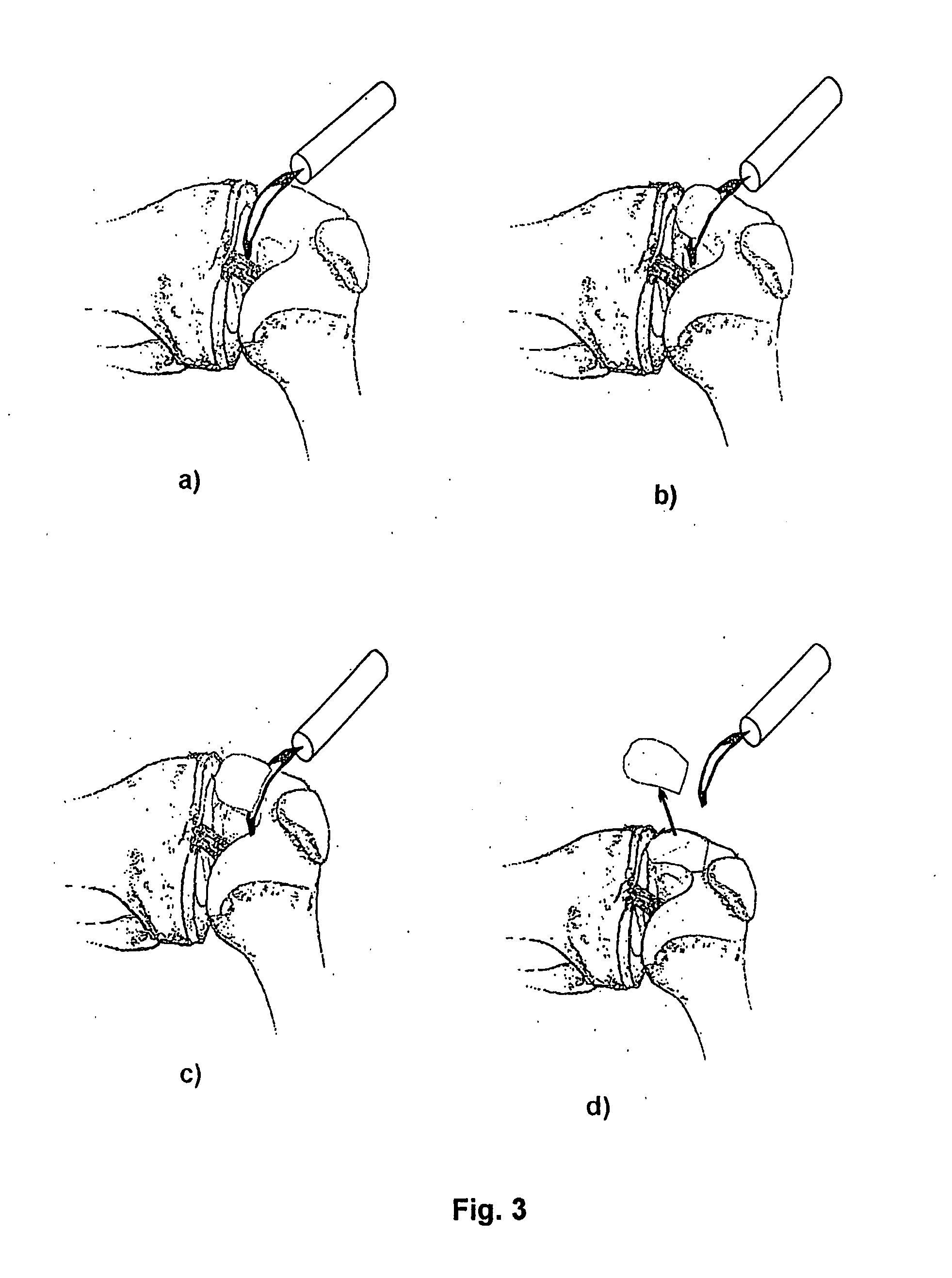
[0040]The cartilage matrix had been harvested in sterile conditions from the knee joint of a cadaver, long before the operation, with the device shown in FIG. 1. The matrix with a surface of 2×3 cm had been provided with incisions on the side facing the bone, with the device shown in FIG. 5. The incisions had been made in two perpendicular directions, wherein the distances between the incisions were 0.5 mm and the thickness of the intact collagen layer was 90 μm. The matrix had than been provided with a sterile packing and stored on a temperature of −80° C.

[0041]The cartilage matrix obtained from a cadaver ...

example 2

[0042]A patient of middle age had ankle complaints. As the result of an examination, it was found that he had focal cartilage defect on the upper surface of her talus. It was decided to perform cartilage substitution by cartilage cell transplantation, therefore bone marrow stem cells were collected for culturing (in cases, when it is not possible, joint cartilage particles may be collected for obtaining cells). The collected cells were delivered to a cell culturing laboratory.

[0043]Prior to the operation, cartilage sample had been harvested in sterile conditions from the knee joint of a cadaver, with the device shown in FIG. 2. The cartilage had been processed with incisions on the side facing the bone, with the device shown in FIG. 5. The incisions had been made in parallel directions, wherein the distances between the incisions were 0.8 mm and the thickness of the intact layer was 120 μm.

[0044]The multiplied cells were centrifuged to the graft provided with incisions, to be captur...

example 3

[0047]During arthroscopy of a women of middle age it was found that she had small cartilage deficiency on the knee joint. Therefore, at the same time, following a small joint exposure, the region of the cartilage deficiency has been cleared up to the healthy cartilage.

[0048]One week before the operation sound articular cartilage had been harvested in sterile conditions from the shoulder joint of a cadaver, with the device shown in FIG. 2. The cartilage had been provided with incisions on the side facing the bone. The incisions had been made in parallel directions, wherein the distances between the incisions were 0.1 mm and the thickness of the intact layer was 500 μm.

[0049]The cartilage had than been stored for one week in sterile conditions, without freezing, on +4° C., until the day of the operation.

[0050]The graft was delivered to the surgeon together with the living cells therein, for operation. A hole was made in the bone below the cartilage (microfracture) and the graft 14, af...


PUM


Abstract

The articular cartilage according to the invention is made of pure cartilage and is provided with incisions (12) on the surface facing the bone. The cartilage cells are preferably seeded on the surface provided with incisions (12). The method for producing the articular cartilage comprises the step of collecting cartilage from joints, wherein pure cartilage is collected without bone, and incisions are made on the surface of the cartilage intended to face the bone. It is preferably fresh frozen until use. The device for harvesting articular cartilage, comprises handle and cutting blade, wherein the cutting blade (4) is curvilinear and is provided with spacer elements (5), meanwhile the device for producing incisions in articular cartilages comprises handle (2) and a bridge (3) connected to said handle (2) and being provided with one or more cutting blade(s) (4). During the method for applying the articular cartilage the articular cartilage is fixed by thin surgical yarn stitches, by fibrin glue or by small anchors (FIG. 8).

Description

FIELD OF THE DISCLOSURE[0001]The present invention relates to articular cartilages for repairing cartilage defects and a method for producing articular cartilage comprising the step of collecting cartilage from joints. Further object of the invention is a device for harvesting articular cartilage, comprising handle and cutting edge as well as another device for producing incisions in articular cartilages.BACKGROUND OF THE INVENTION[0002]Joint cartilage defects or deceases can result in progressive impairment of life quality. The so called biological regeneration methods are more and more applied worldwide, besides the protetical reconstructions. One of these methods is tissue engineering, advancing continuously. Tissue engineering offers a wide field of applications in clinical work, and it seems that this method will be the revolutionary technology for healing, further to the gene-technology. The majority of people above 65 years have joint defects due to the decreased ability of r...

Claims


Application Information

Patent Timeline
IPC IPC(8): A61F2/08A61B17/32
CPCA61B17/00491A61B17/06166A61B17/32A61B17/322A61B17/68A61B2017/00969A61F2002/3096A61F2/3094A61F2/38A61F2/4202A61F2002/30762A61F2002/30764A61F2/30756
Inventor BARDOS, TAMASBELLYEI, ARPADILLES, TAMASNEMETH, PETER
Owner PECSI TUDOMANYEGYETEM
Why Eureka
Try Eureka