
Allergy test based on flow cytometric analysis

a flow cytometric and allergen-induced technology, applied in the field of flow cytometric analysis of allergen-induced basophil activation, can solve the problems of unsuitable routine allergy diagnosis, unspecific test, and low positive reaction ra

Active Publication Date: 2010-09-02
BUHLMANN LAB AG
View PDF2 Cites 17 Cited by
  • Summary
  • Abstract
  • Description
  • Claims
  • Application Information

AI Technical Summary

Problems solved by technology

However, the rate of positive reactions is much lower in non-IgE mediated processes.
This, however, is not suitable for routine allergy diagnosis.
However, this is a very unspecific test, as it does not address the nature of the drug substance potentially causing the anaphylatoxic reaction.
A severe disadvantage of the method described in these latter two reports is the use of labelled anti-IgE antibodies primarily for positive selection of basophils which contrasts somewhat with its simultaneous use for following basophil activation as determined by IgE down-regulation.

Method used


Image

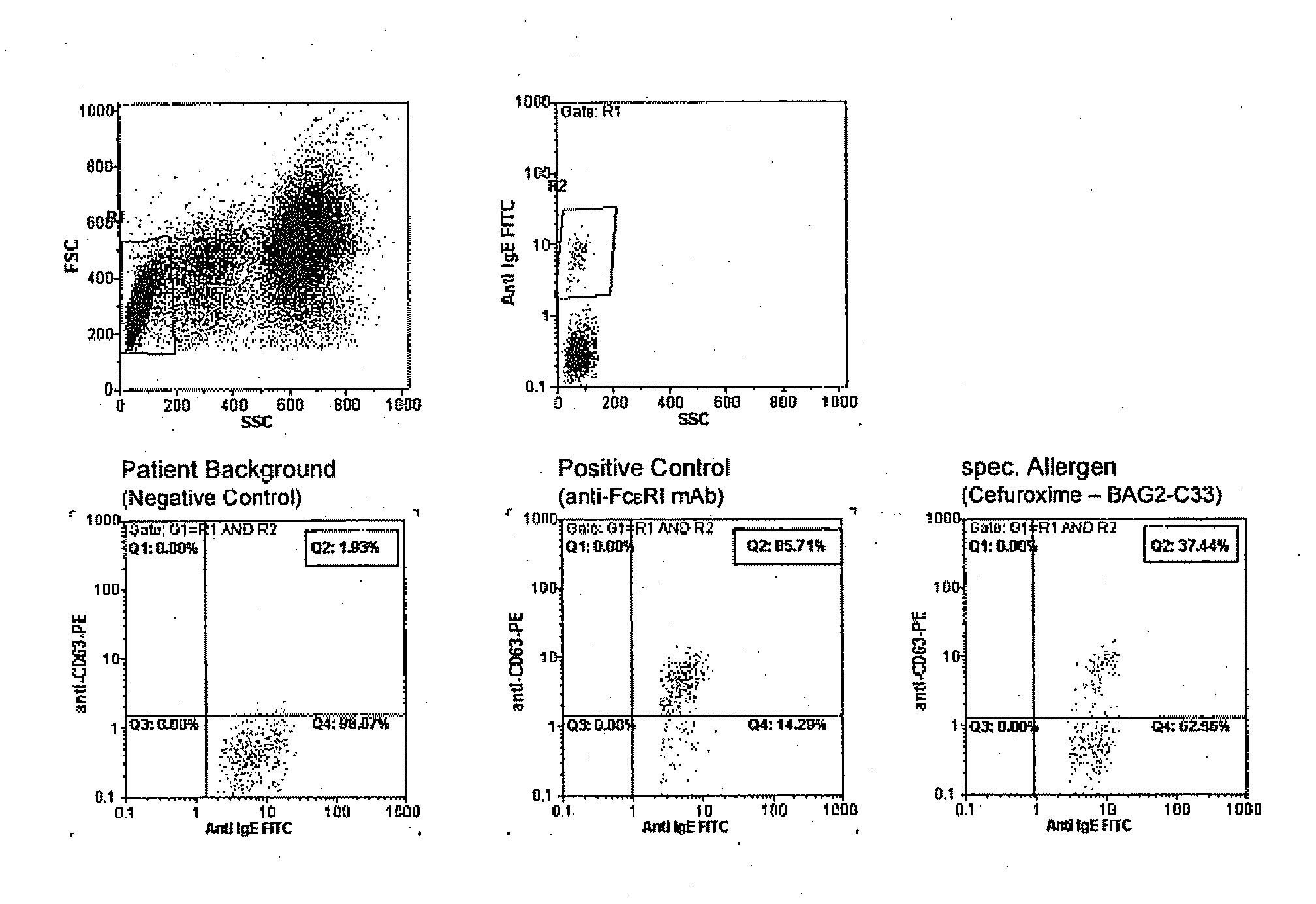
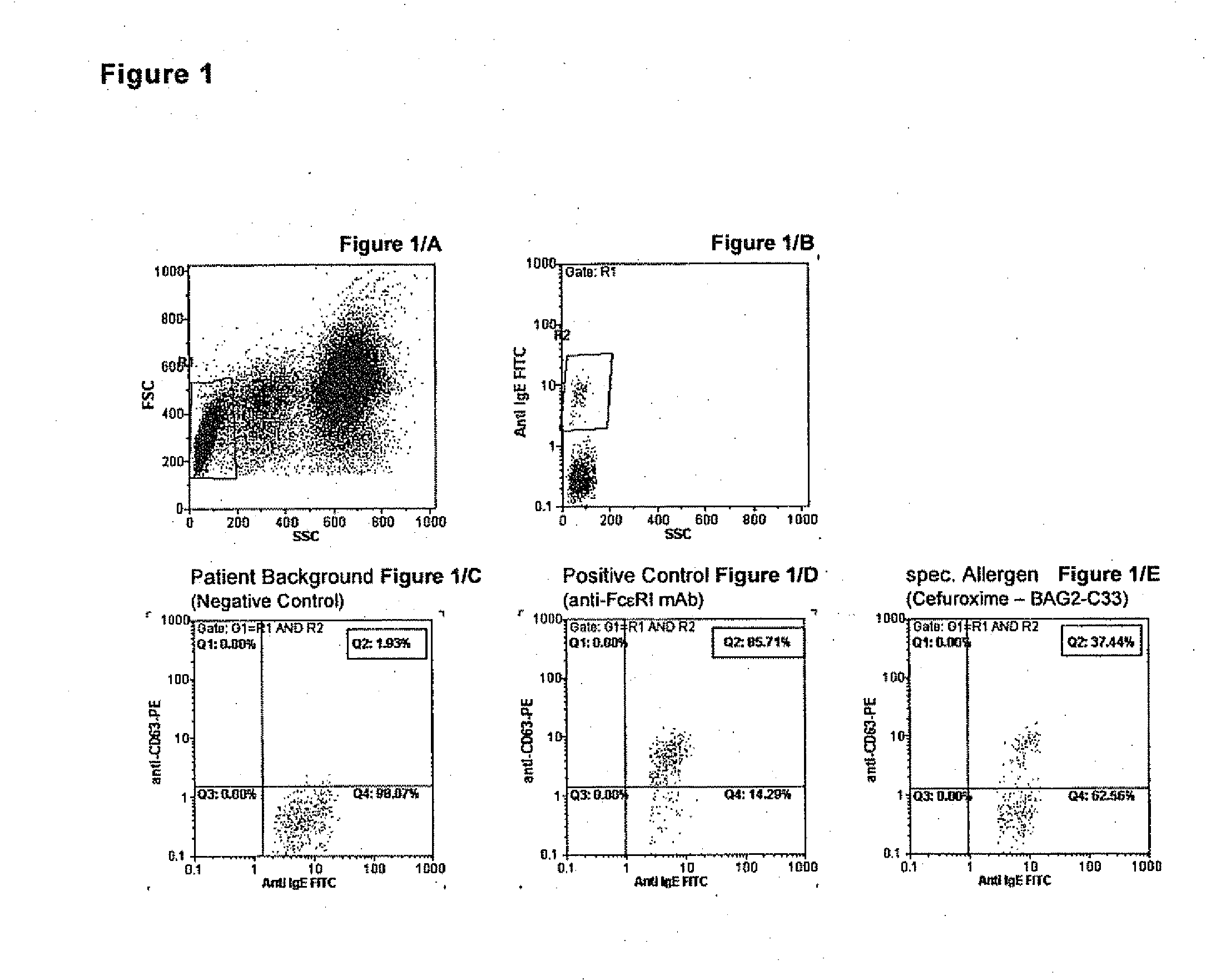
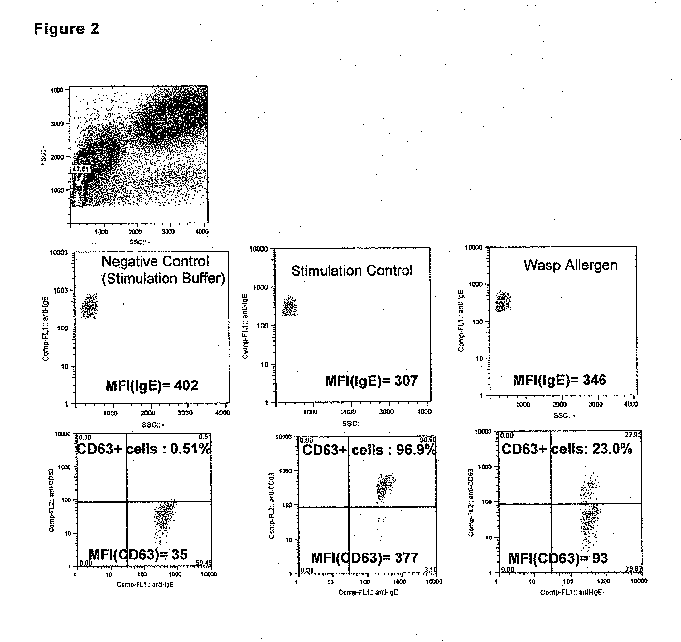
  • Allergy test based on flow cytometric analysis
  • Allergy test based on flow cytometric analysis
  • Allergy test based on flow cytometric analysis

Examples


example 1

EDTA vs. Heparin vs. ACD (Citrate) Blood

[0284]Blood from healthy blood donors or allergic patients was drawn into three different venipuncture tubes containing as anticoagulant either EDTA or Heparin or Citrate (ACD).

[0285]All blood samples were analysed according to Protocol 1 and stimulated with Stimulation Buffer (negative control=NC), anti-FcεRI mAb (positive Stimulation Control=PC) and with allergens as specified in FIGS. 5A, 5C and 5D. The results are expressed in percentage of CD63 expression.

[0286]A direct comparison of the three different blood preparations from a orchard grass (G3) allergic patient is shown in FIG. 5A. Heparin seems to unspecifically stimulate NC which is observed from time to time in other cases as well, whereas this is not seen here and is also not reported in other cases for ACD or EDTA blood, PC and G3 (50 ng / ml) are clearly positive with all blood samples, however EDTA samples give slightly higher stimulation reactions.

[0287]EDTA, ACD or Heparin venip...

example 2

Diluted vs. Purified (Washed) Blood Cells

[0290]The cellular reactivity depends very much on the conditioning of the blood cells before stimulation, i.e. the matrix for the stimulation reaction is very crucial. It must contain heparin, calcium and, preferably, IL-3 (except for specific applications such as the follow up of insect venom immunotherapy), but plasma factors may influence the reactivity of blood basophils, too.

[0291]Therefore, we established two protocols, one with diluted whole blood, wherein the EDTA blood sample is diluted 1 in 4 with cellular incubation buffer (the Stimulation Buffer) and another with purified blood, wherein the basophils were transferred from its plasma matrix to a plasma-free matrix such as the cellular incubation buffer by centrifuging and washing the blood cells. The following illustrative examples show the dramatic influences which the cellular preparation method may have on the reactivity of patients' blood cells in different clinical settings.

[...

example 3

Lysis Procedure

[0296]As just mentioned above, the removal of erythrocytes before analyzing the basophils on a flow cytometer is a prerequisite to correctly gate in resting vs. activated basophils and to get highly informative and quantitative results. In prior art methods, this is, beside the cell stimulation part, the most delicate and most time-consuming step in the entire analysis of quantitative basophil stimulation.

[0297]With the invented stimulation Protocol 2 combined with the CCR3 basophil selection marker it is possible to simplify and speed up or even automatize this process. In FIG. 9A, the detection and gating of basophils using the CCR3-PE selection marker is shown. From the left to the right panel, different erythrocyte lysing procedures are tested (LYR=Lysing Reagent; WB=Wash Buffer). The left panel shows a procedure as used in prior art methods including two centrifugation and one washing steps, the right panel clearly shows that neither a washing nor a centrifugatio...


PUM

PropertyMeasurementUnit
molecular weightaaaaaaaaaa
concentrationaaaaaaaaaa
volumeaaaaaaaaaa

Abstract

The invention pertains to a method for the determination of basophil activation induced by a test substance by flow cytometric measurement of the changes of the mean or median fluorescence intensities (WI) of the basophilic FcεRI receptor present on the cell surface of basophils (MFI-FcεRI) and / or the IgE antibodies bound to the FcεRI receptor (MFI-IgE), and the CD63 antigen exposed on the cell surface of basophils after their activation (MFI-CD63), by means of a mixture of anti-CD63, anti-FcεRI or anti-IgE, and anti-CCR3 antibodies each labelled with a distinct fluorophore, of which at least one antibody acts as a basophil selection marker and at least two antibodies act as basophil activation markers, and bringing the mean fluorescence intensities of the activation markers in correlation to obtain an Activation Index. These methods combining the measurement of an early (such as IgE, FcεRI or CD203c) and a late basophil activation marker (such as CD63), respectively, provide a markedly improved clinical sensitivity in allergy diagnosis over existing methods which consider only one activation marker, such as CD203c or CD63, expressed in percentage of basophil activation. It is also an aspect of the present invention to provide an ex-vivo allergy provocation test comprising the above-mentioned flow cytometric measurement and analysis of the results as well as a test kit for carrying out the test in-vitro.

Description

BACKGROUND[0001]The present invention is directed to a flow cytometric analysis of allergen induced basophil activation. The particular combinations of antibodies labelled with different fluorophores in combination with a new approach to obtain the mean or median fluorescence intensities (MFI) resulting in the provision of an Activation Index which allows for a better correlation of the individual patient results to their clinical history and, therefore, provides a markedly improved clinical sensitivity over existing methods by maintaining a high specificity. Thereby, a novel in vitro allergy test is provided.[0002]Human basophils are one class of leukocytes circulating in the blood stream and belong to the granulocytes. Despite their low abundance in human blood (less than 2% of the leukocyte fraction), they play a central role in allergic hypersensitivity reactions by releasing potent inflammatory mediators. Moreover, basophils represent the major interleukin-4 secreting human cel...

Claims


Application Information

Patent Timeline
Patent Type & Authority Applications(United States)
IPC IPC(8): G01N33/53
CPCG01N33/5047G01N2800/24G01N33/5091
Inventor SAINTE-LAUDY, JEANSCHNEIDER, MICHAELWEBER, JAKOB
Owner BUHLMANN LAB AG
Learn More