

Nuclear magnetic resonance measuring method
a nuclear magnetic resonance and measurement method technology, applied in the field of nuclear magnetic resonance measurement methods, can solve the problems of difficult analysis, obstacle to analysis, and low signal-to-noise ratio, and achieve the effect of reducing contamination signals and high signal-to-noise ratio
- Summary
- Abstract
- Description
- Claims
- Application Information
AI Technical Summary
Benefits of technology
Problems solved by technology
Method used
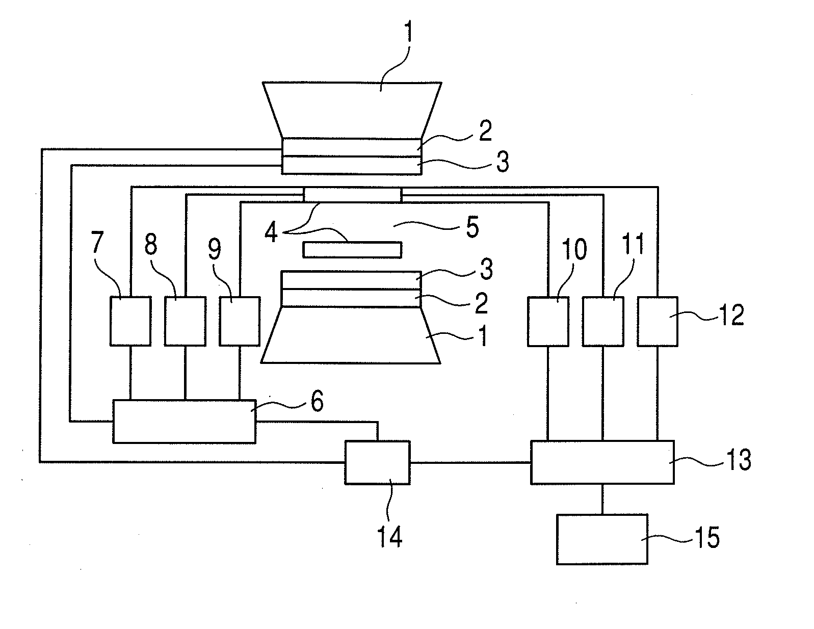
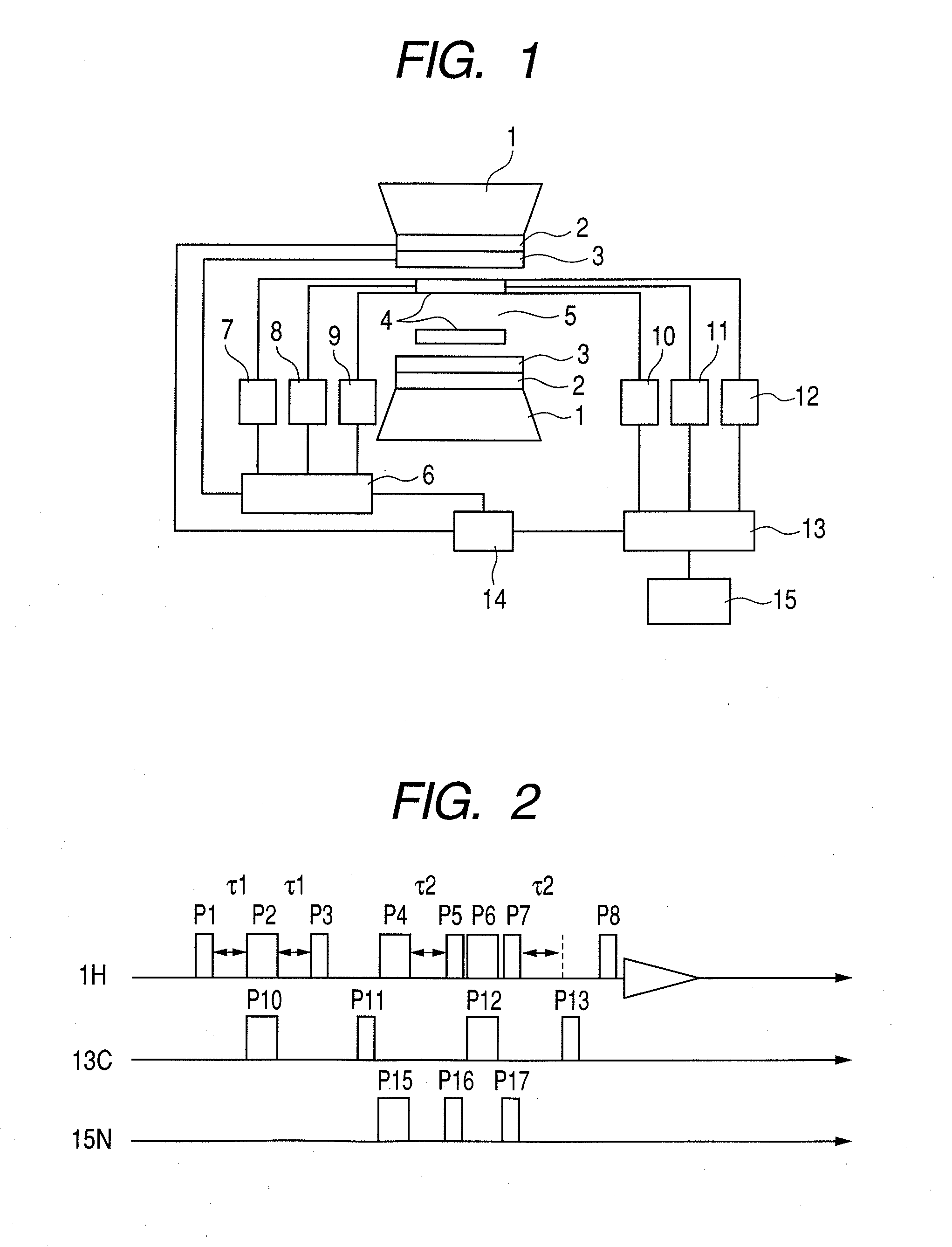
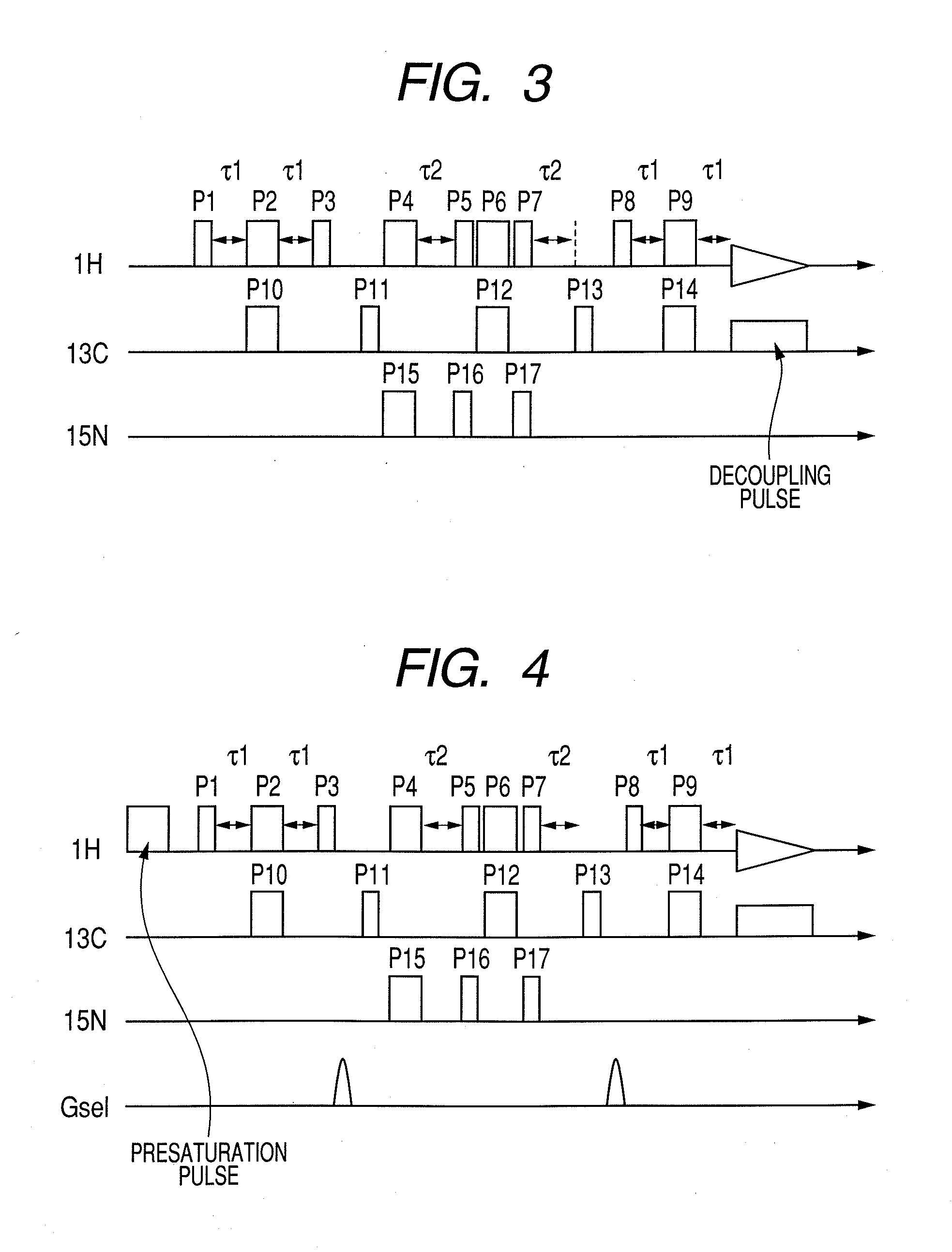
Image

Examples
example
Example 1
[0090]A budding yeast (BY4741 / 4742) was inoculated in 5 mL of a YPD liquid culture medium, and was cultured at 25° C. for two days. Subsequently, 5 mL of the YPD liquid culture medium containing a budding yeast (BY4741 / 4742) was poured into 100 mL of a YPD liquid culture medium containing 5 mg of isotope-labeled choline which was isotope-labeled with 13C and 15N and was cultured at 25° C. until the absorbance by the wavelength of 660 nm reached 1.0 by ABS.
[0091]The culture medium was dispensed into two conical tubes of 50 mL, and the yeast cells were spin down at 1,000×g for 10 minutes and were collected. The yeast cells in each tube were suspended in 50 mL of a phosphoric acid buffer, and were spin down at 1,000×g for 10 minutes. The above described operation was repeated for three times in total, and the cleaned yeast cells were suspended in the smallest amount of YPD (approximately two to three times of pellet capacity) and were subjected to measurement for the signal of...
example 2
Taking in HeLa Cell of 13C3-15N-choline chloride and Confirming by Triple Resonance Nuclear Magnetic Resonance
[0096]At first, 13C3-15N-choline chloride (10 mg namely 69.6 μmol) was dissolved in a DMEM (Dulbecco's Modified Eagle's Medium) culture medium (5 ml), was filtered with the use of a millipore filter having a pore diameter of 220 nm, and was sterilized. The filtrates in respective amounts of 1 ml were added to five pieces of laboratory dishes. DMEM (9 ml) and HeLa cells (approximately 250,000 pieces) were added into each laboratory dish, and then the cells were incubated at 37° C. for five days in the presence of 5% CO2. Then, DMEM was removed, and the HeLa cells on the laboratory dish were cleaned with the use of phosphate buffer PBS(−) (5 ml). Subsequently, trypsin-EDTA (1.5 ml) was added to each laboratory dish, incubated the HeLa cells at room temperature for one minute, and was removed. The HeLa cells having exfoliated from the laboratory dish in each of five pieces of t...
example 3
Taking in HeLa Cell of 13C3-15N-choline chloride and Confirming by Triple Resonance NMR (Part 2)
[0098]The experiment was carried out in a similar way to the above described Example 2, and then the HeLa cells were collected into a tube of 50 ml. The HeLa cells were precipitated by centrifugal separation, and the supernatant was removed. The HeLa cells were subjected to measurement for the signal of 1H nuclear magnetic resonance with the use of the triple resonance method according to the present invention, without being dried (Almost all of the cells were confirmed to be living cells by trypan blue staining carried out after measurement). As a result, a signal attributed to isotope-labeled choline was selectively observed. The result is shown in FIG. 18.
[0099]On the other hand, FIG. 19 shows a measured data of signals of ordinary 1H nuclear magnetic resonance, and FIG. 20 shows a data of measured signals of 1H nuclear magnetic resonance with the use of a double resonance method. In t...
PUM
 Abstract
Description
Claims
Application Information