
Substrate probe, enzyme-activity detection method by a multi-dimensional nuclear magnetic resonance method and enzyme-activity imaging method

a multi-dimensional nuclear magnetic resonance and enzyme activity technology, applied in magnetic measurement, instruments, biochemistry apparatus and processes, etc., can solve the problems of difficult to capture a signal sent from the deep part of a living body, abnormal increase or decrease of enzyme activity, etc., to improve the reliability of enzyme detection

Active Publication Date: 2010-09-30
CANON KK
View PDF5 Cites 17 Cited by
  • Summary
  • Abstract
  • Description
  • Claims
  • Application Information

AI Technical Summary

Benefits of technology

[0010]To enhance the reliability of enzyme detection, the enzyme activity is preferably detected in a system in which the presence of a substrate probe for measuring enzyme activity can be detected and further enzyme activity can be monitored. Furthermore, it is desirable, in view of practice, to use a highly biocompatible probe and a low-invasive detection system.
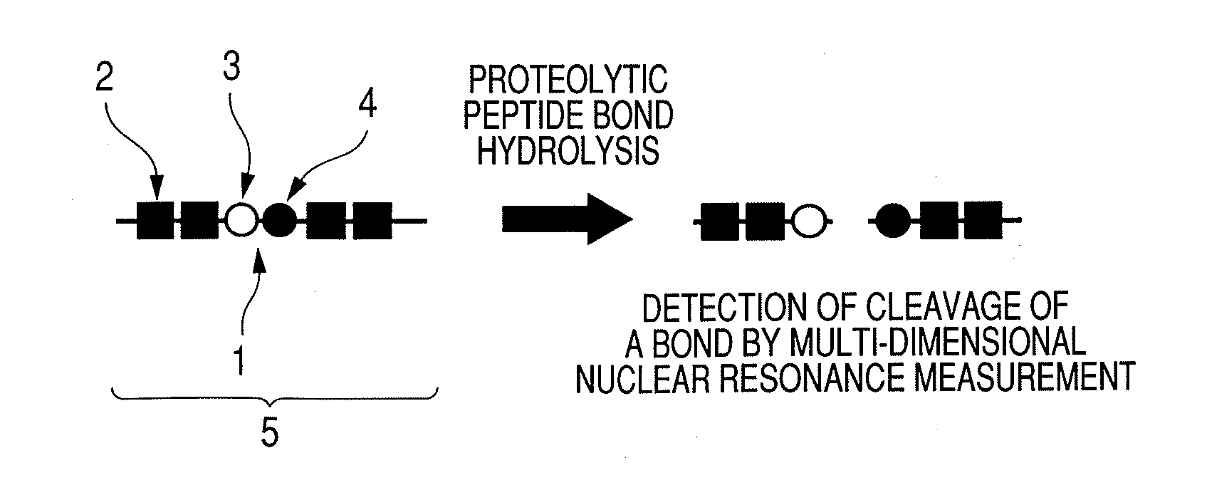
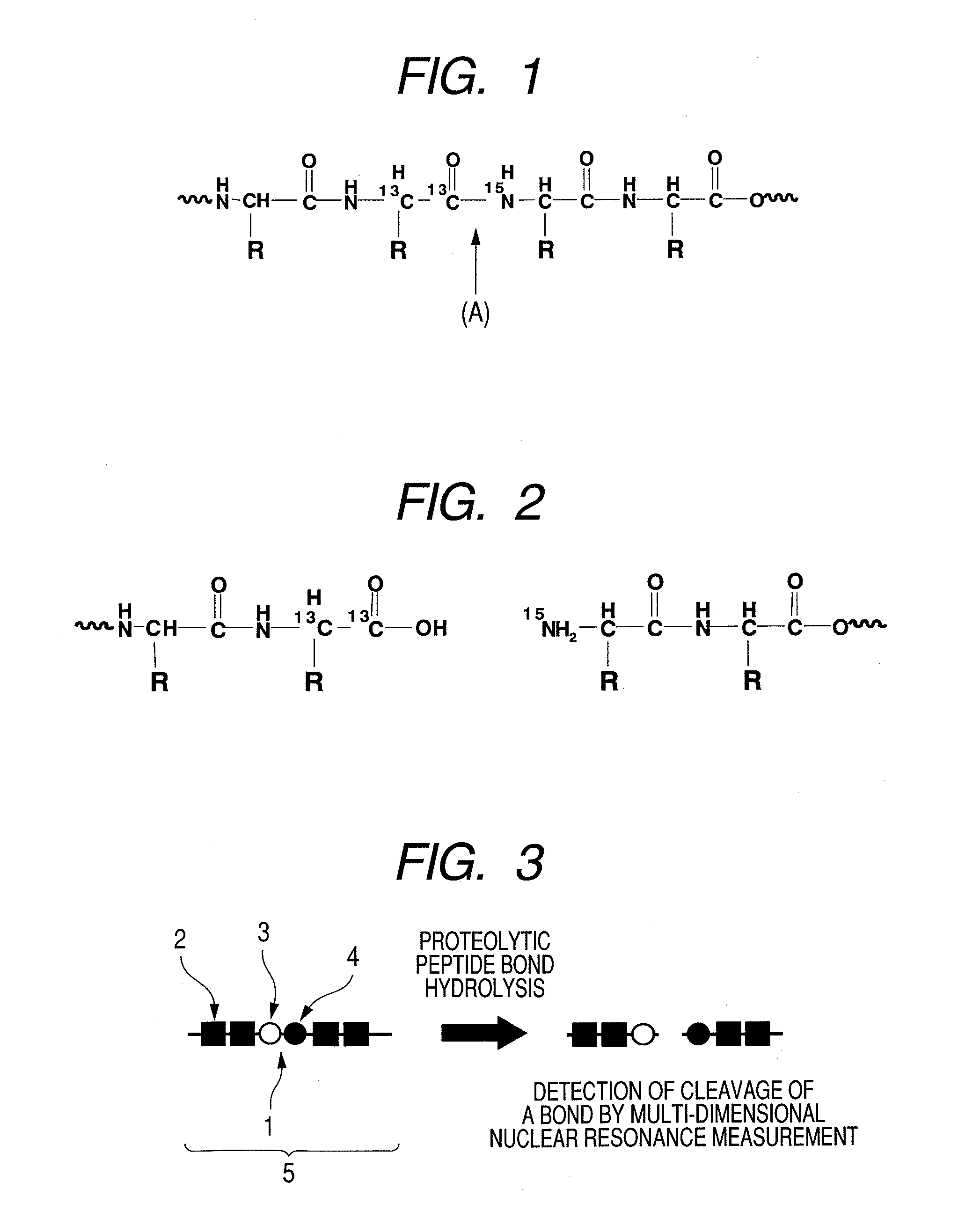
[0012]The present inventors found that the activity of a small amount of enzyme can be detected easily with high sensitivity by employing multi-dimensional nuclear magnetic resonance methods using a substrate probe having a stable isotope(s) labeled at an enzyme recognition site, in combination. Based on the finding, the present invention was accomplished.

Problems solved by technology

It has been found that the localization of enzyme changes and abnormal increase or decrease of enzyme activity occurs in the tissues and cells affected by various diseases.
These conventional techniques have the following problems that have remained unsolved.
First, when an MRI probe is used, degree of relaxation caused by enzymatic activation is relatively low.
When a luminescent probe or a fluorescent probe is used, it is difficult to capture a signal sent from deep part of a living body since permeability of light through the living body is low and light is scattered in the living body.
Another problem resides in the specificity of a probe to a substrate, which decreases due to chemical modification and labeling.
Consequently, the dose of the probe comes to be limited, with the result that a signal / noise ratio decreases when enzyme activity is monitored.
However, up to present, detection of protease activity by a multi-dimensional (three dimensional or more) nuclear magnetic resonance method using a stable-isotope labeled substrate probe has not yet been made and a substrate probe to be used in the method has not been disclosed, either.

Method used


Image

  • Substrate probe, enzyme-activity detection method by a multi-dimensional nuclear magnetic resonance method and enzyme-activity imaging method
  • Substrate probe, enzyme-activity detection method by a multi-dimensional nuclear magnetic resonance method and enzyme-activity imaging method
  • Substrate probe, enzyme-activity detection method by a multi-dimensional nuclear magnetic resonance method and enzyme-activity imaging method

Examples


example 1

Detection of MMP2 Activity

1-1: Synthesis of Isotope-Labeled Substrate Peptide-Probe to MMP2

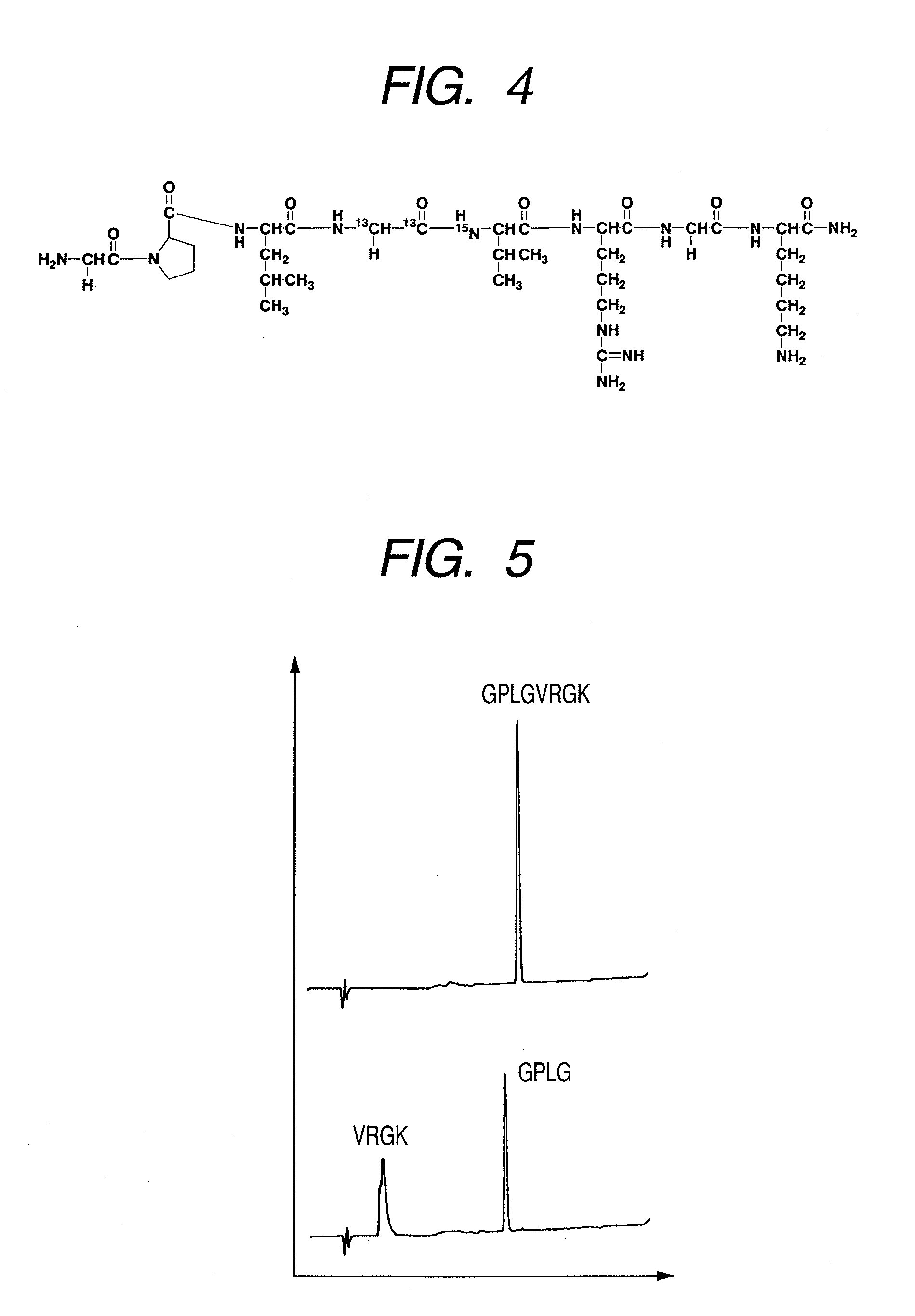
(1) Synthesis of Peptide GPLGVRGK (H-Gly-Pro-Leu-Gly-Val-Arg-Gly-Lys-NH2)

[0090]Peptide GPLGVRGK (H-Gly-Pro-Leu-Gly-Val-Arg-Gly-Lys-NH2: Sequence No. 1) was synthesized by a general Fmoc method. To obtain a peptide having the 4th amino acid (Gly) from the N terminal, labeled with 13C at carbon atoms of the 1-position and the 2-position of the skeleton thereof and the 5th amino acid (Val) from the N terminal, labeled with 15N at nitrogen atom of the skeleton thereof, amino acids (Gly and Val) to be placed to these positions were previously labeled with isotopes and subjected to synthesis. Synthesis was performed by using Rink amide resin (manufactured by Novabiochem) in an amount of 0.2 mmol. The isotope-labeled amino acid reagents (L-Valine-15N,N-FMOC derivative, GLYCINE-13C2, F-MOC derivative) used in synthesis were purchased from ISOTEC. If not otherwise specified, non-isotope labeled amino a...

example 2

Multi-Dimensional Resonance NMR Analysis of Lactic Acid Production by Metabolic Reaction of Pyruvic Acid

example 2-1

Experiment of Culturing HeLa Cells in a Medium Containing 13C-Labeled Pyruvic Acid

[0106]13C-labeled pyruvic acid was dissolved in a DMEM (+FBS) solution at 7 mM (30 ml) and the solution was sterilized by filtration. Three dishes were prepared in which a DMEM (+FBS) solution (9 ml) containing 13C-labeled pyruvic acid was poured. A DMEM (+FBS, +AB) solution (1 ml) containing Hela cells (700,000 cells / ml) was added to each of the dishes and cultured for 3 days under the conditions of 37° C., 5% CO2.

[0107]After the culture solution was collected, each dish was washed with a PBS solution (3 ml). The washing solution was also collected together with the culture solution. Thereafter, a trypsin-EDTA solution (1.5 ml) was added and cells were removed from the dishes by a cell scraper. After that, this solution was collected in another tube. Washing was performed again by the PBS solution (3 ml) and collected together with a cell solution. The culture solution and cell solution thus collected...


PUM


Abstract

Substrate probe capable of detecting enzyme activity with high accuracy and a method for detecting the enzyme activity by a multi nuclear magnetic resonance method using the substrate probe. Multi-dimensional nuclear magnetic resonance is performed by using a substrate probe, which is used for measuring enzyme activity by a multi-dimensional nuclear magnetic resonance method and characterized by containing a enzyme recognition site that is selectively recognized by an active-state enzyme, as at least one constitutional unit, and a group to which at least three nuclear magnetic resonance active nuclei each having a nuclear spin and a different resonance frequency are connected, being present specifically to the enzyme recognition, thereby detecting presence of the substrate probe and the enzyme activity. Alternatively, imaging of the enzyme activity is performed by a multi-dimensional nuclear resonance imaging method.

Description

TECHNICAL FIELD[0001]The present invention relates to a substrate probe for measuring enzyme activity. The present invention also relates to a method for measuring enzyme activity by a multi-dimensional nuclear magnetic resonance method using the probe and a method for imaging enzyme activity by multi-dimensional nuclear magnetic resonance imaging.BACKGROUND ART[0002]Enzymes are important proteins which catalyze chemical reactions in a living body. The localization and activities thereof are strictly controlled. It has been found that the localization of enzyme changes and abnormal increase or decrease of enzyme activity occurs in the tissues and cells affected by various diseases.[0003]In tumor cells, the glycolysis using glucose as a raw material plays an important role in producing energy. Particularly in tumor cells present in a low-oxygen environment, the glycolysis acts as a major energy supply source. In the glycolysis, various enzymes act. For example, lactic dehydrogenase i...

Claims


Application Information

Patent Timeline
IPC IPC(8): C12Q1/54C12Q1/32C12Q1/37
CPCG01N24/088C12Q1/54G01R33/4633C12Q1/32C12Q1/34G01R33/281G01R33/465G01R33/483G01R33/5601Y02P20/582
Inventor YAMAUCHI, FUMIOYOSHIMURA, KIMIHIROKUGE, KATSUAKIYANO, TETSUYASHIRAKAWA, MASAHIROAOYAMA, YASUHIROSANDO, SHINSUKE
Owner CANON KK
Learn More