Discovery and validation of cancer biomarkers using a protein analysis methodology to analyze specimens

a protein analysis and specimen technology, applied in the field of gene and proteomics, can solve the problems of cell death, inability to detect cancer biomarkers, and inability to detect cancer biomarkers, and achieve the effect of optimizing the selection of therapeutic agents

Inactive Publication Date: 2010-10-14
THE BOARD OF TRUSTEES OF THE LELAND STANFORD JUNIOR UNIV
View PDF16 Cites 5 Cited by
  • Summary
  • Abstract
  • Description
  • Claims
  • Application Information

AI Technical Summary

Benefits of technology

[0043]The information obtained from NIA is used to monitor treatment, modify therapeutic regimens, and to further optimize the selection of therapeutic agents. With this approach, therapeutic and/or diagnostic regimens can be individualized and tailored according to the data obtained at different times over the course of treatment.
[0044]It is to be understood that this invention is not limited to the particular methodology, protocols, cell lines, animal species or genera, and reagents described, as such may vary. It is also to be understood that the terminology used herein is for the purpose of describing particular embodiments only, and is not intended to limit the scope of the present invention, which will be limited only by the appended claims.
[0045]As used herein the singular forms “a”, “and”, and “the” include plural referents unless the context clearly dictates otherwise. Thus, for example, reference to “a compound” includes a plurality of such compounds and reference to “the agent” includes reference to one or more agents and equivalents thereof known to those skilled in the art, and so forth. All technical and scientific terms used herein have the same meaning as commonly understood to one of ordinary skill in the art to which this invention belongs unless clearly indicated otherwise.
[0046]Glycosylation. Among the post-translational modifications that can be probed, are protein specific glycoslyation. Membrane associated carbohydrate is exclusively in the form of oliogsaccharides covalently attached to proteins forming glycoproteins, and to a lesser extent covalently attached to lipid forming the glycolipids. Glycoproteins consist of proteins covalently linked to carbohydrate. The predominant sugars found in glycoproteins are glucose, galactose, mannose, fucose, GalNAc, GlcNAc and NANA. The distinction between proteoglyca

Problems solved by technology

Once the disease has progressed to locally advanced cancer or metastatic disease, these therapies are less successful.
With over 2000 human genes predicted to code for kinases and the potential for each kinase to act on multiple targets, signaling networks are

Method used


Image

  • Discovery and validation of cancer biomarkers using a protein analysis methodology to analyze specimens
  • Discovery and validation of cancer biomarkers using a protein analysis methodology to analyze specimens
  • Discovery and validation of cancer biomarkers using a protein analysis methodology to analyze specimens

Examples


example 1

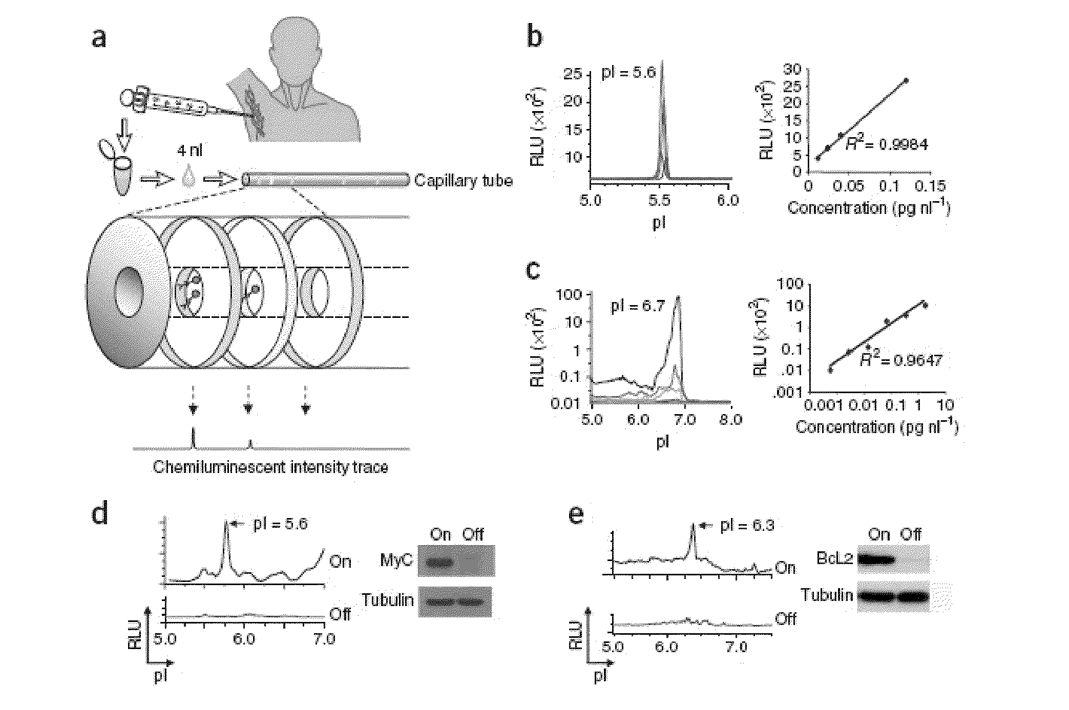
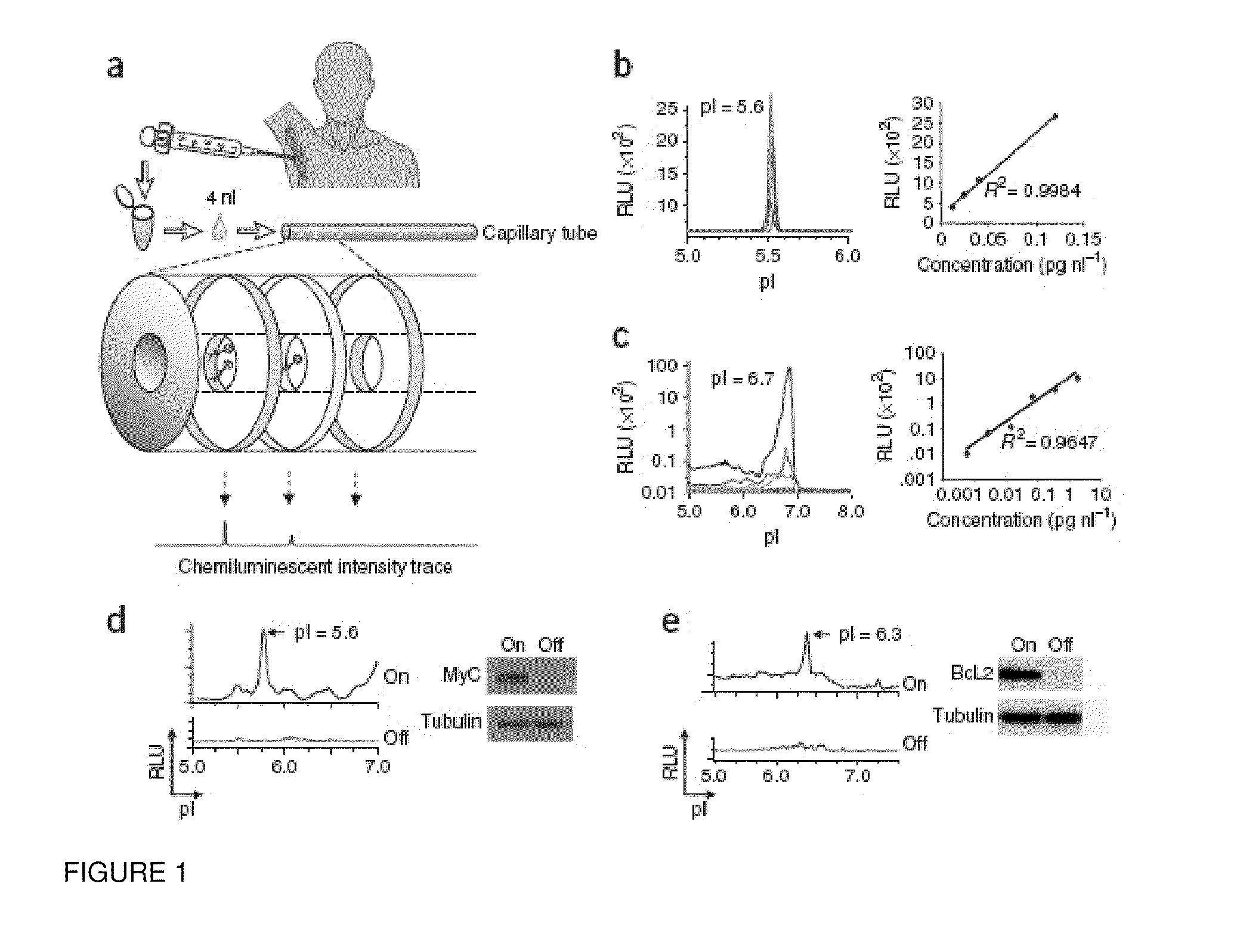
[0075]In order to accurately measure oncoprotein expression and activation in limited clinical specimens, we have developed a nano-fluidic proteomic immunoassay detection method (NIA) that combines isoelectric protein focusing and antibody detection. Here we demonstrate that we can use this new technique to quantitate oncoprotein expression and phosphorylation in clinical specimens to precisely measure specific changes in phospho-isomers of oncoproteins in vitro and in vivo.

Results

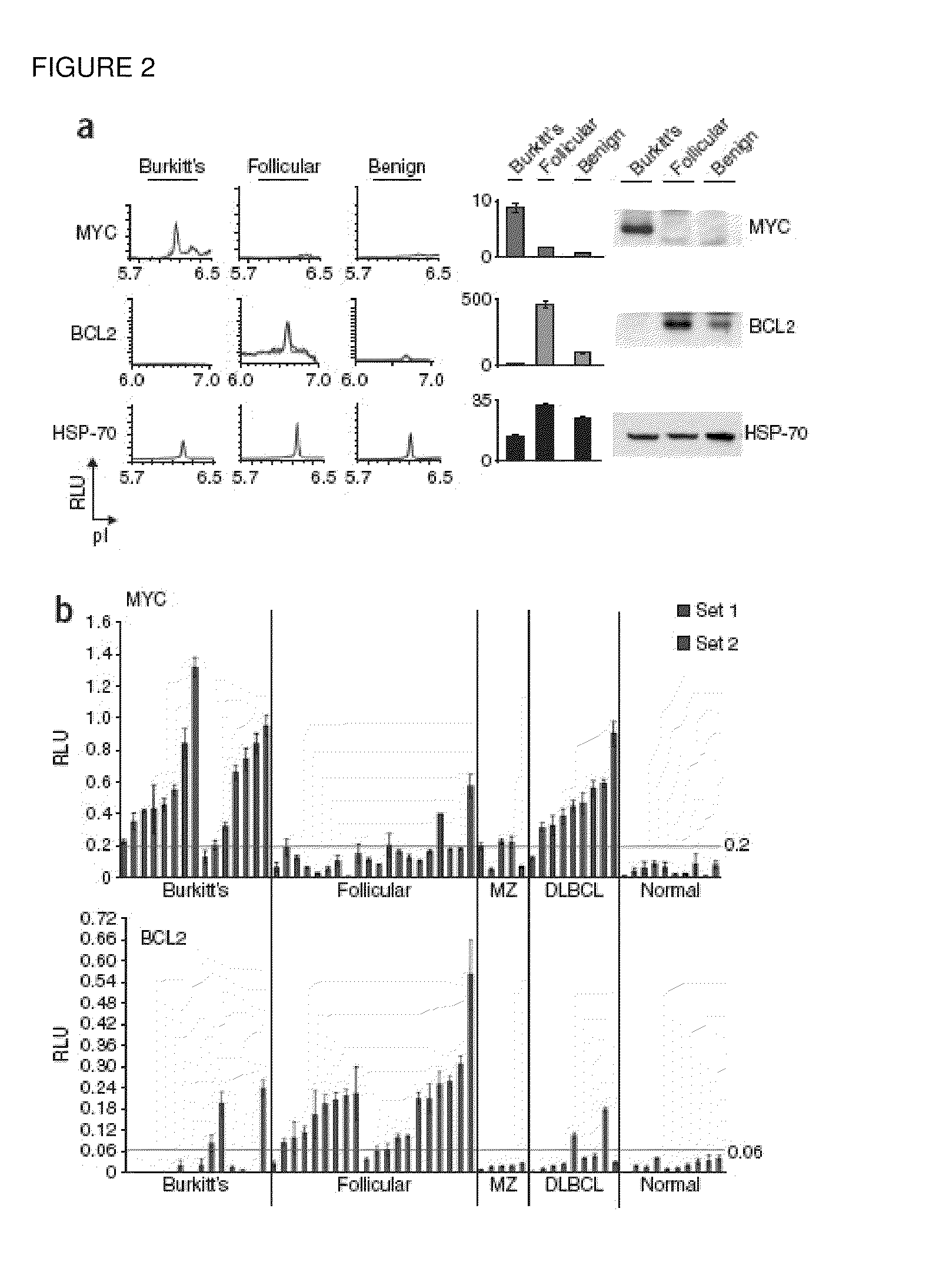
[0076]NIA Detection of Oncoprotein Expression in Clinical Specimens. NIA incorporates isoelectric focusing of proteins, followed by antibody detection of specific epitopes with chemiluminescence. The chemiluminescent signal is rendered as a chemiluminescence isoelectropherogram (trace) of “relative luminescence units (RLU)” on the y-axis vs. isoelectric point (pI) on the x-axis (FIG. 1a). As little as 2 picograms of MYC could be detected using 4 nanoliters of recombinant protein per capillary (final capill...

example 2

Nanoscale Quantification of Phosphorylated and Unphosphorylated ERK and MEK Isoforms Differentiates Tumor and Non-Tumor Clinical Specimens

[0108]We have developed the use of a highly sensitive microfluidic nano-immunoassay system (NIA) to perform detailed analysis of ERK and MEK activation in hematopoietic and solid tumors. We described above the use of NIA for measurement of proteins in as little as 4 nL of lysate from lymphoma and leukemia specimens. Now, we present results measuring specific isoforms of MAPK proteins in fine needle aspirates (FNAs) from patients with solid tumors and in blood buffy coats from patients with Myelo-Dysplastic Syndrome (MDS).

[0109]Using a single antibody that recognizes both the phosphorylated and unphosphorylated isoforms of ERK, we can determine levels of each ERK isoform, and also percent phosphorylation of ERK. NIA revealed that different tumor types could be distinguished based upon differing patterns of ERK isoforms. To determine if NIA can meas...


PUM


Abstract

Methods are provided for the analysis, including the serial analysis, of very small samples of tissue. The methods utilize a nanofluidic proteomic immunoassay (NIA) to quantify total and low-abundance protein isoforms in a small amount of lysate. NIA detection accurately measure oncoprotein expression and activation in limited clinical specimens, including isoforms that differ in post-translational modifications, such as phosphorylation, and the like. The NIA detection method combines isoelectric protein focusing and antibody detection in a nanofluidic system.

Description

BACKGROUND OF THE INVENTION[0001]The recent explosion of information in the fields of genomics and proteomics has provided a rich ground for the discovery of molecular targets against which therapeutic and / or diagnostic agents can be directed. Tissues for potential target discovery may include tumors and other malignant growths, or infected or inflamed tissues. For example, methods have been described for gene expression profiling of tumor cells (see any one of Ono et al. (2000) Cancer Res. 60(18):5007-11; Svaren et al. (2000) J Biol Chem.; or Forozan et al. (2000) Cancer Res. 60(16):4519-25 for examples). Similarly, proteomics has been used to profile the protein expression in tumor samples (see Minowa et al. (2000) Electrophoresis 21(9):1782-6; Cole et al. (2000) Electrophoresis 21(9):1772-81; Simpson et al. (2000) Electrophoresis 21(9):1707-32); etc.[0002]Cancer is caused by multiple genetic events that result in the activation of proto-oncogenes and / or the inactivation of tumor ...

Claims


Application Information

Patent Timeline
IPC IPC(8): G01N33/50
CPCG01N33/57407B82Y15/00G01N33/5748G01N2333/82G01N2333/912G01N2570/00
Inventor FELSHER, DEAN W.FAN, ALICE
Owner THE BOARD OF TRUSTEES OF THE LELAND STANFORD JUNIOR UNIV
Why Eureka
Try Eureka