Nanoparticles for cancer sonodynamic and photodynamic therapy

a cancer and photodynamic technology, applied in the field of nanoparticles comprising a cancer therapeutic agent, can solve the problems of nausea, vomiting, hair loss, unsatisfactory systemic reactions, etc., and achieve the effects of preventing exposure, similar cytotoxic effects, and increasing tumor burden

Inactive Publication Date: 2010-10-14
INTELLIGENTNANO +1
View PDF2 Cites 23 Cited by
  • Summary
  • Abstract
  • Description
  • Claims
  • Application Information

AI Technical Summary

Benefits of technology

[0079]The effect of nanoparticle size upon cytotoxicity was determined by testing SL052-NPs having different sizes, 131 nm, 150 nm and 247 nm (FIG. 5C). Serial dilutions of each sample of SL052-NPs were made (concentrations of 120 μg / ml, 60 μg / ml, 30 μg / ml, 15 μg / ml, and 7.5 μg / ml). The cells were treated with SL052-NPs at 37° C. for two hours and then ultrasound at an intensity of 0.56 w / cm2 for two minutes. The IC50 results show that SL052-NPs, having a concentration between 9.5 μg / ml and 36 μg / ml and sizes between 131 nm and 247 nm, display similar cytotoxic effects upon application of ultrasound. Line A in FIG. 5C shows 131 nm results, line B shows 150 nm results, while line C shows 247 nm results.
[0080]Male Balb / c mice were obtained from Charles River Laboratories International, Inc. (Wilmington, Mass.) and allowed to acclimatize for two weeks prior to testing. All mice had a bilateral flank implant of 1×10⁶ EMT-6 murine mammary tumor on each side. The tumors were allowed to grow to at least 5 mm in diameter before drug and light treatment. All treated mice received an intravenous tail vein injection of either 2 mg / kg, 4 mg / kg or 6 mg / kg nanoparticle solution. The injection contained 40 μL of the nanoparticle solution plus 60 μL of sterile saline to yield a 100 μl total volume injection. Two groups of nanoparticle formulation injected animals were used. The first group was treated with light four hours after injection. The second group was treated with light twenty-four hours after injection. The light treatment for both groups was 100 J / cm2 at a wavelength of 650 nm with a fluency rate of 200 mW from a HPD diode laser via a 400 nm fiber. The tumor was measured and the longest axis was used to calculate the light spot size. The animals were anaesthetized and then draped during the light treatment except for the tumor area to prevent exposure in case of uptake by normal tissues. Once treatment was completed, the animals were left to recover and returned to their cages. The tumor response was monitored daily by caliper measurements of the tumor length, width and thickness. The following formula was used to calculate the tumor volume:
[0081]All tumor-bearing mice, which were treated with SL052-NPs at doses of 2 or 6 mg / kg and exposed to light, became tumor-free after light treatment after a 4 hour incubation period, except for one mouse which had been treated with a 6 mg / kg dose of SL052-NPs and light. The mouse was ill on day one post-treatment and died on day two. The cause of death was inconclusive from a gross postmortem examination. Mice treated with light 24 hours after injection of SL052 NPs remained healthy, but none of the animals had tumor ablation and had to be euthanized due to increasing tumor burden.
[0082]In a further experiment, mice bearing the original tumor were treated with a 4 mg / kg dose of SL052-NPs, with PDT after a 4 hour incubation period (FIG. 6B). After two weeks, the treated mice became tumor-free. In mice bearing both original and migrated tumors, the PDT was administrated at four hours post-treatment with SL052-NPs only to the original tumor. Without being bound to theory, re-growth of tumors in these mice may have been due to tumor-implanted cells which migrated away from the treatment site and were subsequently not PDT-treated, resulting in delayed but steady tumor re-growth.

Problems solved by technology

However, both radiation and chemotherapy may harm healthy cells, resulting in undesirable systemic reactions including malaise, fatigue, loss of appetite, nausea, vomiting, headache, pain and hair loss.
Photodynamic therapy is mainly limited to superficial and / or small lesions since light cannot penetrate through more than about one centimetre of tissue.
However, hypocrellins are strongly hydrophobic (i.e., not water-soluble), making them problematic for clinical applications.
Hypocrellins modified in these manners exhibit low cellular uptake and poor biological compatibility; for example, the compound in Wang et al.

Method used


Image

  • Nanoparticles for cancer sonodynamic and photodynamic therapy
  • Nanoparticles for cancer sonodynamic and photodynamic therapy
  • Nanoparticles for cancer sonodynamic and photodynamic therapy

Examples


example 1

Formation of SL052—PVP Nanoparticles

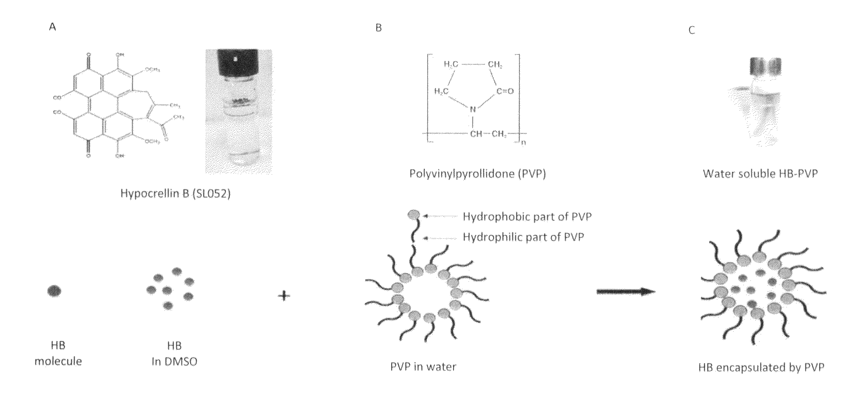
[0055]Polyvinylpyrollidone (PVP) (average molecular weight of 40,000) was purchased from Sigma Aldrich Canada Ltd. (Oakville, Canada), and an exemplary hypocrellin-B derivative (designated as “SL052”) was provided by Quest PharmaTech Inc. (Edmonton, Canada). The precipitation method was used to prepare SL052-NPS. Briefly, 1.5 mL of 0.5% (7.5 mg / ml) PVP aqueous solution was added to 6 mL of water with mixing at room temperature. After ten minutes, 1.59 mL of 4.6 mM SL052 in dimethylsulfoxide (DMSO, Fisher Scientific) was added to the mixture. The resulting solution was stirred for ten minutes under darkness to yield a nanodispersion with a nanoparticle size of 136 nm. The SL052-NPS were deep blue in color and water-soluble (FIG. 1C).

example 2

Formation of Fluorescent SL052—PVP Nanoparticles

[0056]Fluorescent SL052 nanoparticles are formed by adding 1.5 mL of 0.5% PVP aqueous solution to 6 mL of water with mixing at room temperature. After ten minutes, 1.59 mL of 4.6 mM SL052 and 0.1 mM fluorescein isothiocyanate in DMSO was added to the mixture. The resulting solution was stirred for ten minutes under darkness to yield a nanodispersion.

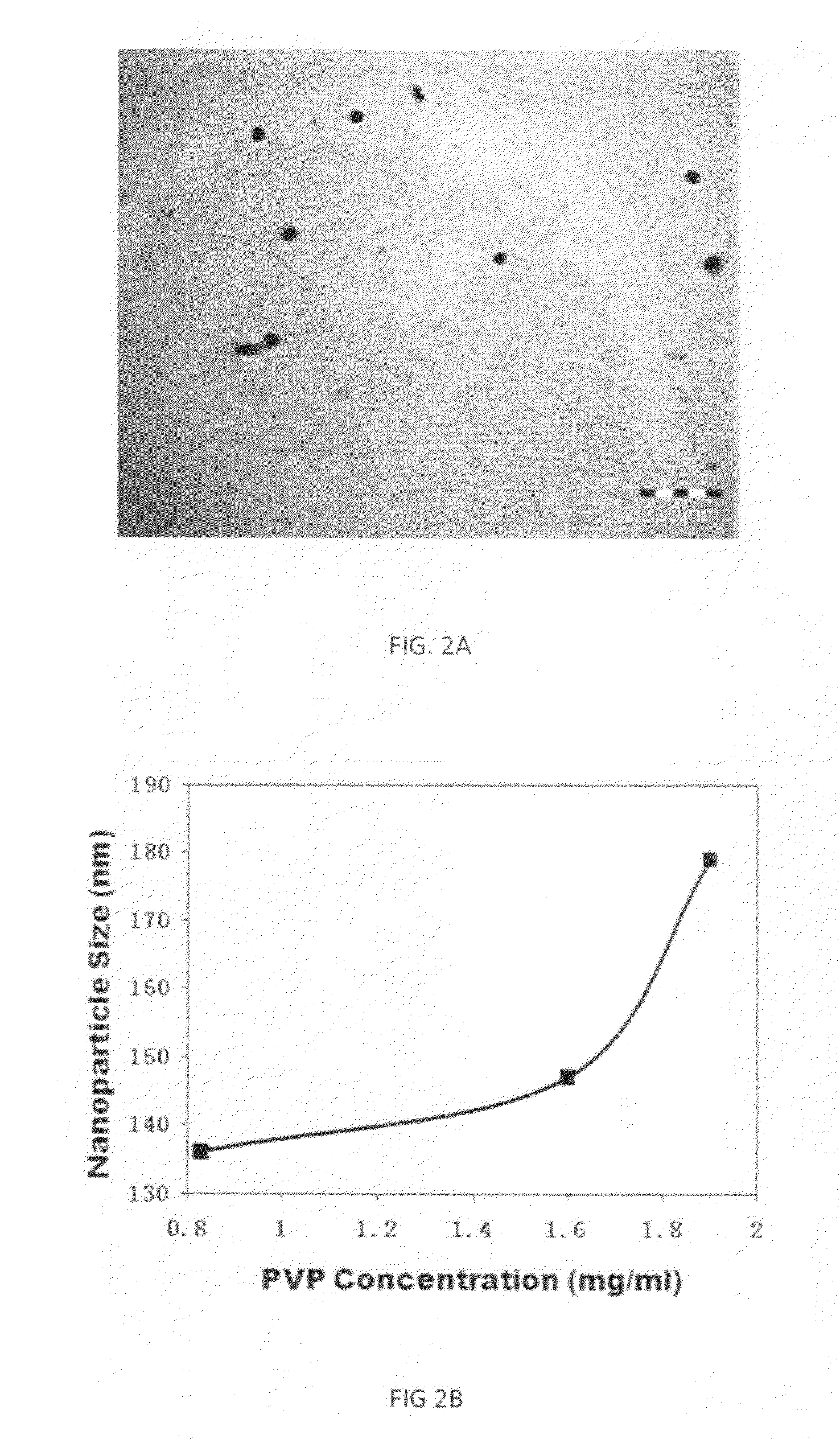
[0057]SL052-NPs labelled with fluorescein isothiocyanate were used to treat cells for two hours with 6.25 mg / ml, 12.5 μg / ml, or 25 μg / ml SL052-NPs. Confocal microscopy confirmed that as the concentration of SL052-NPs increased, more SL052-NPs entered into cells, as may be seen in FIG. 2A.

example 3

Determination of the Structures of the SL052-NPS

[0058]The structures of the SL052-NPS were determined by transmission electron microscopy (TEM). SL052-NPS were negatively stained with phosphotungstic acid and observed using TEM to confirm their spherical structure (FIG. 2A).


PUM

PropertyMeasurementUnit
concentrationaaaaaaaaaa
sizeaaaaaaaaaa

Abstract

The present invention is directed to nanoparticles comprising a cancer therapeutic, pharmaceutical compositions comprising same, and methods for using same for drug delivery and ultrasound or light-based treatment of cancer.

Description

CROSS REFERENCE TO RELATED APPLICATION[0001]This application claims the priority benefit of U.S. Provisional Application No. 61 / 167,403 filed on Apr. 7, 2009 entitled “Nanoparticles for Cancer Sonodynamic and Photodynamic Therapy,” the contents of which are incorporated herein by reference.FIELD OF THE INVENTION[0002]This invention relates to nanoparticles comprising a cancer therapeutic agent, pharmaceutical compositions comprising same, and methods for using same for drug delivery and sonodynamic or photodynamic treatment of cancer.BACKGROUND OF THE INVENTION[0003]Radiation therapy and chemotherapy are conventional treatments for cancer. Radiation therapy involves delivery of an optimal dose of either particulate or electromagnetic radiation to a particular area of the body with minimal damage to normal tissues. The source of radiation may be outside the body of the patient or may be an isotope implanted or instilled into the body. Chemotherapy involves treatment by chemical agent...

Claims


Application Information

Patent Timeline
Patent Type & Authority Applications(United States)
IPC IPC(8): A61M37/00A61K9/14A61K31/498A61K39/44A61K51/12A61P35/00
CPCA61K9/0009A61K9/1075A61K41/0057A61K31/498A61K41/0033A61K9/5138A61P35/00
Inventor MADIYALAKAN, RAGUPATHYXING, JAMESWOO, THOMASSWANSON, ERIC
Owner INTELLIGENTNANO
Why Eureka
Try Eureka