Process for sterilizing acellular soft tissue with irradiation

a technology of acellular soft tissue and irradiation, which is applied in the field of soft tissue treatment, can solve the problems of narrow therapeutic window between adequate immunosuppression and toxicity, infection threat, and significant risk of rejection in transplantation

Inactive Publication Date: 2010-11-18
MUSCULOSKELETAL TRANSPLANT FOUND INC
View PDF5 Cites 6 Cited by
  • Summary
  • Abstract
  • Description
  • Claims
  • Application Information

AI Technical Summary

Benefits of technology

[0020]It is still another object of the invention to provide acellular sterilized dermis which can be stored for long periods of time for later use by a surgeon for implantation into a human being.

Problems solved by technology

However, problems exist when there is a transfer of biological material from one individual to another Tissue rejection is a significant risk associated with transplantation, even with a good histocompatability match.
These immunosuppressive drugs however, have a narrow therapeutic window between adequate immunosuppression and toxicity.
Prolonged immunosuppression can weaken the immune system, which can lead to a threat of infection.
It can thus be seen that the prior art processes require extensive chemical treatment with a multitude of process steps in an attempt to obtain an acellular sterilized soft tissue specimen which has limited shelf life.

Method used


Image

  • Process for sterilizing acellular soft tissue with irradiation

Examples


example 1

Sterilization of Dermis with E-beam Irradiation

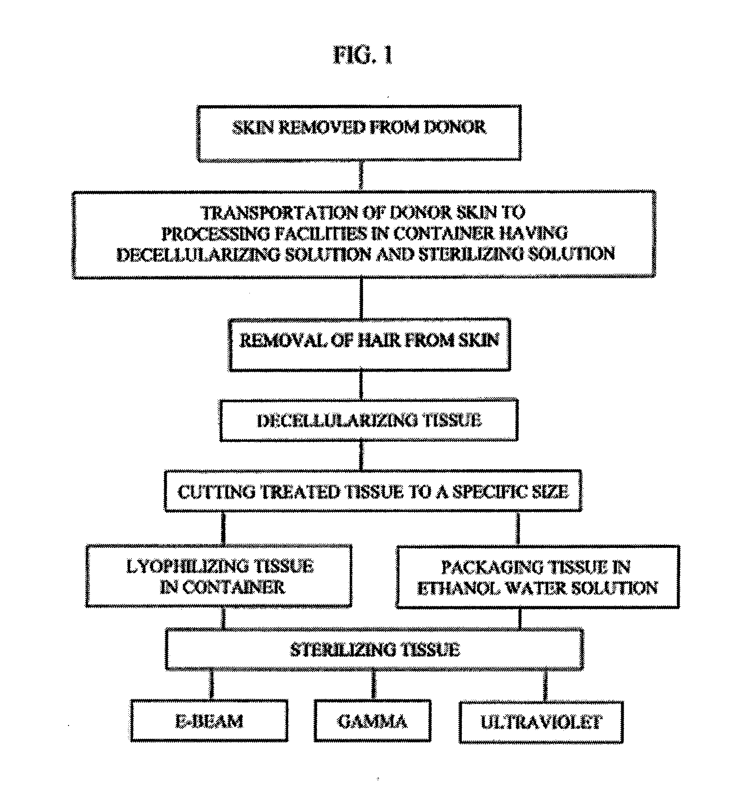
[0032]The tissue which has been previously obtained from a donor is shipped from the donor site in a container having a sterilization solution mixed with a decellularizing solution such as sodium chloride and then frozen.

[0033]The donor tissue is then thawed and then rinsed to maintain moisture. The thawed tissue is processed by removing hair and is then decellularized using 1M NaCI and 0.1% of Triton X-100. If desired at the time of decellularization one or more of the following protease inhibitors may be added; Aminoethylbenzenesulfonyl fluoride HCL (serine proteases) (25-100 μm, Aprotinin (broad spectrum, serine proteases) (7.5-30 μm), Protease Inhibitor E-64 (cysteine proteases) (0.05-0.20 μm), Leupeptin, Hemisulfate (cysteine proteases) (0.05-0.20 μm), EDTA, Disodium (0.025-0.10 μm), and trypsin-like proteases, Pepstatin A (Aspartic Proteases), Marmistat (MMP2). The tissue is processed and decellularized and is inspected for visual...

example 2

Sterilization of Dermis with Gamma Irradiation

[0042]The tissue which has been previously obtained from a donor is shipped from the donor site in a container having a sterilization solution mixed with a decellularizing solution such as sodium chloride and then frozen.

[0043]The donor tissue is then thawed and then rinsed to maintain moisture. The thawed tissue is processed by removing hair and is then decellularized using 1M NaCl and 0.1% of Triton X-100. If desired at the time of decellularization one or more of the following protease inhibitors may be added; Aminoethylbenzenesulfonyl fluoride HCL (serine proteases) (25-100 Aprotinin (broad spectrum, serine proteases) (7.5-30 μm), Protease Inhibitor E-64 (cysteine proteases) (0.05-0.20 μm), Leupeptin, Hemisulfate (cysteine proteases) (0.05-0.20 μm), EDTA, Disodium (0.025-0.10 μm), and trypsin-like proteases, Pepstatin A (Aspartic Proteases), Marmistat (MMP2). The tissue is processed and decellularized and is inspected for visual defe...

example 3

Sterilization of Dermis with Ultraviolet Light

[0053]The tissue which has been previously obtained from a donor is shipped from the donor site in a container having a sterilization solution mixed with a decellularizing solution such as sodium chloride and then frozen.

[0054]The donor tissue is then thawed and then rinsed to maintain moisture. The thawed tissue is processed by removing hair and is then decellularized using 1M NaCl and 0.1% of Triton X100. If desired at the time of decellularization one or more of the following protease inhibitors may be added; Aminoethylbenzenesulfonyl fluoride HCL (serine proteases) (25-100 μm, Aprotinin (broad spectrum, serine proteases) (7.5-30 μm), Protease Inhibitor E-64 (cysteine proteases) (0.05-0.20 μm), Leupeptin, Hemisulfate (cysteine proteases) (0.05-0.20 μm), EDTA, Disodium (0.025-0.10 μm), and trypsin-like proteases, Pepstatin A (Aspartic Proteases) Marmistat (MMP2). The tissue is processed and decellularized and is inspected for visual de...


PUM


Abstract

The present invention relates to a process for preparing skin removed from a human donor, removal of cellular components and sterilizing the decellularized skin. The process comprises the following steps:
    • (1) decellularizing the skin including soaking the tissue in a detergent and rinsing same with sterile water;
    • (2) sterilizing the skin with electronic beam irradiation, gamma irradiation or ultraviolet light for a time period to achieve a concentration to maximize sterilization; and
    • (3) processing the tissue by cutting the tissue to a designated size.

Description

RELATED APPLICATIONS[0001]This application claims the benefit of priority of U.S. Provisional Application No. 60 / 929,082 filed Jun. 12, 2007.[0002]The foregoing applications, and all documents cited therein or during their prosecution (“application cited documents”) and all documents cited or referenced in the application cited documents, and all documents cited or referenced herein (“herein cited documents”), and all documents cited or referenced in herein cited documents, together with any manufacturer's instructions, descriptions, product specifications, and product sheets for any products mentioned herein or in any document incorporated by reference herein, are hereby incorporated herein by reference, and may be employed in the practice of the invention.FIELD OF INVENTION[0003]The present invention is generally directed toward methods of treatment of soft tissue including decellularizing and sterilization of the soft tissue by irradiation for implantation into another human bein...

Claims


Application Information

Patent Timeline
Patent Type & Authority Applications(United States)
IPC IPC(8): A01N1/02
CPCA61L2/0011A61L2/0035A61L27/362A61L2430/40A61L27/3691A61L27/60A61L27/3683
Inventor NGO, MANH-DANSTUART, JEFFREY
Owner MUSCULOSKELETAL TRANSPLANT FOUND INC
Why Eureka
Try Eureka