Method for nonlinear imaging of ultrasound contrast agents at high frequencies

a contrast agent and ultrasound technology, applied in the field of nonlinear imaging of ultrasound contrast agents at high frequencies, can solve the problems of poor contrast between microbubbles and surrounding tissue, difficult visualization of microbubbles, and reduced image quality outside of fixed focus, so as to improve the sensitivity of microbubble contrast agents

Inactive Publication Date: 2010-11-25
VISUALSONICS
View PDF6 Cites 11 Cited by
  • Summary
  • Abstract
  • Description
  • Claims
  • Application Information

AI Technical Summary

Benefits of technology

[0006]The invention provides ultrasound devices and methods that provide improved sensitivity to microbubble contrast agents. The invention uses multiple transmitted ultrasound pulses, which allows

Problems solved by technology

A disadvantage with this approach, however, is that in many cases the ultrasound echoes from tissue have a comparable magnitude to microbubbles, resulting in poor contrast between the microbubbles and the surrounding tissue.
This effect can make visualization of the microbubbles difficult, even after post-processing.
These characteristics can result in a large varia

Method used


Image

  • Method for nonlinear imaging of ultrasound contrast agents at high frequencies
  • Method for nonlinear imaging of ultrasound contrast agents at high frequencies
  • Method for nonlinear imaging of ultrasound contrast agents at high frequencies

Examples


example 1

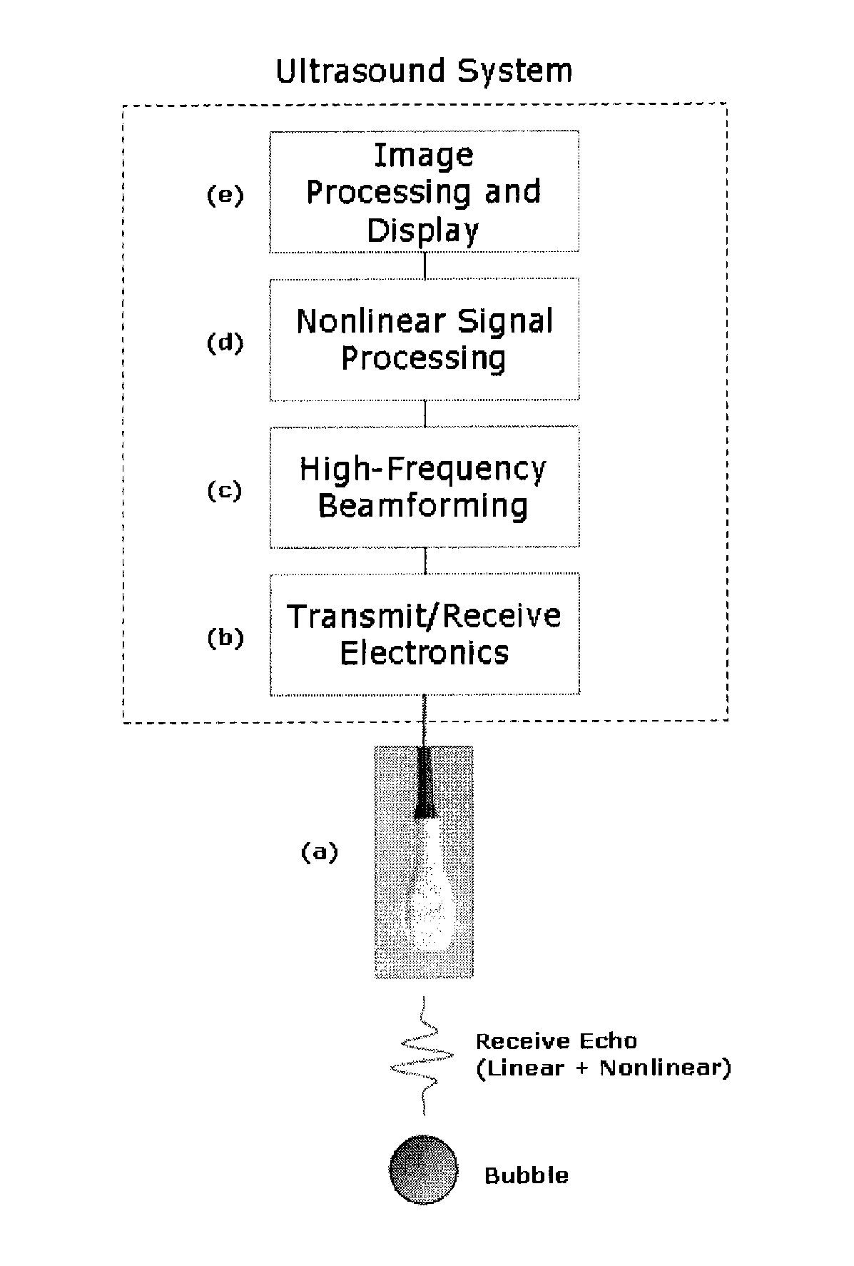
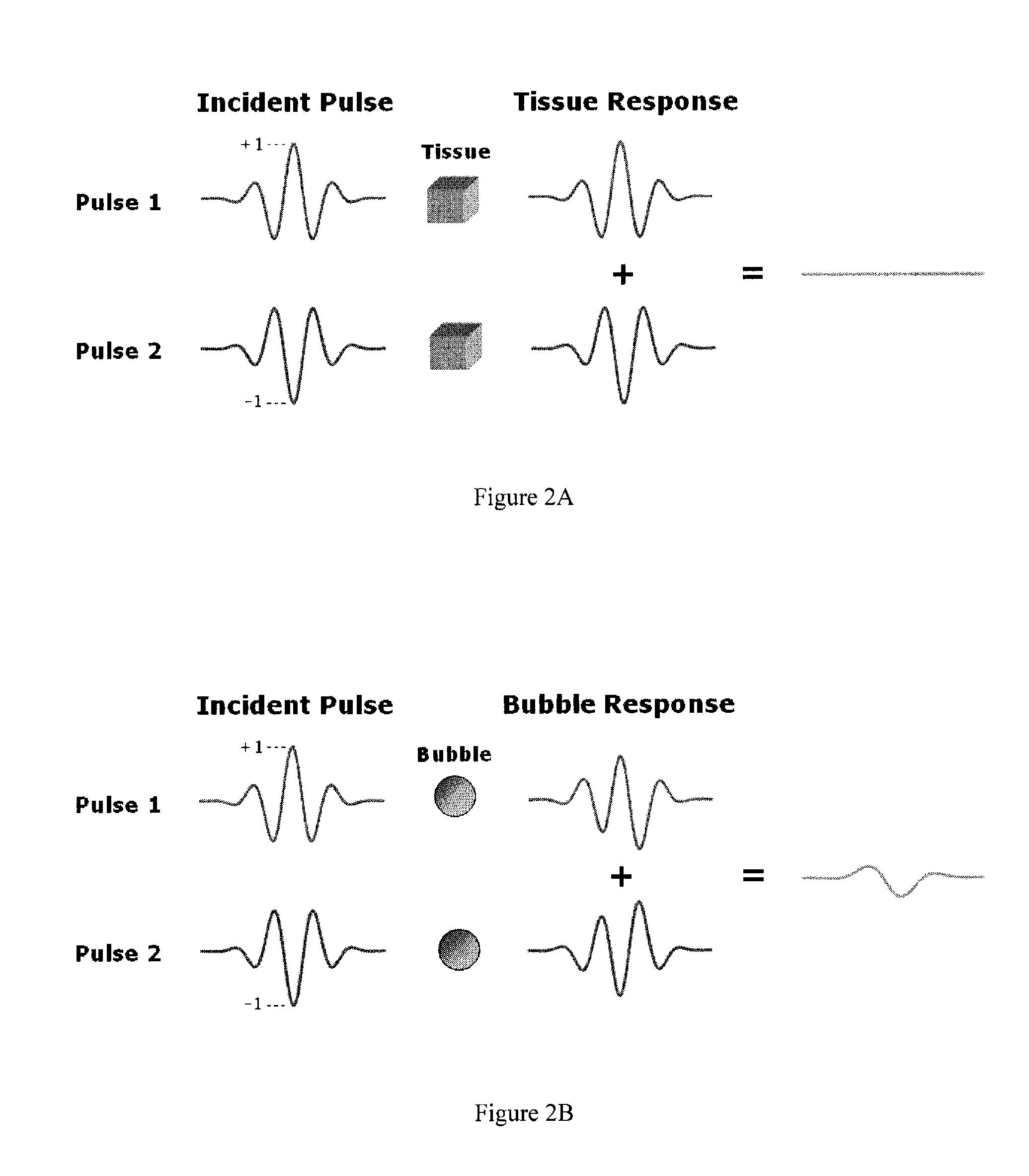
[0073]FIG. 4 shows data collected with a 21 MHz linear array (MS-250, VisualSonics, Toronto) at a transmit frequency of 24 MHz. The array was connected to a VisualSonics Vevo 2100 micro-ultrasound imaging system. The system is capable of beamforming 64 channels of data. The resulting summation from the 64 channels can be recorded digitally in baseband quadrature format and offloaded from the system for processing and analysis. The data are from MicroMarker (VisualSonics, Toronto) high frequency contrast agent flowing through a tissue-mimicking medium, using either phase inversion or amplitude scaling. FIG. 3 is a frequency plot of received ultrasound echoes, with all curves referenced to the raw unprocessed data (not shown). As shown in FIG. 4, both phase inversion and amplitude scaling detect nonlinear subharmonic energy at 12 MHz. In the case of amplitude scaling, additional nonlinear energy is detected at the fundamental frequency (24 MHz). In addition, phase inversion is better ...

example 2

[0075]An adult female mouse was administered a single 50-μl bolus of MicroMarker contrast agent (1.2·×107 bubbles per bolus) and imaged with amplitude scaling at 18 MHz using a Vevo 2100 ultrasound imagining platform (VisualSonics). The nonlinear contrast agent signal (right) is shown simultaneously with B-Mode images (left) in FIG. 6. The sequence of images shows the contrast enhancement attributable to the bolus over time. The scan plane was oriented from the dorsal side of the mouse, through a long section of the kidney.


PUM


Abstract

This invention employs multiple ultrasound pulse firings of either alternating phase and/or amplitude to detect nonlinear fundamental and subharmonic signals from microbubble contrast agents within living tissue, at high frequencies (≧15 MHz), e.g., with a linear array transducer. It can be shown that the contrast-to-tissue ratio (CTR) decreases with increasing ultrasound frequency because of nonlinear ultrasound propagation in tissue. However, using the subharmonic signal in addition to the nonlinear fundamental harmonic component, rather than the conventional second harmonic used at lower frequencies, provides appreciable signal strength to overcome the limitations of nonlinear tissue propagation. Additionally, the method provides for the ability to switch, at some desired frequency above 20 MHz, into a purely alternating phase inversion acquisition, in combination with bandpass filtering of the subharmonic frequency band, minimizing the losses in CTR as the frequency increases. This maintains contrast sensitivity for more limited fields of view, as penetration depth will be limited at higher frequencies. Thus, within the same micro-ultrasound imaging system, many applications of microbubble detection can be achieved with a wide range of frequencies that covers both resolution and sensitivity requirements.

Description

CROSS-REFERENCE TO RELATED APPLICATIONS[0001]This application claims benefit of U.S. Provisional Application No. 61 / 170,451, filed Apr. 17, 2009, which is hereby incorporated by reference.BACKGROUND OF THE INVENTION[0002]The invention relates to the field of nonlinear ultrasound imaging.[0003]Understanding the patterns of blood flow in the microcirculation is a powerful tool for evaluating the differences between normal and pathological tissues. In addition to visualizing and quantifying blood flow in the microcirculation, targeting microbubbles to cellular receptors and detecting them with ultrasound can give valuable insight about the molecular state of small animal models of human disease. Microbubble contrast agents have been used in ultrasound imaging as a means of improving the visualization of blood flow with respect to the surrounding tissue beyond the sensitivity of Power and Color Doppler imaging. These micron-sized particles (˜1-10 μm, on the order of the size of a red bl...

Claims


Application Information

Patent Timeline
IPC IPC(8): A61B8/14
CPCA61B8/06A61B8/13A61B8/14A61B8/481G01S15/8959G01S7/52038G01S7/52039G01S15/108G01S15/8956A61B8/543
Inventor NEEDLES, ANDREWMEHI, JAMES I.HIRSON, DESMOND
Owner VISUALSONICS
Why Eureka
Try Eureka