Method and system for vascular elastography
a vascular elastography and elastography technology, applied in the field of vascular tissue characterization, can solve the problems of difficult interpretation of motion parameters, potential limitations of non-invasive characterization of vessel walls with extracorporal ultrasound probes, and plaque rupture and thrombosis, and achieve the effect of non-invasive mapping the elastic properties of vessels
- Summary
- Abstract
- Description
- Claims
- Application Information
AI Technical Summary
Benefits of technology
Problems solved by technology
Method used
Image
Examples
first embodiment
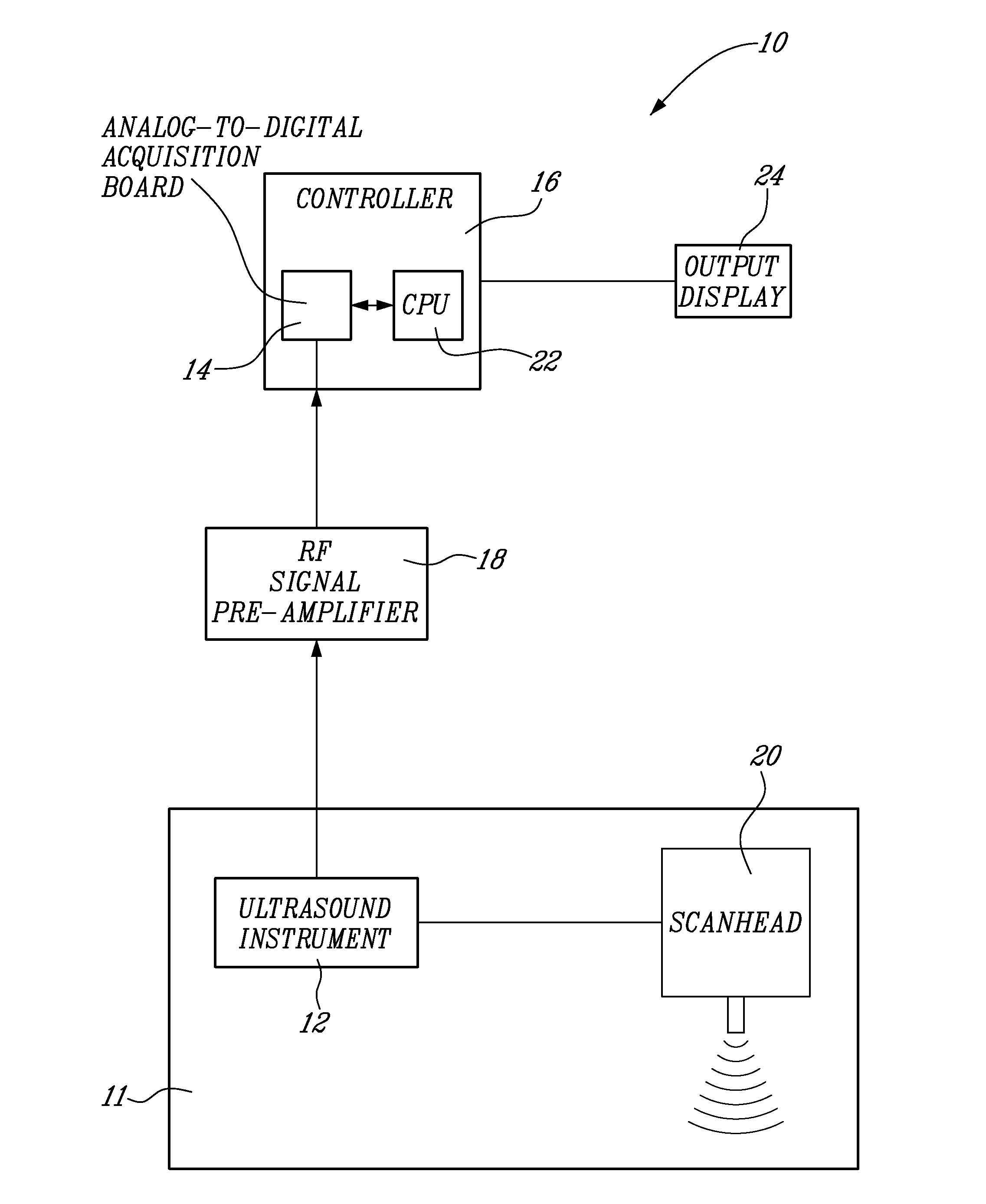
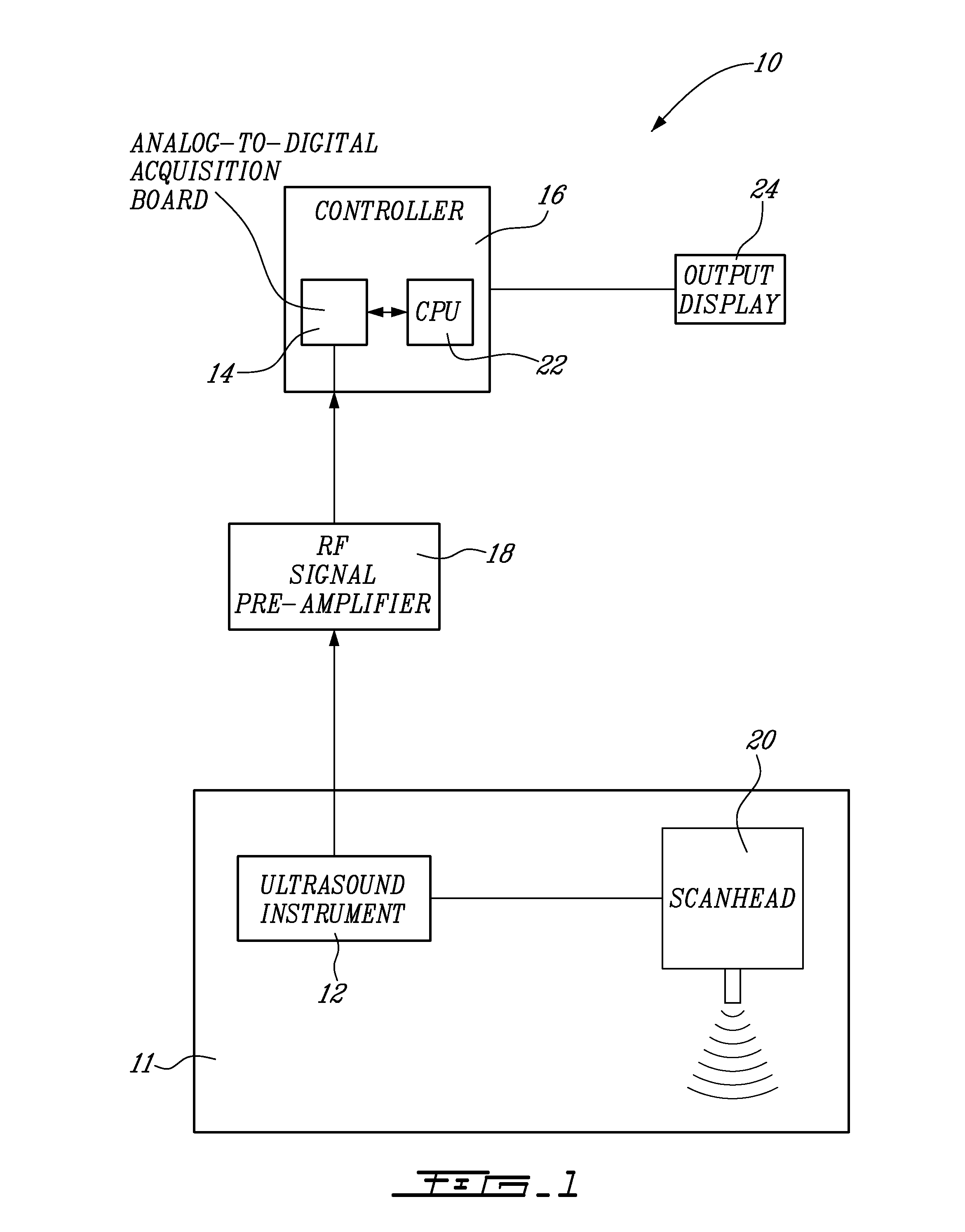
[0058]A system 10 for vascular elastography according to a first aspect of the present invention will now be described with reference to FIG. 1. More specifically, the system 10 allows for non-invasively characterizing arteries. Whereas not restricted to, this system allows predicting risks of vascular tissue rupture due to the presence of atherosclerotic plaques and potentially vascular aneurysms. Since vascular tissue rupture due to atherosclerotic plaques and aneurysms is believed to be well known in the art, it will not be described herein in more detail.
[0059]The system 10 comprises an ultrasound system 11 including an ultrasound instrument 12 provided with a scanhead 20 including an ultrasound transducer. The instrument 12 is coupled to an analog-to-digital acquisition board 14 of a controller 16 via a radio-frequency (RF) pre-amplifier 18.
[0060]For NIVE, the ultrasound instrument 12 is configured for extracorporal measurement, while for MicroNive, it is in the form of an ultr...
third embodiment
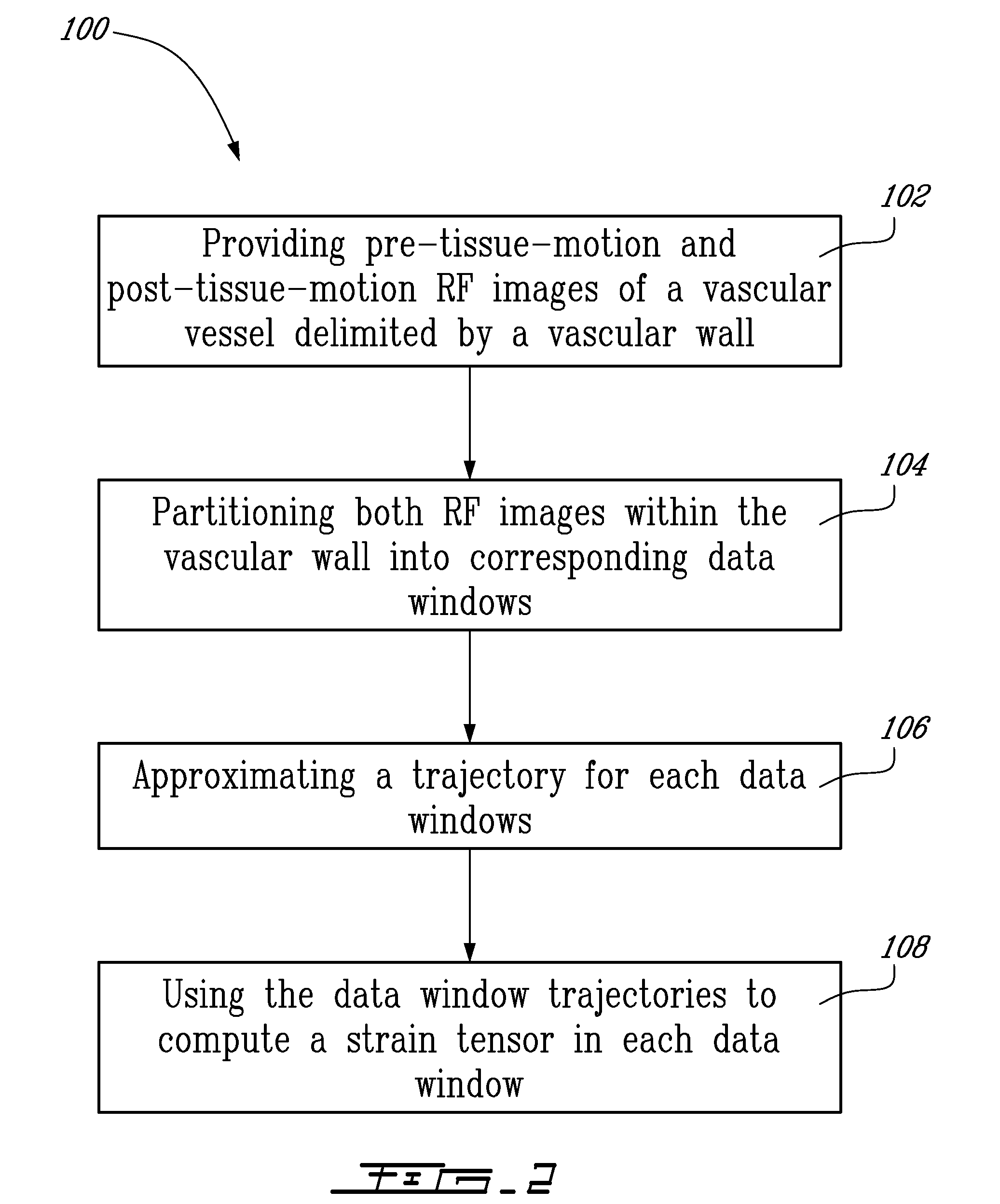
[0133]A method for endovascular elastography (EVE) according to a third illustrative embodiment of the second aspect of the present invention will now be described. Since the method according to this third embodiment is similar to the method illustrated in FIG. 2, and for concision purposes, only the differences between these two methods will be described herein.
[0134]The first step of the method is to acquire intravascular RF images using a catheter. Following the example of IVUS, and as schematically illustrated in FIG. 15, a transducer is placed at the tip of the catheter and cross-sectional imaging of a vessel is generated by sequentially sweeping the ultrasound beam over a 360° angle. It is to be noted that, in the ideal situation illustrated in FIG. 15, the ultrasound beam runs parallel with the vascular tissue motion, i.e. in the (r,φ) coordinate system.
[0135]Mechanical parameters (radial strain, in this case) are then estimated from analyzing the kinematics of the vascular t...
second embodiment
[0151]The method for endovascular elastography according to the present invention has also been validated in vitro using a fresh excised human carotid artery. The experimental set-up 50 used in the validation is illustrated in FIG. 23. The set-up 50 includes a system 52 for endovascular elastography according to the first aspect of the present invention.
[0152]The system 52 comprises an ultrasound scanner 54 in the form of a CVIS (ClearView, CardioVascular Imaging System Inc.) ultrasound scanner, working with a 30 MHz mechanical rotating single-element transducer (not shown), a digital oscilloscope 56, more specifically the model 9374L from LECROY, and a pressuring system 58.
[0153]The extremities 60-62 of an artery 64 are fixed to two rigid sheaths by watertight connectors 66, separated according to the original longitudinal dimension of the vessel 64 before excision. The intravascular, catheter 68, part of the system 52 was introduced through the proximal sheath into the lumen of th...
PUM
 Abstract
Description
Claims
Application Information
 - R&D
- Intellectual Property
- Life Sciences
- Materials
- Tech Scout
- Unparalleled Data Quality
- Higher Quality Content
- 60% Fewer Hallucinations
Browse by: Latest US Patents, China's latest patents, Technical Efficacy Thesaurus, Application Domain, Technology Topic, Popular Technical Reports.
© 2025 PatSnap. All rights reserved.Legal|Privacy policy|Modern Slavery Act Transparency Statement|Sitemap|About US| Contact US: help@patsnap.com


