Myostatin inhibitor enhancement of musculoskeletal repair

a myostatin inhibitor and musculoskeletal technology, applied in the field of muscle physiology and therapeutics, can solve the problems of insufficient novel strategies to utilize myostatin inhibitor enhancement, time-consuming treatment and care of these patients, etc., and achieve the effects of enhancing muscle regeneration, enhancing muscle regeneration, and improving fracture healing

Inactive Publication Date: 2010-12-30
GEORGIA HEALTH SCI UNIV RES INST
View PDF9 Cites 36 Cited by
  • Summary
  • Abstract
  • Description
  • Claims
  • Application Information

AI Technical Summary

Benefits of technology

[0012]Myostatin (GDF-8) is known as a potent inhibitor of muscle growth and development, and myostatin is also expressed early in the fracture healing process. The invention demonstrated that a myostatin inhibitor, a recombinant myostatin propeptide, enhanced the repair and regeneration of both muscle and bone in cases of deep penetrant injury. Blocking myostatin signaling in the injured limb improves fracture healing and enhances muscle regeneration. These data suggest that myostatin inhibitors are effective for improving wound repair in cases of orthopedic trauma and extremity injury.

Problems solved by technology

Approximately 2 million cases of delayed and nonunion fractures occur annually in the United States, and the treatment and care of these patients requires considerable time and cost.
Specifically, the prior art is deficient in novel strategies to utilize myostatin inhibitor enhancement of musculoskeletal repair.

Method used


Image

  • Myostatin inhibitor enhancement of musculoskeletal repair
  • Myostatin inhibitor enhancement of musculoskeletal repair
  • Myostatin inhibitor enhancement of musculoskeletal repair

Examples


example 1

Animals, Treatments, & Surgical Procedures

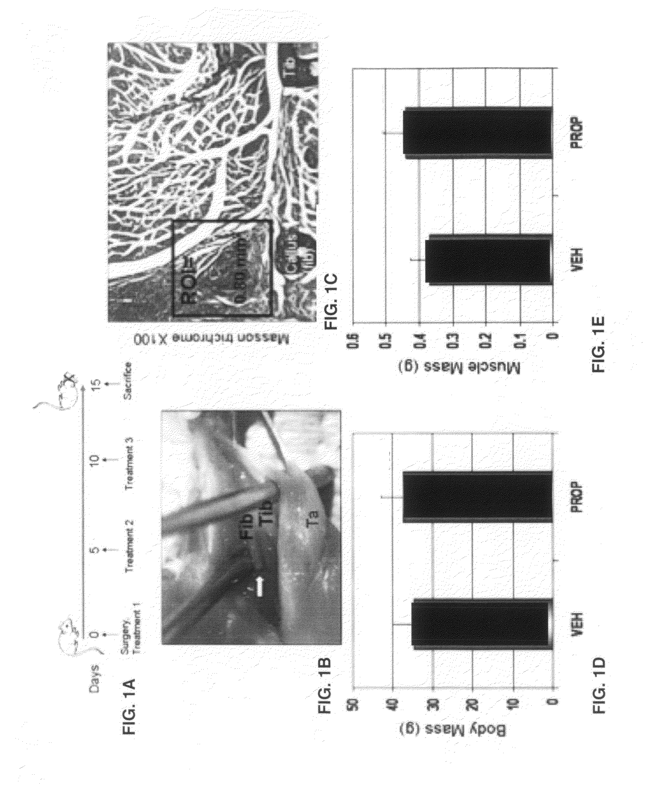
[0034]An initial dose-response study was performed to evaluate efficacy of the myostatin inhibitor in male CD-1 mice four months of age. Mice were treated with the propeptide at 0 mg / kg, 10 mg / kg, 20 mg / kg, or 50 mg / kg at day 0, day 5, and 10 and then sacrificed one week after the last treatment (FIG. 1A).

[0035]Results showed that three injections of the propeptide over a 15 day treatment period increased fore- and hindlimb muscle mass by 10% at the 10 mg / kg dose and increase muscle mass by more than 15% at the 20 mg / kg dose. The 50 mg / kg dose did not increase muscle mass beyond the increase observed in the 20 mg / kg group and so the 20 mg / kg dose was used. Adult CD-1 mice were separated into two groups: those receiving the propeptide (PRO) or saline (VEH). Each treatment group included 10-12 male and 10-12 female mice, for a total of 20-24 mice per treatment group. Fibula osteotomy was performed on the left leg under isoflurane anesthesia as...

example 2

MicroCT, Histology, & Histomorphometry

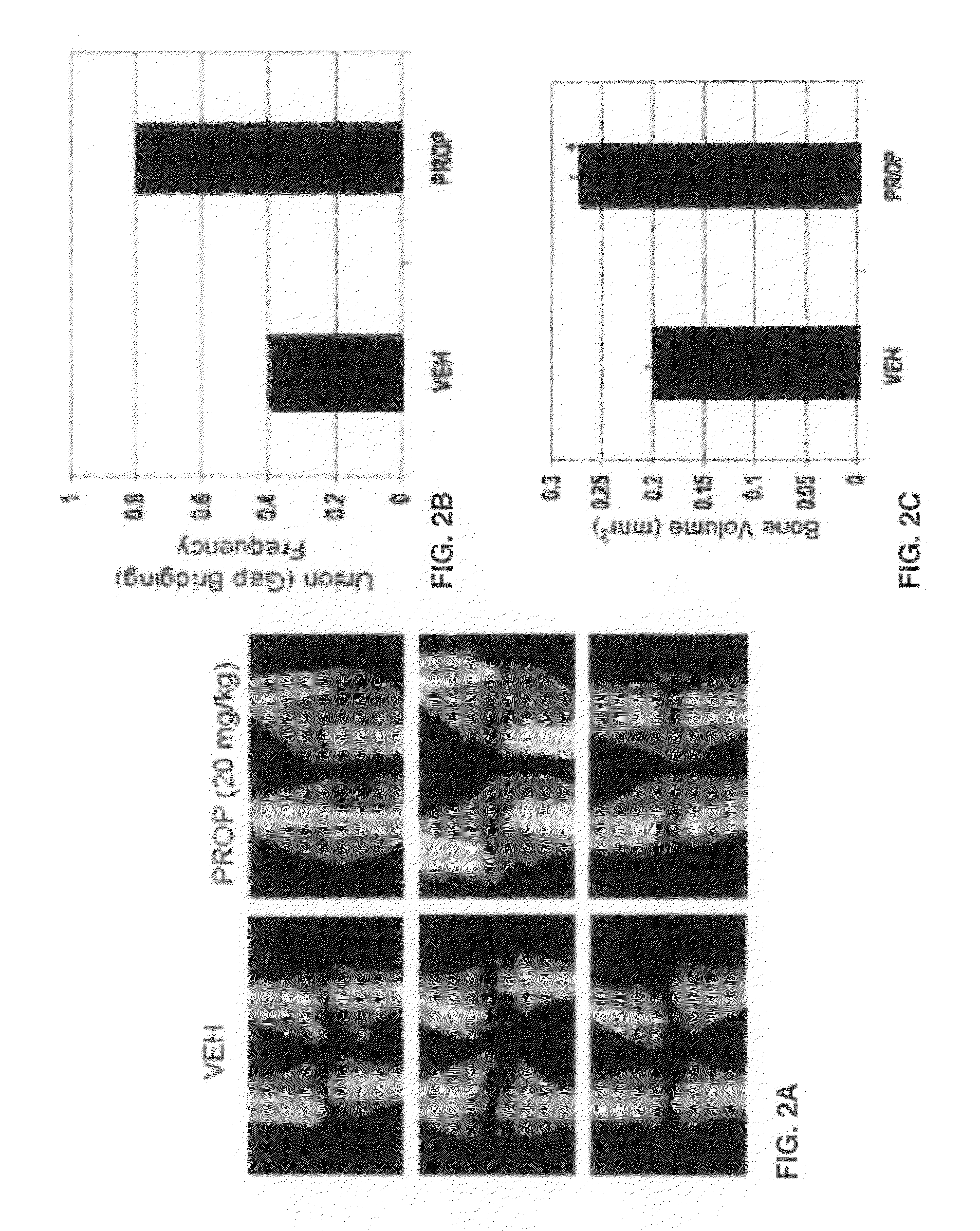
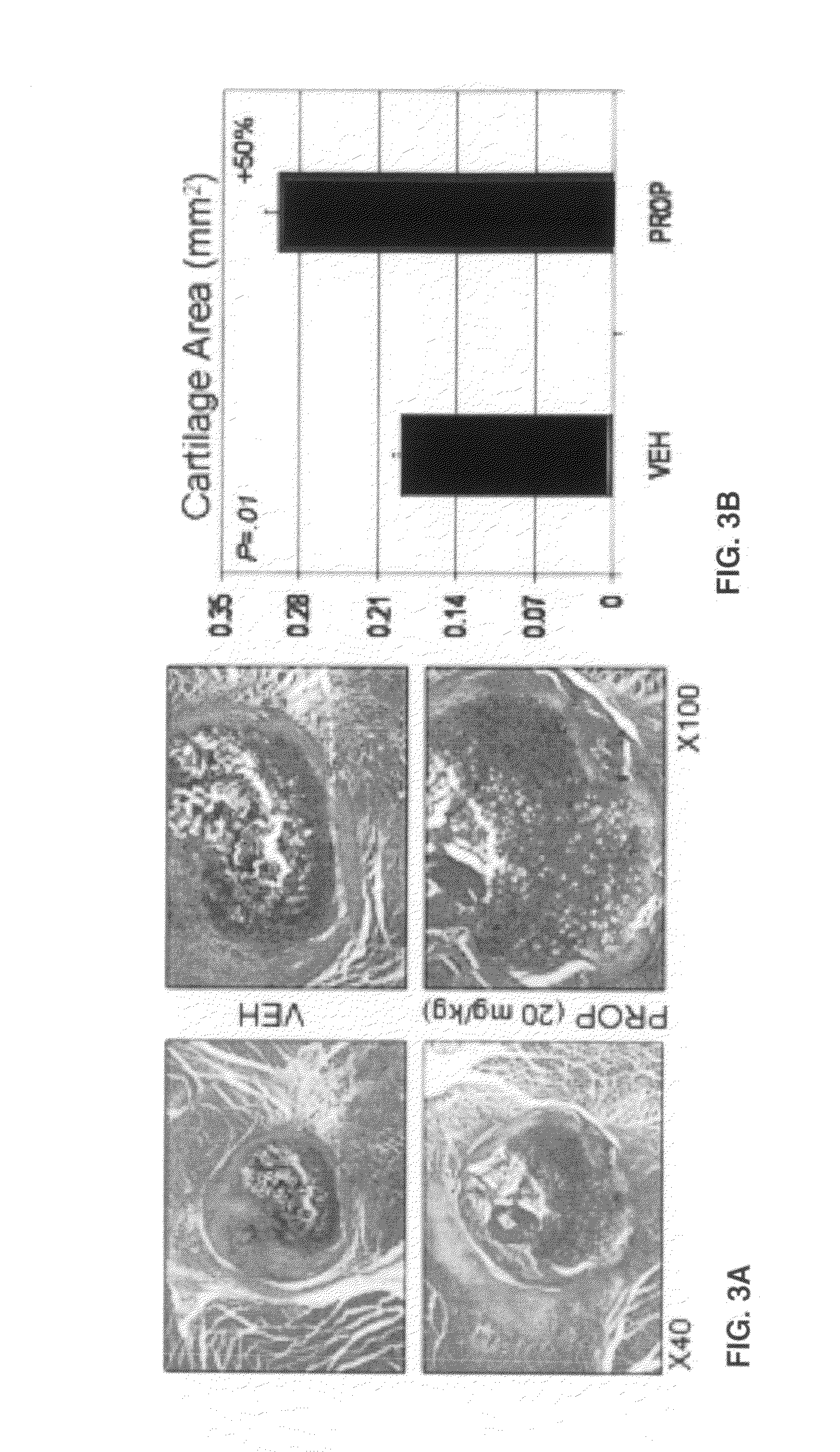
[0036]Intact legs with surrounding muscle were first imaged using a FAXITRON small-animal x-ray cabinet at 35 kVP, 2.5 mA for 45 seconds to verify that the fibula osteotomy was successful and that the tibia was not damaged. Specimens were sent to the Savannah River Site National Laboratory (Aiken, S.C.) for micro-computed tomography using a 160 kV micro-focus X-ray machine (Kevex Inc., Model 16010), a four-axis positioning system (New England Affiliated Technologies series 300), and an amorphous silicon imager (Varian Inc, Paxscan 4030) at 12 micron resolution. Measurements of total callus volume and callus bone mineral density were calculated 0.5 mm either side of the callus center. MicroCT images were then scored by a technician blind to the treatments as either having bone crossing the osteotomy site (‘bridged’) or showing no bone crossing the fracture gap ‘unbridged’). Specimens were then decalcified using EDTA, embedded in paraffin, and sec...

example 3

Statistical Analysis

[0037]Experiments were performed in two blocks, with osteotomy performed in half the mice (n=20-24) for Block 1 and then a second group of 20-24 mice included for Block 2 approximately six weeks later. Single-factor ANOVA was used to detect significant effects of treatment and block on the outcome measures described above. Chi-square test was used to test for differences between treatment groups in the frequency of bridged or unbridged osteotomy sites.


PUM

PropertyMeasurementUnit
particle sizeaaaaaaaaaa
timeaaaaaaaaaa
timeaaaaaaaaaa

Abstract

The methods and compositions of this invention provide a means of regenerating injured musculoskeletal tissue by inhibition of myostatin function. The invention provides methods of treating a nonunion fracture in an individual comprising delivering to the fracture via a delivery system comprising biodegradable hydrogel, a pharmacological amount of myostatin propeptide effective to inhibit myostatin function. The invention also teaches compositions useful for the treatment of non-union fracture in an individual, said compositions comprising a pharmacological amount of myostatin propeptide effective to inhibit myostatin function; and a delivery system for delivering said myostatin propeptide to said fracture, wherein the delivery system comprises a biodegradable hydrogel or biodegradable nanobeads.

Description

CROSS-REFERENCE TO RELATED APPLICATION[0001]This nonprovisional application claims benefit of priority under 35 U.S.C. §119(e) of provisional U.S. Ser. No. 61 / 269,421, filed Jun. 25, 2009, now abandoned, the entirety of which is hereby incorporated by reference.FEDERAL FUNDING LEGEND[0002]This invention was created in part using funds from the federal government under grant AR49717 from the National Institutes of Health and Dept of Defense (Office of Naval Research) under grant N000140810197. Consequently, the federal government has certain rights in this invention.BACKGROUND OF THE INVENTION[0003]1. Field of the Invention[0004]The present invention relates to the fields of muscle physiology and therapeutics. Specifically, the present invention relates to myostatin inhibitor enhancement of musculoskeletal repair.[0005]2. Description of the Related Art[0006]Approximately 2 million cases of delayed and nonunion fractures occur annually in the United States, and the treatment and care ...

Claims


Application Information

Patent Timeline
Patent Type & Authority Applications(United States)
IPC IPC(8): A61K38/18A61K9/14A61P19/04
CPCA61K9/0019A61K9/0092A61K9/06A61K38/179A61K38/18A61K47/20A61K47/42A61K9/5153A61P19/00A61P19/04
Inventor HAMRICK, MARK W.
Owner GEORGIA HEALTH SCI UNIV RES INST
Why Eureka
Try Eureka