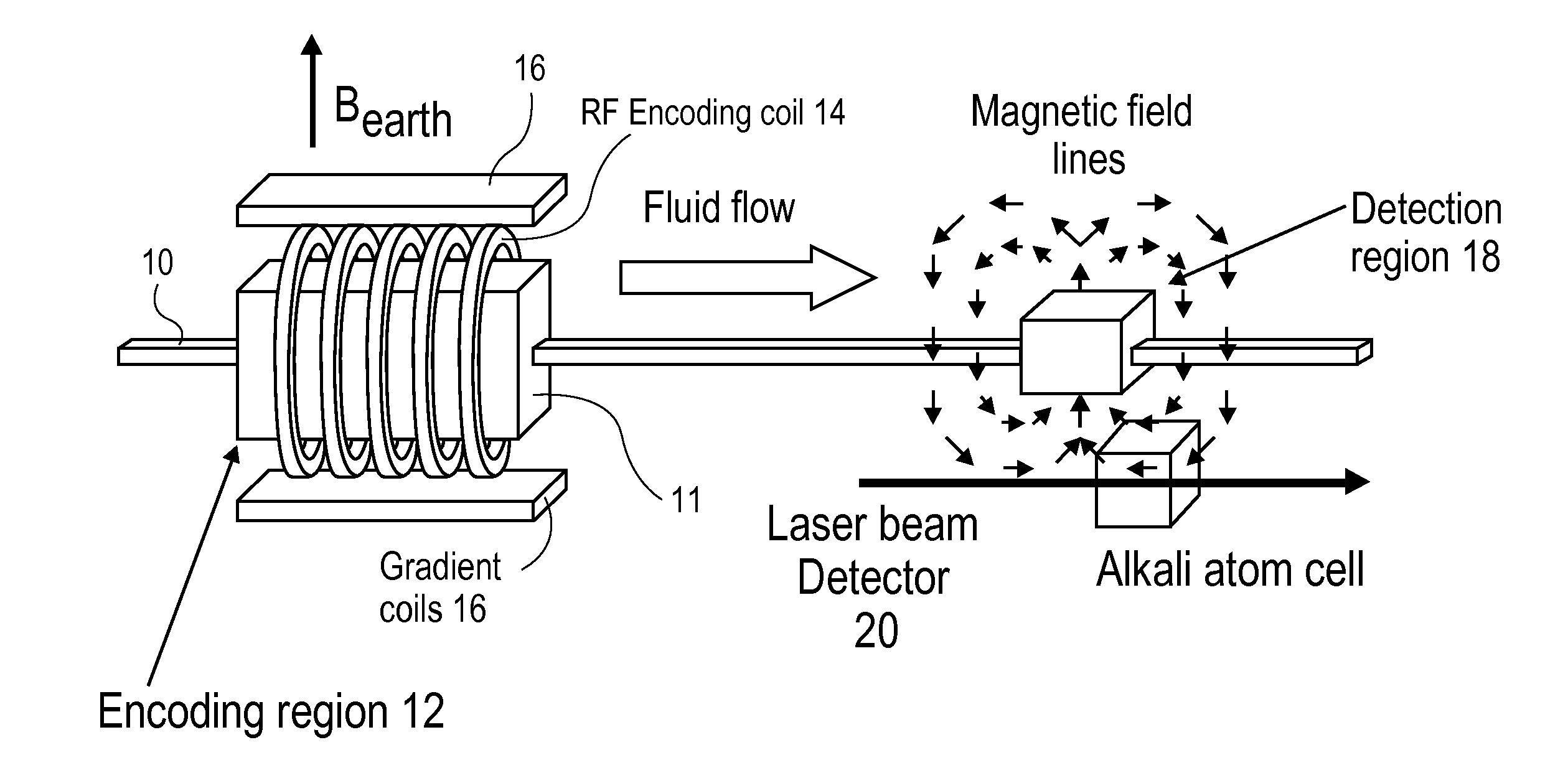
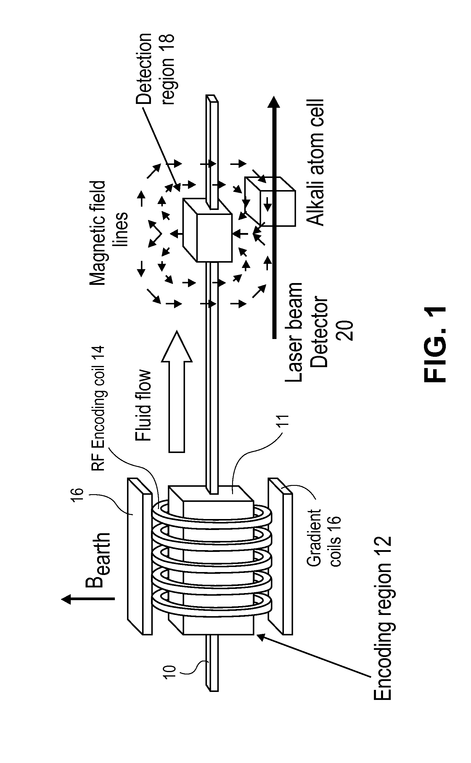
[0009]By way of this invention, a means has been found to address both the MRI sensitivity issue as well as the cost and portability issue. Thus as opposed to conventional NMR /
MRI imaging, where the
filling factor is low when detecting fluctuations in nuclear
magnetization measured in deep-seated tissues distant from the sensor, in the instant invention the nuclear
spins in a region are excited with RF (or AF) pulses, encoded using
chemical shift and / or imaging gradients (as longitudinal
magnetization, i.e., along the applied
magnetic field), and the
spins then physically transferred to a separate region where
magnetization is read out. This transport, which may occur over a period of 1 to several seconds, is possible because of the storage step. Because there is a much more effective
coupling of the
spins at the
detector in the optimized detection region, where the
filling factor is much greater, the sensitivity of afforded detection is much higher. Moreover, if
magnetic flux detection is used at the
detector, an acquisition-bandwidth
advantage is obtained due to the longer T1 (
longitudinal relaxation time) values compared to T2 (
transverse relaxation time) values. By repeated cycles of
frequency encoding and readout, the frequencies (for
spectroscopy) or the
spatial distribution of spins (for imaging) can be deduced. In addition, by using
highly sensitive detectors it is possible to do low field imaging, with the attendant
elimination of the need for the
high field superconducting magnets of conventional MRI.
[0011]In a second embodiment, one more suitable for portable NMR applications, the blood is prepolarized in a region upstream of the brain, such as the heart using a permanent
magnet such as a Halbach magnet array. In such a magnet, the
magnetic field is strongly focused over a
region of interest, resulting in maximal flux over that region, and minimal losses of flux to the regions outside the desired region. In this embodiment, the magnet being a permanent or electro magnet, the encoding step is performed at the head of the subject in the presence of the constant
magnetic field, thus eliminating the need for a
superconducting magnet. A typical RF head coil arrangement may be used for encoding. In this mode, encoding can be non selective (without any gradient applied), or selective, with one or more dedicated surface coils placed strategically as to cover a desired region. In this embodiment employing small permanent magnets, a degree of portability is afforded not otherwise possible.
[0013]Accordingly
highly sensitive detectors are required. In an embodiment of the invention,
laser-based atomic magnetometers that dramatically outperform conventional
inductive coupling sensors at low frequencies can be used. Alternatively, anisotropic magnetoresistive (AMR) sensors or any other sensitive
magnetometer can also be used. In addition to the benefits of cost, size and portability, low field imaging is potentially much more benign to patients with metallic inclusions such as shrapnel, and / or artificial joint implants.

