Biosynthetic cartilaginous matrix and methods for their production

a biosynthetic cartilaginous matrix and biosynthetic technology, applied in the direction of biocide, plant growth regulators, skeletal/connective tissue cells, etc., can solve the problems of expensive procedures and extensive medical procedures

Inactive Publication Date: 2011-01-20
COLOPLAST AS +1
View PDF22 Cites 9 Cited by
  • Summary
  • Abstract
  • Description
  • Claims
  • Application Information

AI Technical Summary

Problems solved by technology

Such procedures are costly, and require extensive medical procedures such as rehabilitation and physical therapy.
Methods using scaffold technologies of various forms, where the scaffold (with, or without cells grown in the scaffold) is inserted into the defect, have suffered from difficulties in performing the cell implantation procedure solely guided by arthroscopy.

Method used


Image

  • Biosynthetic cartilaginous matrix and methods for their production
  • Biosynthetic cartilaginous matrix and methods for their production
  • Biosynthetic cartilaginous matrix and methods for their production

Examples


example 1

In Vitro Study of Chondrogenesis of hAC-Loaded MPEG-PLGA Scaffolds

These in vitro studies were done in order to evaluate the degree of chondrogenic matrix synthesis in a 3-dimensional scaffold system base on a polymer part and a cellular / hydrogel part. The results from this study will indicate whether the tested scaffold system could be a candidate in an in vivo cartilage repair study.

The scaffold system tested in this in vitro study is composed of three major parts:1. The polymer part: MPEG-PLGA (methoxypolyetheleneglycol-block-poly(lactide-co-glycolide)), Coloplast NS.2. The cellular part; human articular chondrocytes (hACs)3. The hydrogel part; Fibrin-based gel.

The MPEG-PLGA polymer is able to absorb liquid due to its hydrophillic characteristics and in this way a cellular suspension can be distributed into the scaffold structure. The cellular part used for this in vitro study is normal hACs of low passages, which is an important parameter affecting the degree of matrix synthesis ...

example 2

Degradation of Scaffolds of MPEG-PLGA 2.000-30.000 and EDC Cross-Linked Gelatine in Wound Exudate, 10% FCS in DMEM, FCS and PBS. A 14-Day Study

The present example demonstrates the degradation of MPEG-PLGA and EDC cross-linked gelatine, when incubated in wound exudates, medium, serum and PBS at 37° C. for up to 14 days.

Material and Methods

Eight mm biopsies were punched out of MPEG-PLGA and EDC cross-linked gelatine (150506E) scaffold and placed in it's own well in a 48 well plate. The scaffolds were covered by 1 ml of respectively wound exudates (debrided ischemic diabetic leg ulcer with low elastase level, collected using VAC therapy, properly corresponding to acute wound exudates), medium (10% Fetal Calf Serum (FCS) in Dulbecco's Modified Eagle's Medium (DMEM)), FCS and PBS pH 7.4. The scaffolds were tested in duplicates.

The plates were incubated for 1, 3, 8 and 14 days at 37° C. RH 50% after which the scaffolds were placed on a glass plate and photographed.

Result and Conclusion as...

example 3

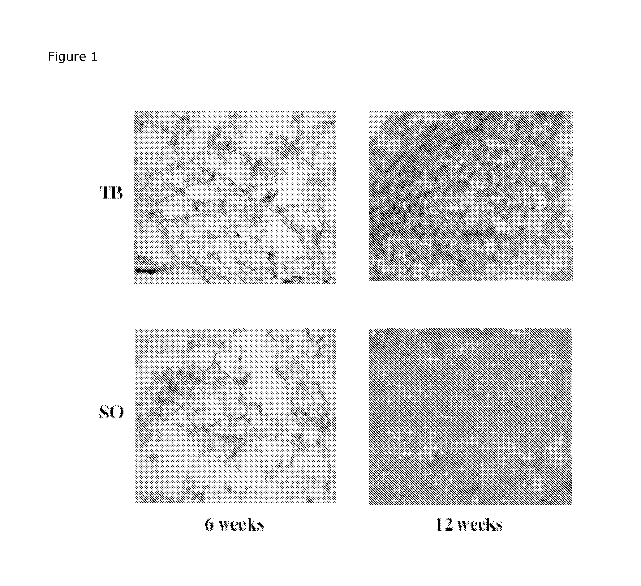
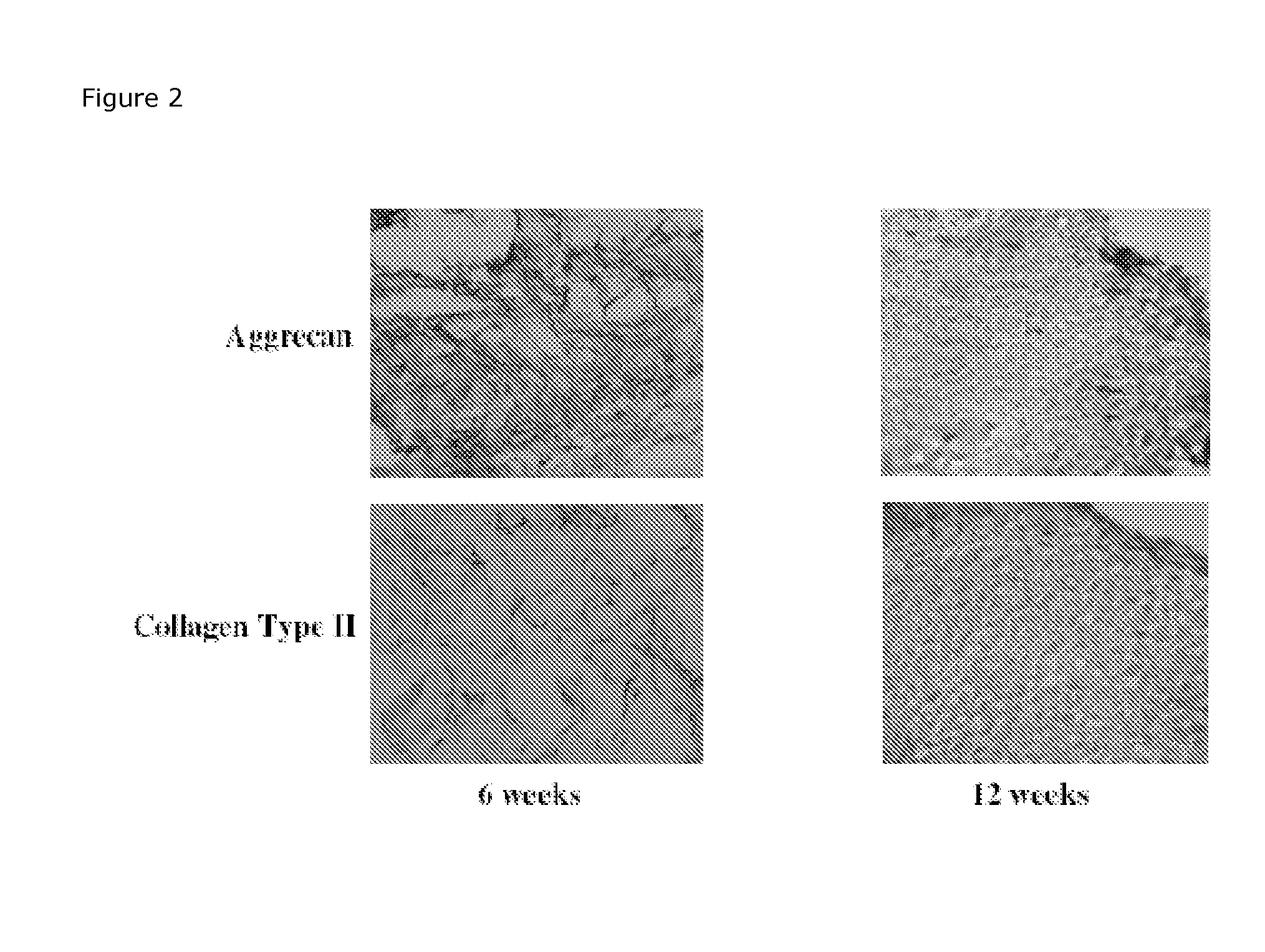
Determination of Remaining Scaffold Material in In Vitro Cultured Cartilage

Preparation of Scaffolds of Mpeg-Plga: Metoxy-Polyethylene Glycol—Poly(Lactide-Co-glycolide) (Mn 2.000-30.000, L:G 1:1) are dissolved in 1,4-dioxane to solutions containing 4%. Ten ml of the solution are poured into a 7.3×7.3 cm mould and frozen at −5° C. and lyophilised at −20° C. for 5 h and 20° C. for approx 15 h. The samples should afterwards be placed in draw (hydraulic pump) in desiccators for 24 h.

Scaffolds of MPEG-PLGA will be cultivated with chondocytes to produce in vitro cartilage as described previously. After cultivation will the scaffolds be placed in Lillys fixative for 3 days before the scaffolds are embedded in paraffin and sectioning into 8 μm slices.

An appropriate histological staining technique like Meyer's haematoxylin erosion (HE), Masson's trichrome or similar will be used to stain the new tissue but not the scaffold material. Digital images (10× and 20× magnifications) will be taken as...


PUM

PropertyMeasurementUnit
molecular weightaaaaaaaaaa

Abstract

An isolated, acellular biosynthetic cartilaginous matrix substantially devoid of synthetic biodegradable scaffold structure is provided. Through a method with the steps of a) contacting in vitro a population of chondrogenic cells with a synthetic biodegradable scaffold; b) culturing in vitro for a period of time said chondrogenic cells within said synthetic biodegradable scaffold so that the chondrogenic cells produce a biosynthetic cartilaginous matrix; c) substantially removing any antigen derived from said chondrogenic cells a matrix suitable of implantation into a living individual mammal, such as a human being is obtained.

Description

FIELD OF THE INVENTIONThe present invention relates to a biosynthetic cartilage, methods for the in vitro preparation of such cartilage suitable for in situ cartilage repair, as well as methods of treatment.BACKGROUND OF THE INVENTIONTissue engineering methods using cell transplantation are known, and for example, may involve for instance open joint surgery (e.g. open knee surgery) and, in case of joint surgery, extensive periods of relative disability for the patient to recuperate in order to ensure that optimal results are achieved. Such procedures are costly, and require extensive medical procedures such as rehabilitation and physical therapy.Methods using scaffold technologies of various forms, where the scaffold (with, or without cells grown in the scaffold) is inserted into the defect, have suffered from difficulties in performing the cell implantation procedure solely guided by arthroscopy.Arthroscopic Autologous Cell Implantation (called AACI or ACI using minor surgical inte...

Claims


Application Information

Patent Timeline
Patent Type & Authority Applications(United States)
IPC IPC(8): A61K9/00C12N5/077A61K35/12A61P43/00
CPCA61L27/3817A61L27/3852A61L2300/414A61L27/58A61L27/54A61L27/3895A61L2430/06A61P43/00
Inventor EVERLAND, HANNESAMUELSEN, PETERVANGE, JAKOBCLAUSEN, CHRISTIAN
Owner COLOPLAST AS
Why Eureka
Try Eureka