Post-exposure prophylaxis and treatment of infections
- Summary
- Abstract
- Description
- Claims
- Application Information
AI Technical Summary
Benefits of technology
Problems solved by technology
Method used
Image
Examples
example 1
Reagents and Antibodies
[0131]Cell culture reagents were obtained from Cellgro (Herndon., Va.).
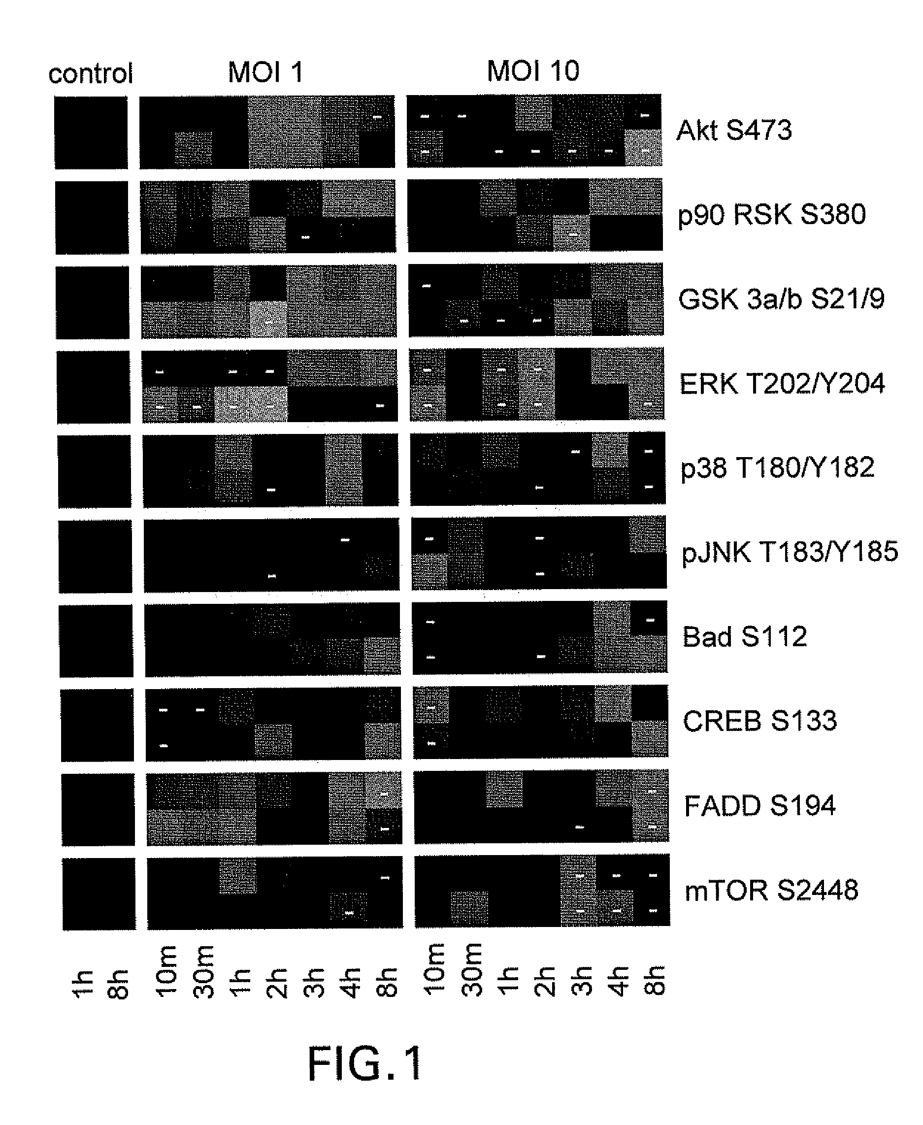
[0132]Antibodies against total and phosphorylated forms of the following proteins used for reverse phase protein microarray and Western blot analyses were obtained from Cell Signaling Technology (Beverly, Mass.).
[0133]Antibodies (identified by their specificities) were used at the following dilutions:
TABLE 11:20 for p70 S6 Kinase (Thr389)1:50 for c-Abl (Thr735), Stat5 (Tyr694), and 4E-BP1 (Ser65)1:100 for Akt (Ser473), MEK1 / 2 (Ser 217 / 221), pIKBa (Ser32 / Ser36),Bad (Ser112, 155 and 136), 4E-BP1 (Thr70), GSK-3α / β(Ser21 / 9), CREB (Ser 133), Stat3 (Ser727, Tyr705), Jak1(Tyr1022 / 1023), FAK (Tyr576 / 577), Etk (Tyr 40), Elk-1 (Ser383),and MARCKS (Ser152 / 156)1:200 for mTOR (Ser2448), eNOS (Ser1177), Pyk2 (Tyr402), FADD(Ser194), Stat6 (Tyr641) and Bcl-2 Ser701:250 for p38 (Thr180 / Tyr182), IL-1β-cleaved (Asp116); 1:400 forp90RSK (Ser380); 1:500 for PKC-δ (Thr505), Src (Tyr 527),PKC-α / β (Thr638 / 641), PK...
example 2
Challenge of Lung Epithelial Cells with Spores
[0138]HSAECs were grown in Ham's F12 media supplemented with non-essential amino acids, pyruvate, β-mercaptoethanol, and 10% FCS.
[0139]Confluent HSAECs (seeded at 106 / well in 12-well plates) were starved in the same media as above but containing 1% FCS for 16 hours and then challenged with spores. As shown in the figures, as described below, cells were cultured for up to 12 hours after challenge. Supernatants were removed, and cells were lysed and immediately boiled for 10 min in 100 μl of a 1:1 mixture of T-PER Reagent (Pierce, Rockford, Ill.) and 2× Tris-glycine SDS sample buffer (Novex / Invitrogen) in presence of 2.5% β-mercaptoethanol and protease inhibitors. Lysed samples were stored at −80° C. prior to use.
example 3
Slide Printing and Staining
[0140]Nine nl of each sample were arrayed by a direct contact pin arrayer (Aushon Biosystems, Burlington, Mass.) onto nitrocellulose slides (Whatman, Mass.). Samples were printed in duplicate and in five-point 1:2 dilution curves to ensure a linear detection range for the antibody concentrations used. Slides were kept at −20° C. before analysis with antibodies.
[0141]To estimate total protein content, selected slides were stained with Sypro Ruby Protein Blot Stain (Molecular Probes, Eugene, Oreg.) and visualized on a Fluorchemk imaging system (Alpha Innotech, San Leandro, Calif.).
[0142]Antibodies were pre-validated for specificity by western blotting and peptide competition.
[0143]Slides were stained with pre-validated specific antibodies on an automated slide stainer (Dako, Carpinteria, Calif.) using a biotin-linked peroxidase catalyzed signal amplification. The arrayed slides were placed into 1× Re-Blot solution (Chemicon, Temecula, Calif.) for 15 min, was...
PUM
| Property | Measurement | Unit |
|---|---|---|
| Antimicrobial properties | aaaaa | aaaaa |
Abstract
Description
Claims
Application Information
 - R&D
- Intellectual Property
- Life Sciences
- Materials
- Tech Scout
- Unparalleled Data Quality
- Higher Quality Content
- 60% Fewer Hallucinations
Browse by: Latest US Patents, China's latest patents, Technical Efficacy Thesaurus, Application Domain, Technology Topic, Popular Technical Reports.
© 2025 PatSnap. All rights reserved.Legal|Privacy policy|Modern Slavery Act Transparency Statement|Sitemap|About US| Contact US: help@patsnap.com


