Silica-based material for detection and isolation of chitin and chitin-containing microorganisms

Inactive Publication Date: 2011-03-03
UNIV OF SOUTH FLORIDA
View PDF0 Cites 3 Cited by
  • Summary
  • Abstract
  • Description
  • Claims
  • Application Information

AI Technical Summary

Benefits of technology

[0008]In addition, the subject invention provides rapid, specific, sensitive, accurate and convenient methods for detection, isolation, and purification of chitin, chitin derivatives and chitin-containing microorganisms. The methods are especially useful for separation and/or removal of such microorganisms from

Problems solved by technology

Despite its adverse public health impacts, there lacks an accurate, sensitive, and convenient clinical assay capable of detecting microsporidia infection.
As a result, a myriad of diseases associated with microsporidia remain vastly under-diagnosed.
Serological tests produce high false-positive results in immuno-deficient individuals, who are often incapable of eliciting adequate and specific immune responses.
Histochemical staining methods also produce high false-positive results in detecting microsporidia infection, since the commonly used dyes such as Calcofluor white, Fungi-fluor and Uvitex 2B stain a variety of fungi species indiscriminately.
While tra

Method used


Image

  • Silica-based material for detection and isolation of chitin and chitin-containing microorganisms
  • Silica-based material for detection and isolation of chitin and chitin-containing microorganisms
  • Silica-based material for detection and isolation of chitin and chitin-containing microorganisms

Examples


example 1

Preparation of Chitin-Binding Glass

[0097]This Example illustrates methods for preparing chitin-binding glass, which can be used for isolation, purification, removal and / or detection of chitin-containing microorganisms. In an embodiment, chromatographic columns for isolation of chitin-containing microorganism are prepared as follows. Briefly, glass beads purchased from Sigma Chemical Company were washed and boiled in deionized water, and then soaked in 10% acetic acid. The acid-treated beads were then used to pack chromatographic columns. In another embodiment, chromatographic columns useful for detection of chitin-containing microorganisms were prepared by packing glass wool (also known as glass fiber or fiberglass; purchased from Pyrex) into chromatographic columns, followed by soaking the column packed with glass wool in the column in 10% acetic acid.

example 2

Isolation of Chitin-Containing Microorganisms by Chitin-Binding Glass

[0098]This Example illustrates the isolation of chitin-containing nicroorganisms by chromatographic columns packed with glass beads. Briefly, samples believed to contain chitin-binding microorganisms are diluted at about 0.1:10 to about 1:10 in binding solution, and loaded onto the chitin-binding column packed with glass beads. The column is washed with 3 column volumes of phosphate buffered saline (PBS) or PBS containing about 0.05% to about 2% Tween (PBS-T) at pH 7.5. Chitin-containing microorganisms are eluted from the column by applying 3 column volumes of elution buffer (pH 8), consisting of 20 mM Tris, 2 mM EDTA. The elute is centrifuged at 12,000×g for 15 min or until a pellet is formed. The supernatant is discarded, and chitin-containing microorganisms are isolated by recovering the pellet. Optionally, the pellet may be resuspended in PBS to form spore suspension.

example 3

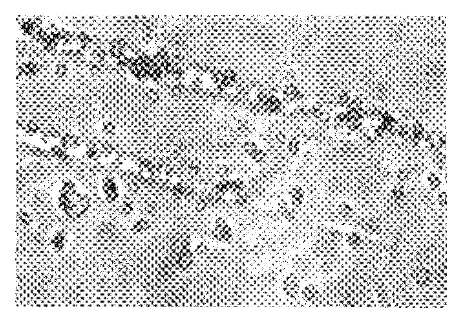
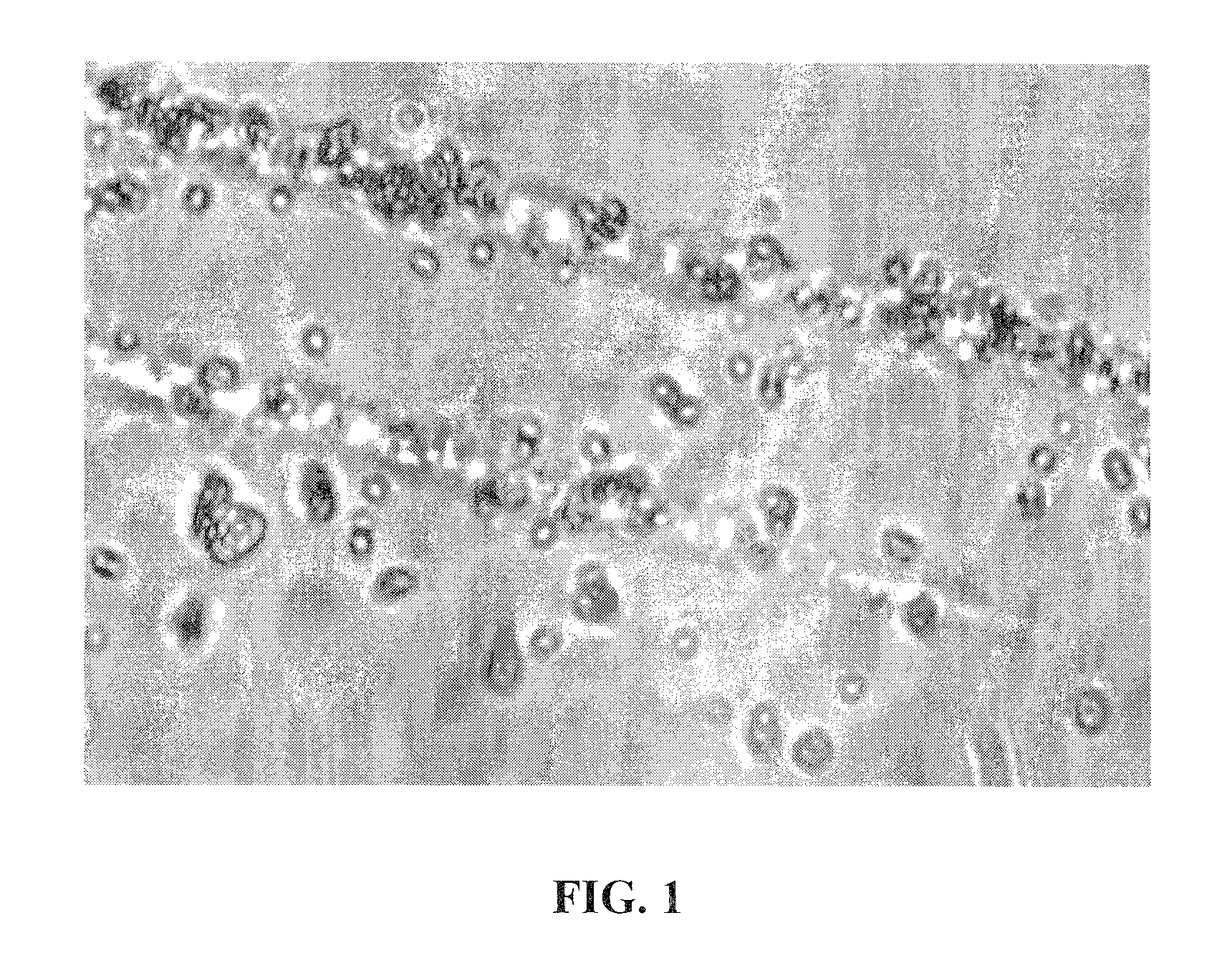
Detection of Chitin-Containing Microorganisms

[0099]This Example illustrates detection of chitin-containing microorganisms such as microsporidia using in-situ immunochemical staining of spores bound to chitin-binding columns packed with glass fiber. Briefly, spores of chitin-containing microorganisms are isolated by using chitin-binding glass column packed with glass beads, as described in Example 2. The spore suspensions are loaded onto the chitin-binding glass fiber column. The column is washed with 3 column volumes PBS containing about 0.05% to about 2% Tween 20, followed by 3 column volumes of PBS-T. The column is then incubated with 1 ml primary antibody (antibody to microsporidia), diluted 1:100-1:1,000 (dilution depends on antibody concentration) in PBS-T. The column is then sequentially washed with 3 column volumes of PBS containing 10 mM EDTA, followed by 3 column volumes of PBS-T. The column is then incubated with 1 ml alkaline phosphatase-conjugated secondary antibody dilu...


PUM

PropertyMeasurementUnit
Alkalinityaaaaaaaaaa

Abstract

The subject invention provides silica-based material that has high affinity to chitin, chitin derivatives and chitin-containing microorganisms at an acidic pH. In an embodiment, the silica-based material surface comprises glass. Also provided are methods for preparing the subject silica-based chitin-binding material. In addition, the subject invention provides rapid, specific, sensitive, accurate and convenient methods for detection, isolation and purification of chitin, chitin derivatives and chitin-containing microorganisms.

Description

CROSS-REFERENCE TO RELATED APPLICATION[0001]The subject application claims the benefit of U.S. Provisional application Ser. No. 61 / 236,989, filed Aug. 26, 2009, which is incorporated herein by reference in its entirety, including any figures, tables, or drawings.GOVERNMENTAL SUPPORT[0002]This invention was made with government support under National Institutes of Health and National Institute of Dental and Craniofacial Research under grant numbers R21DE015127 and R21DE014296. The government has certain rights in the invention.BACKGROUND OF THE INVENTION[0003]Fungi are ubiquitous opportunistic parasites that infect a variety of species, including humans. Microsporidia, a phylum of unicellular fungi that include over 1200 species, infect a range of hosts. Species of the genus Encephalitozoon can disseminate in the host body and infect organs such as intestine, lung, kidney, brain, sinuses, muscles and eyes, and shed spores in feces, tissue, saliva, ocular and nasal discharged fluids, ...

Claims


Application Information

Patent Timeline
IPC IPC(8): G01N33/569C12N1/00C08B37/08C12N1/14C12M1/00G01N30/50G01N30/60
CPCC08B37/003Y10T29/4998B01J20/283G01N33/56961G01N33/5308G01N2400/28C08B37/0003
Inventor DAO, MY LIEN
Owner UNIV OF SOUTH FLORIDA
Why Eureka
Try Eureka