
Method for multi-scale meshing of branching biological structures

a biological structure and multi-scale technology, applied in the field of mathematical representation of the structure and function of branching biological structures, can solve the problems of inability to meet the needs of modern technology, the best image system is of little use, and the structure such as the lung is vast and complicated, so as to facilitate specifying boundary conditions, improve computation speed, and speed up the effect of mesh generation

Inactive Publication Date: 2011-04-21
UNIV OF IOWA RES FOUND +1
View PDF12 Cites 106 Cited by
  • Summary
  • Abstract
  • Description
  • Claims
  • Application Information

AI Technical Summary

Benefits of technology

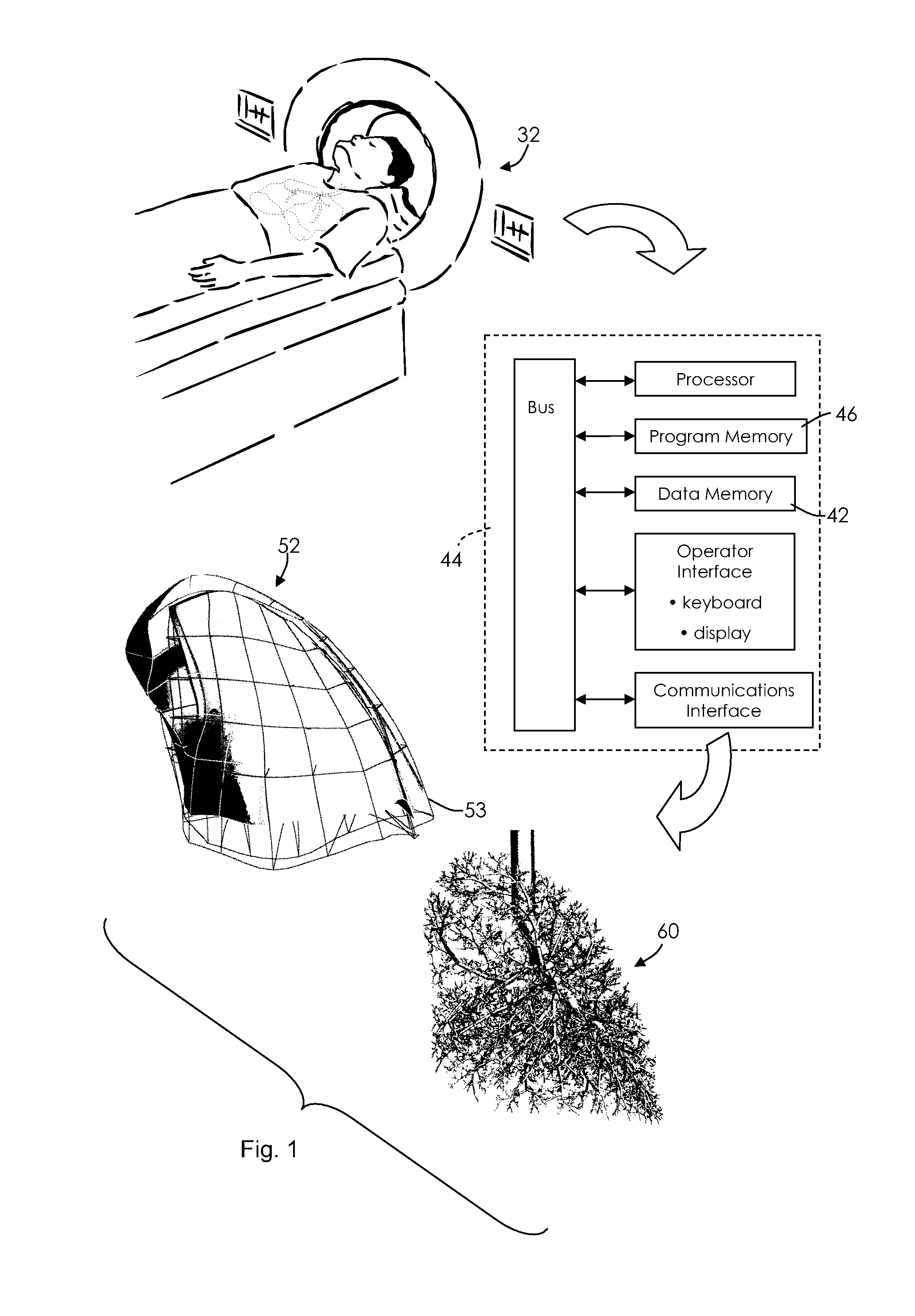
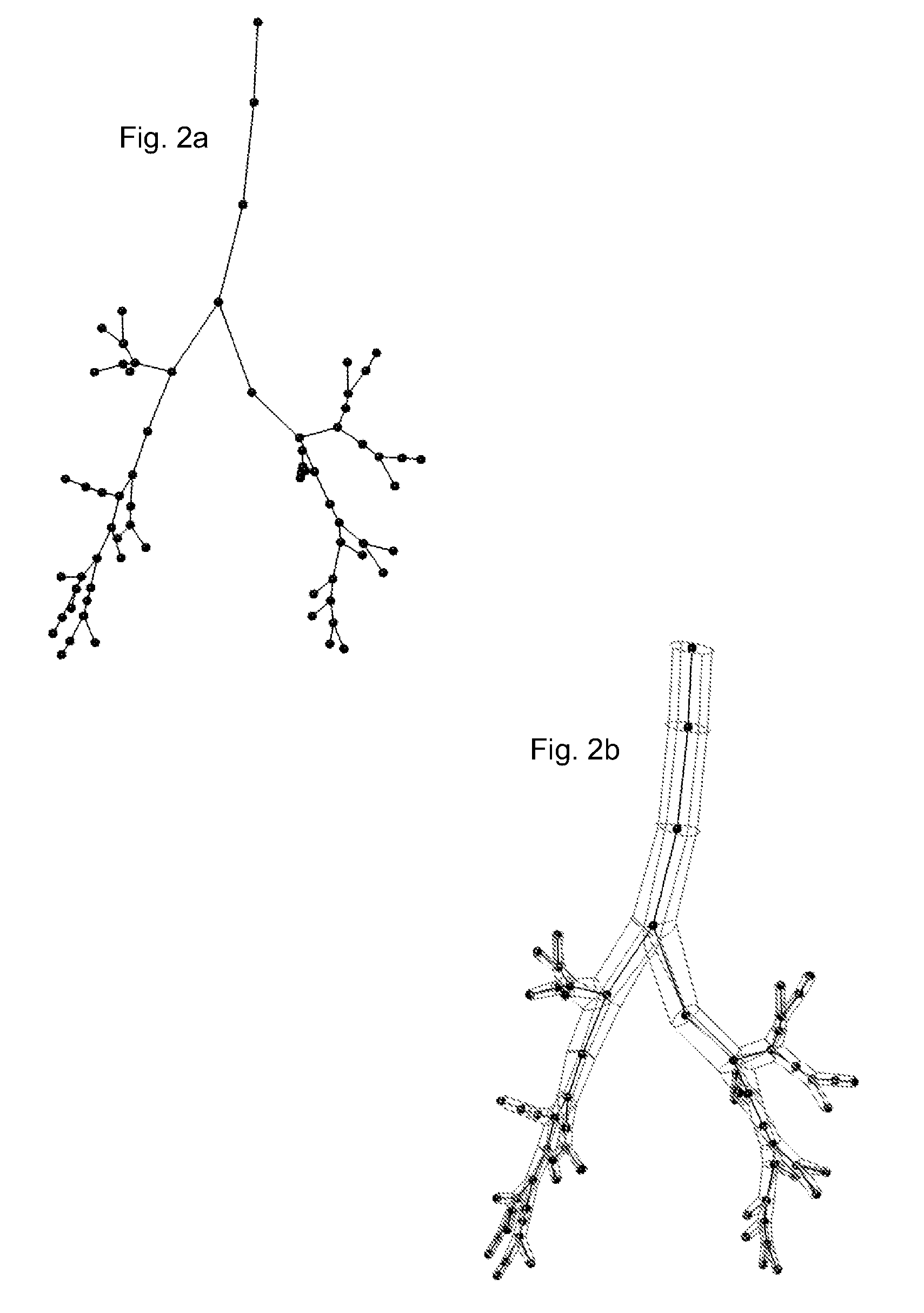
[0016]The method uses the center line locations and measurements of airway diameter from the CT images (or other image source), including estimated diameters for the one dimensionally modeled airways, to create an initial estimation of a branching mesh, using cylinders to model the elongations of the branches or passageways. A two dimensional (2D) surface mesh is derived to lie over the central scaffold, and a smooth transition structure is inferred at each bifurcation (each branch division). In this way the surface of the entire airway tree can be modeled. Some of the airways, preferably the uppermost airways, are defined by geometry and measurements taken direct from the CT, MR or other imaging input. The structural details defining these airways can be more or less extensive, with accuracy depending on the application of the model. Furthermore, data can be collected that encompasses any number of divisions and passageways. However with each branching, the number of passageways is multiplied and the size of the passageways is reduced. One aspect of the technique is to define airways that are lower (more distal) in the branching pattern, at some level, with geometry controlled by the shape of the lung itself, using modeling rather than measurement or a hybrid of modeling and measurement of at least exemplary biological structural units.
[0022]The full combined mesh is translated into a format that is suitable for CFD simulation. The knowledge of tree connectivity from the scaffold models as described, allows us to define triangles on the perimeters of the domain. This facilitates specifying boundary conditions for simulation, namely at the perimeters.
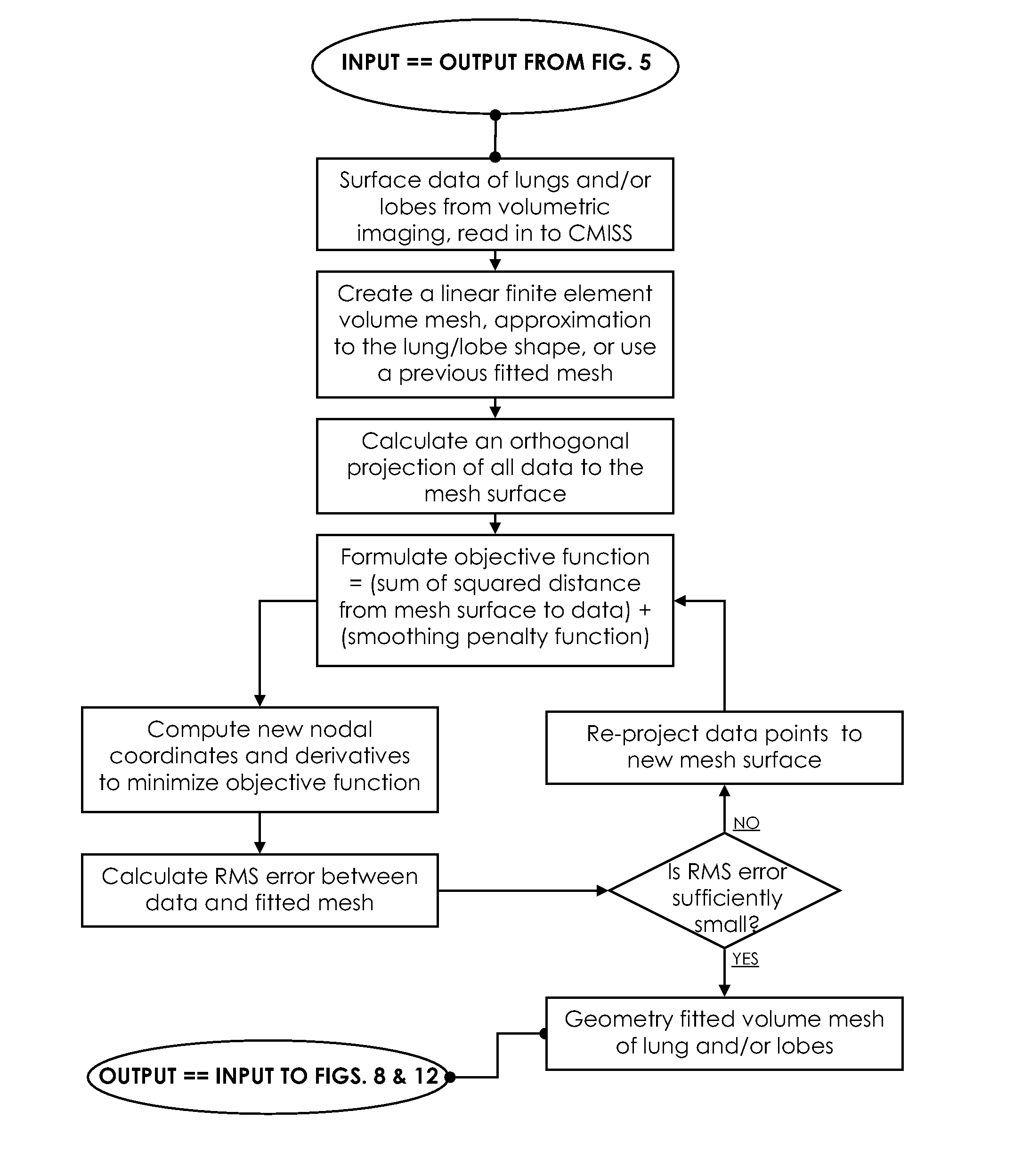
[0023]Computational fluid dynamics has been used with software meshing for simulating flow patterns and the like, but conventional mesh techniques do not cope well with the complexity of an airway or pulmonary vascular tree. The branching structure of these biological systems introduces challenges. However, it is an aspect of the present disclosure that the definition and calculation of center lines is performed as part of an analysis of the geometry of the airway tree. An inner scaffold is created that ultimately controls the CFD mesh generation. This improves the speed of computation, perhaps completing a useful mesh in a time on the order of hours (for 16 generations of branches), compared with the potential approach of first meshing the airway surface geometry, then filling the surface with a volume mesh, which may take weeks or months. It is an aspect of the scaffold technique that an initial coarse or simplified structure is determined, just from branching characteristics. Then, geometry-fitting captures anatomical detail.
[0024]Much of the tedious labor-intensive process of manual editing of the tree is avoided. CFD mesh generation is rapid because by using the center line scaffold, the tree up can be subdivided into smaller sections that are meshed independently—so can be computed on multiple processors simultaneously—then joined back together to make the final mesh. A further aspect is use of a 1D center line tree that is ‘grown’ into an imaging-based definition of the lung geometry as an extra scaffold to extend the CFD mesh into regions of the lung that cannot be imaged.

Problems solved by technology

Although imaging systems are sophisticated and capable, the best image system is of little use if its sole function is to show that a branching biological structure such as a lung is vast and complicated.
This is not possible with today's technology.
If the enabling technology did exist, it would still be a formidable computational job to measure all the lung's bronchial airways to the extent necessary to infer pressure and flow comprehensively throughout the lung.

Method used


Image

  • Method for multi-scale meshing of branching biological structures
  • Method for multi-scale meshing of branching biological structures
  • Method for multi-scale meshing of branching biological structures

Examples


Embodiment Construction

[0047]A biological branching structure such as a human lung or lobe, comprises larger and smaller passageways affecting the characteristics of the organ for its nominal functions, including respiration, heat exchange, evaporation, deposition of airborne particles, discharge of more or less viscous material and particulates with coughing, allergic reactions, inflammation and other specific functions and effects. It is desirable to study the relationship of structure and function and to exploit the conclusions that can be gleaned in that way.

[0048]With computed tomography or magnetic resonance imaging, it is possible to measure the air passages in a local area of the lung or other branching structure. Although there are relatively few passageways at the trachea, bronchi and proximal branching passageways, every generation of branching divisions multiplies the number of passageways. After a number of generations of branching, it becomes a formidable task to measure and account for the ...


PUM


Abstract

A structural and functional model for a lung or similar organ is virtually defined by encoding aspects of branching passageways. Larger passageways that are visible in medical images are surface mesh fitted to the anatomical surface geometry. Smaller distal passageways, beyond a given number of branch generations, are modeled by inference as linear passages with nominal diameters and branching characteristics, virtually filling the space within the outer envelope of the organ. The model encodes finite volumetric elements for elasticity and compliance in passageway walls, and for local pressure and flow conditions in passageway lumens during respiration. The modeling can assess organ performance, help to plan surgery or therapy, determine likely particle deposition, assess respiratory pharmaceutical dosing, and otherwise represent structural and functional organ parameters.

Description

CROSS REFERENCE TO RELATED APPLICATION[0001]This application claims the priority of U.S. provisional patent application Ser. No. 60 / 987,844, filed Nov. 14, 2007, the content of which application is hereby incorporated by reference in its entirety.STATEMENT REGARDING GOVERNMENT SPONSORED RESEARCH OR DEVELOPMENT[0002]The subject matter of this disclosure was supported in part by National Institutes of Health (NIH) Bioengineering Research Partnership Grant R01-HL-064368, NIH Grant R01-HL-064368, and NIH Grant R01-EB-005823.BACKGROUND OF THE INVENTION[0003]1. Field of the Invention[0004]The invention concerns methods for mathematically representing the structure and function of branching biological structures in data processing models, and programmed systems for applying the models in investigational, diagnostic, therapeutic and similar activities.[0005]2. Related Art[0006]It is possible using magnetic resonance imaging, computed tomography and similar image data collection and image da...

Claims


Application Information

Patent Timeline
IPC IPC(8): G06F7/60G06G7/60
CPCG06F17/5018G06F2217/16G06T7/0012G06T17/20G06T2207/10081G06F19/3437G06T2207/20068G06T2207/30061G06T2210/24G06T2210/41G06T2207/10088G06F30/23G06F2111/10G16H50/50
Inventor TAWHAI, MERRYN H.HOFFMAN, ERIC A.LIN, CHING-LONG
Owner UNIV OF IOWA RES FOUND
Learn More