

Genetic alteration associated with pre-malignant cancer
a technology of premalignant breast cancer and gene modification, applied in the direction of tumor/cancer cells, drug compositions, instruments, etc., can solve the problems of inability to quickly assess the therapeutic efficacy of strategies for arresting breast cancer at the pre-invasive stage, little is known about, etc., to prevent or limit the progression inhibit the growth of cells, and prevent or limit the effect of pre-malignant breast lesion progression
- Summary
- Abstract
- Description
- Claims
- Application Information
AI Technical Summary
Benefits of technology
Problems solved by technology
Method used
Image

Examples
example 1
Malignant and Invasive Cancer Cells from DCIS Lesions
[0085]It has been discovered that human living DCIS lesions contain cells with the ability to grow as invasive tumors in mouse xenografts.
[0086]Tumor Transplantation: Breast ductal tissue was incubated with EGF, insulin, and Estrogen in RPMI1640 for 4-12 hours prior to transplantation into the mammary fat pad of NOD / SCID mice (Jackson Labs / Harlan). Tumors that appeared within 2 months of transplantation were excised (Table 1). A portion was saved for in vitro cultivation and the remainder was transplanted for propagation and phenotype analysis.
Results
[0087]
TABLE 1Breast xenograft characteristics and tumor generation.XenograftMouse IDTissue typeERPRHer2Tumor793Breast normalno395DCISyes398DCISyes581DCISyes876DCISyes079DCISPos (50%)Pos (50%)yes080DCISPos (50%)Pos (50%)yes081DCISPos (50%)Pos (50%)yes082DCISPos (50%)Pos (50%)yes379 / 579DCISyes396DCISyes783 / 763DCISPos (50%)Pos (50%)yes791DCISyes792DCISPos (>90%)Pos (>90%)yes794DCISno795D...
example 2
DCIS Cells Propagated In Vitro
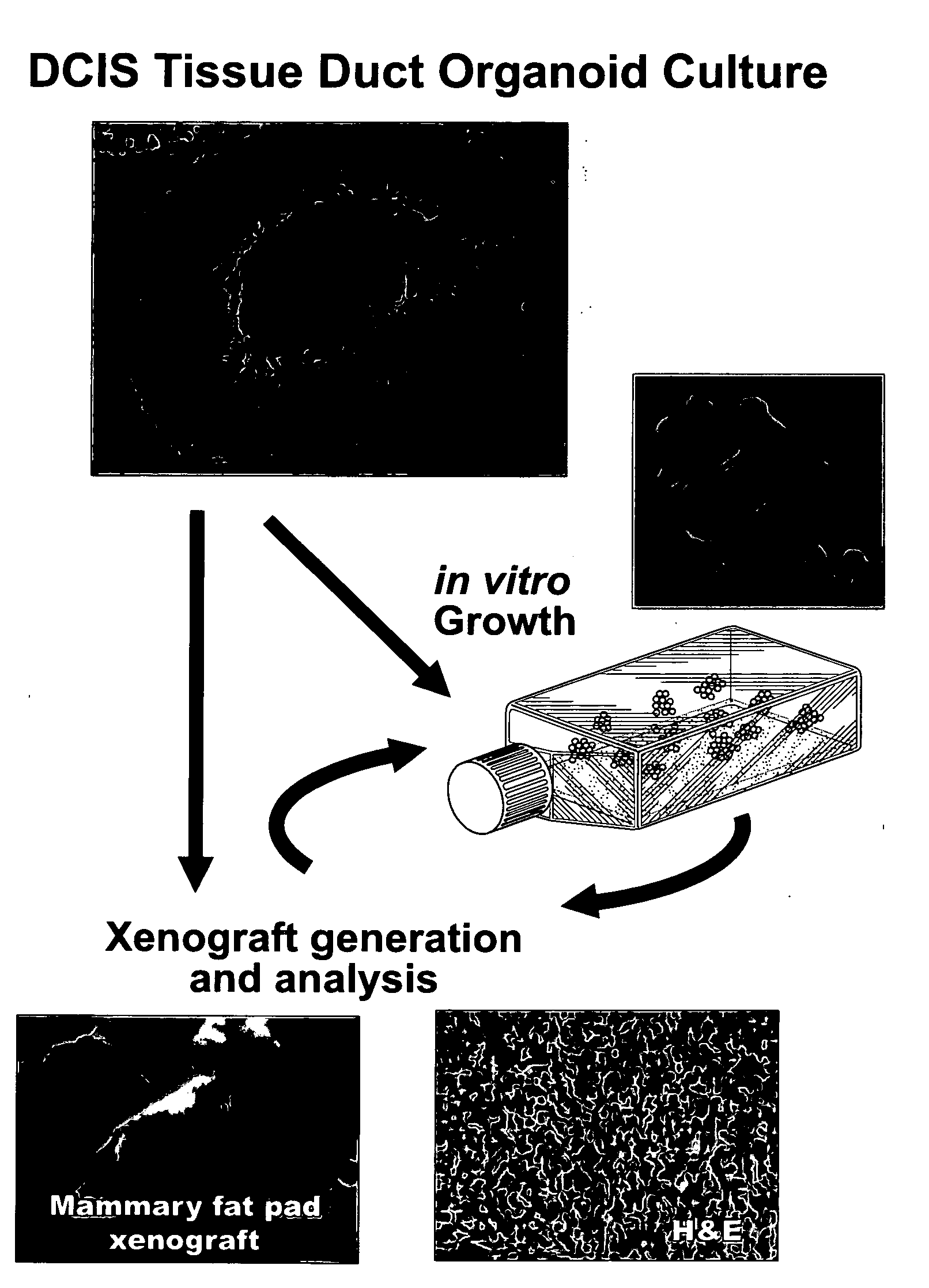
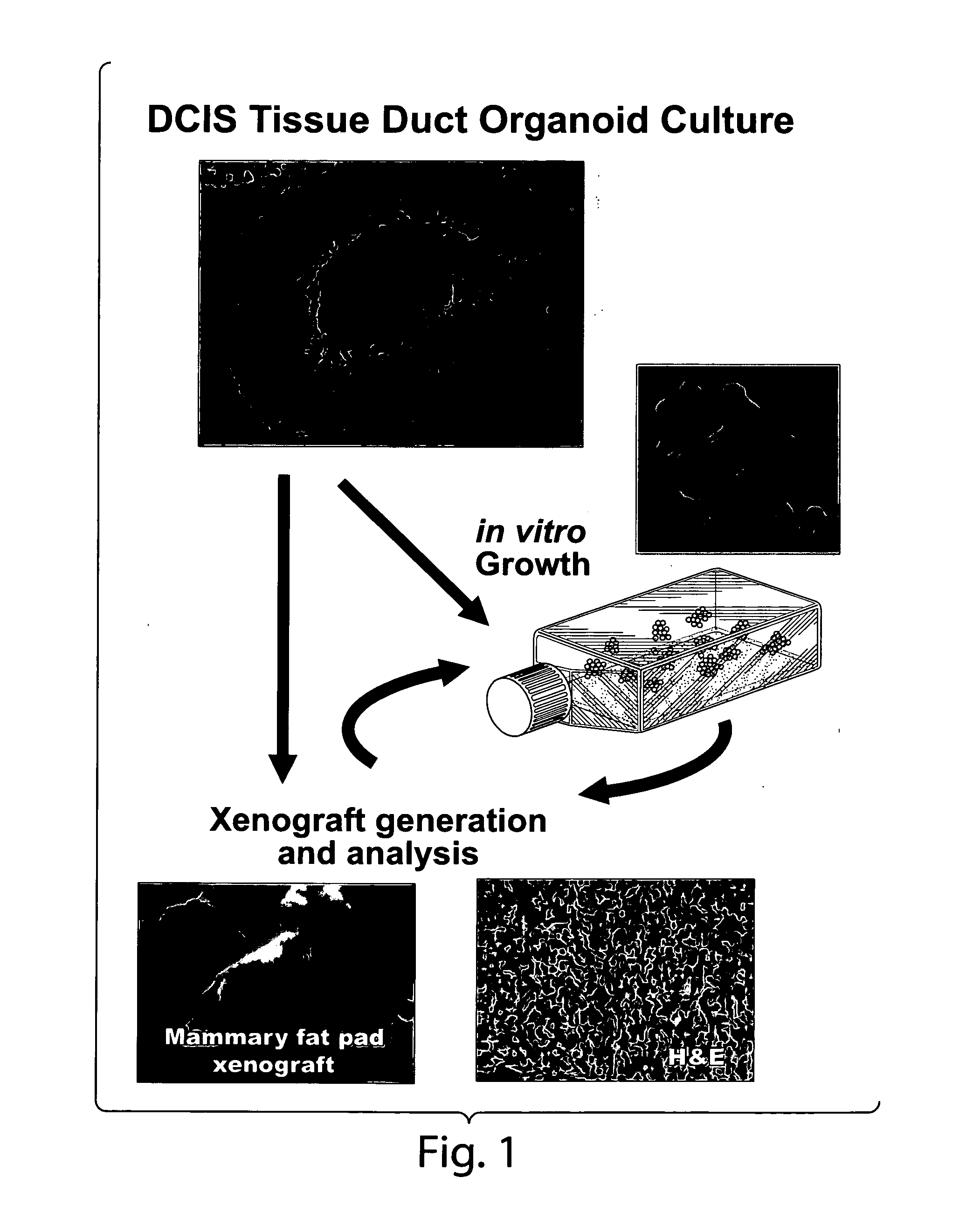
[0089]No information exists concerning the existence of tumorigenic / malignant precursor cells within living human pre-invasive lesions such as Ductal Carcinoma in situ (DCIS). It has been discovered, however, that malignant precursor cells exist in DCIS lesions and can propagate in vitro and in vivo (FIGS. 1-4).
Methods
[0090]Reverse Phase Protein Array Analysis. Human DCIS breast cells were cultured in minimal medium supplemented with EGF and Insulin in the presence of Streptomycin and Gentamicin. Cells with distinct morphologies were removed by aspiration and mechanical disruption (scraping), spun at 1000 rpm for 5 min, medium was removed and the cell pellet was lysed in TPER, 2× Tris-glycine sample buffer with 10% TCEP Bond Breaker. Reverse phase protein microarrays were printed with an Aushon 2470 arrayer.
[0091]Staining and Analysis. Slides were stained with 65 antibodies against phosphorylated proteins involved in pro-survival, growth regulation and ...
example 3
Further Testing of the Malignant and Invasive Properties of DCIS Cells
[0097]Further testing can be performed to examine the malignant and invasive properties of the DCIS malignant precursor cells, such as in vivo invasion and metastasis testing using xenotransplantation and signaling pathway profiling.
Organ Culture and Microdissection Technology
[0098]Organ Culture. Organ cultures consist of isolated cut segments of breast duct organoids less than 5 mm in length that have an exposed duct lumen. The tissue microenvironment is modeled by the addition of adipose tissue and stroma from the local patient donor lesion. The serum free medium is supplemented with insulin, EGF, and Estrogen. In addition, the serum free medium can be supplemented with basement membrane extracts. As shown in Examples 1 and 2, outgrowth of invasive cells can occur in 2 to 4 weeks.
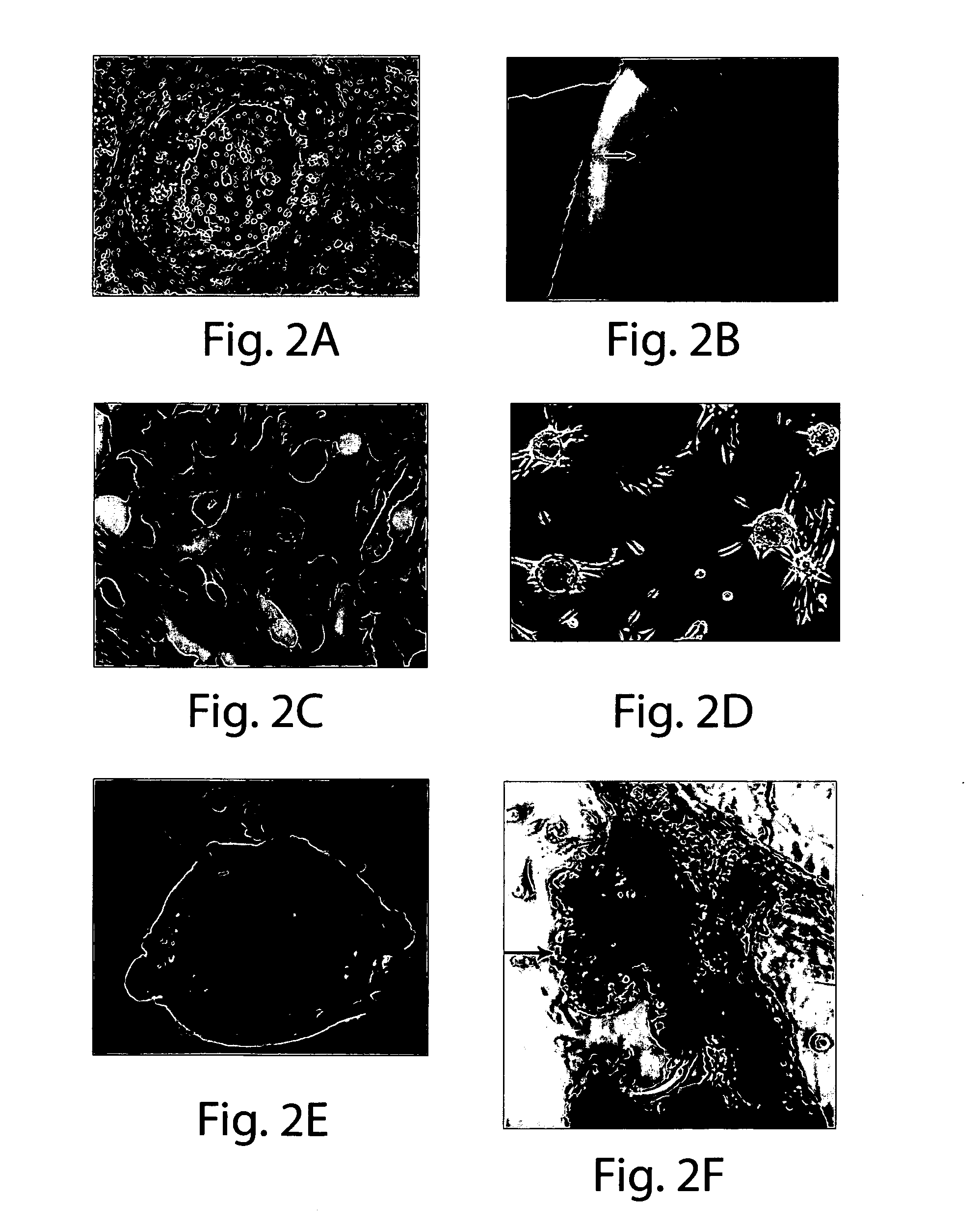
[0099]DCIS morphologic subtypes. As described in Examples 1 and 2, the DCIS outgrowths in organ culture have a distinct set of morphol...
PUM
| Property | Measurement | Unit |
|---|---|---|
| diameter | aaaaa | aaaaa |
| diameter | aaaaa | aaaaa |
| length | aaaaa | aaaaa |
Abstract
Description
Claims
Application Information