Diagnosis/Treatment Option for Head-and-Neck Tumor Using Micro-RNA as Biomarker

a head and neck tumor and expression profile technology, applied in the field of head and neck tumor expression profiles, can solve the problems of difficult to distinguish tumor from benign disease, difficult to determine the progression and invasion degree of the tumor, and not enabling the prediction of the prognosis, etc., to achieve rapid and accurate determination of the presence, and efficient screening

Inactive Publication Date: 2011-06-16
SAITO KOICHIRO +1
View PDF2 Cites 9 Cited by
  • Summary
  • Abstract
  • Description
  • Claims
  • Application Information

AI Technical Summary

Benefits of technology

[0029]The method for the determination of head-and-neck tumor and the method for using a microRNA as a biomarker for head-and-neck tumor according to the present invention enable the head-and-neck tumor to be rapidly and accurately determined. The kit for the determination of head-and-neck tumor and the method for using a microRNA for the determination of head-and-neck tumor according to the present invention also enable the rapid and accurate determination of the presence of the head-and-neck tumor. In addition, the method for screening therapeutic agents for head-and-neck tumor enables the therapeutic agents for head-and-neck tumor to be efficiently screened. Particularly, a microRNA (for example, miR-196a or the like) whose concentration in the blood of a subject also changes when a subject suffers from head-and-neck tumor is excellent especially in terms of speeding-up and simplifying the method for use as a biomarker for head-and-neck tumor, the method for the determin...

Problems solved by technology

However, for the diagnosis of head-and-neck tumor, the imaging tests cannot qualitatively diagnose the tumor and in many instances make it difficult to differentiate the tumor from benign disease.
The tissue diagnosis by biopsy, which is used as a definite diagnosis, also sometimes makes the determination of progression and invasion degrees of the tumor difficult as well as at present not enabling the prediction of prognosis thereof even when the basic hematoxylin-eosin (HE) stain is used in combination with various immunostainings.
Although several factors for prediction of onset and prognosis of the tumor are reported (P...

Method used


Image

  • Diagnosis/Treatment Option for Head-and-Neck Tumor Using Micro-RNA as Biomarker
  • Diagnosis/Treatment Option for Head-and-Neck Tumor Using Micro-RNA as Biomarker
  • Diagnosis/Treatment Option for Head-and-Neck Tumor Using Micro-RNA as Biomarker

Examples


example 1

Collection of Sample Tissue—1

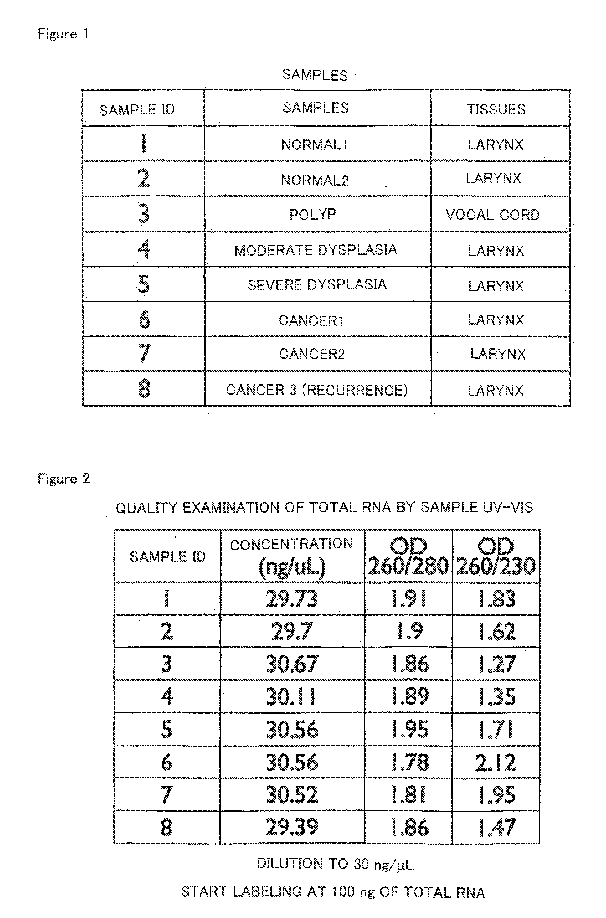
[0085]Under the approval of the ethics committees of Keio University School of Medicine and Sano Kosei General Hospital, patients visiting the otorhinolaryngology clinic of Keio University Hospital or Sano Kosei General Hospital were selected and used as persons to be asked for sample donation. Then, after obtaining informed consent from the persons, 8 sample tissues (“SAMPLE ID 1 to 8” in FIG. 1) were collected from 6 of the persons. In this respect, the sample ID 1 and 6 were those from the same patient, and the sample ID 2 and 7 were also those from the same patient. The tissue types of these 8 sample tissues (the article “TISSUES” in FIG. 1) consisted of two types: larynx and vocal cord; the tissue conditions (the article “SAMPLES” in FIG. 1) consisted of conditions: normal, polyp, moderate dysplasia, severe dysplasia and cancer conditions; and the types of the cancers consisted of two types: incipient cancer (incipient) for cancers 1 to 2 and recurr...

example 2

Extraction of RNA from Sample Tissue and Qualitative Evaluation Thereof—1

[0087]RNA was extracted from each tissue frozen in Example 1 in order to use in a microarray to be described later. Specifically, total RNA comprising microRNAs was extracted from each of the above tissues using mirVanamiRNA Isolation Kit (from Applied Biosystems) according to the appended protocol.

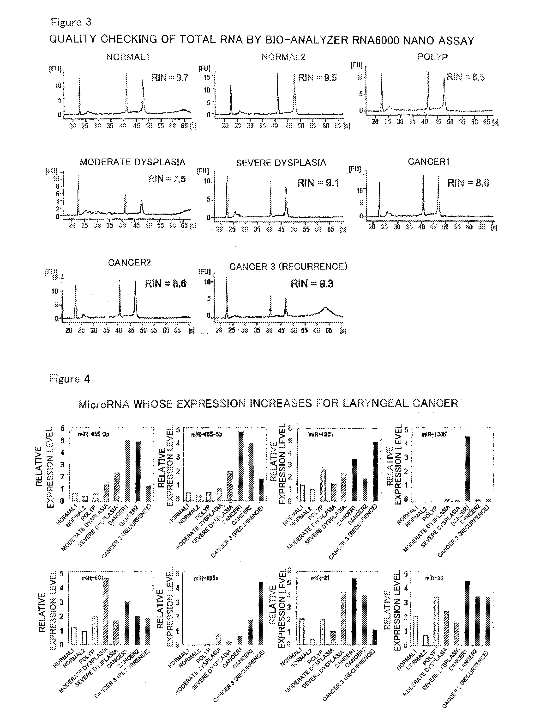
[0088]Then, the extracted RNA was subjected to qualitative evaluation in order to make sure that sufficient accuracy would be obtained in the microarray to be described later. Specifically, the resultant RNA was adjusted to a concentration of about 30 ng / μL using distilled water, and OD260 / 280 (the numerical value obtained by dividing the measured value of OD260 by the measured value of OD280) and the like were measured using a spectrophotometer for calculation. The results are shown in FIG. 2. As shown in FIG. 2, each OD260 / 280 fell in the range of about 1.7 to 2.0, indicating that each RNA was little contaminated w...

example 3

Expression Analysis of microRNAs in Each Tissue—1

[0089]Using Agilent Human miRNA V2 (from Agilent Technologies), 723 human microRNAs were subjected to exhaustive analysis. The above-described Agilent Human miRNA V2 microarray comprises DNA sequences complementary to nucleotide sequences represented by SEQ ID NOs:1 to 11 as probes for miR-455-3p, miR-455-5p, miR-130b, miR-130b*, miR-801, miR-196a, miR-21, miR-31, miR-133b, miR-145 and miR-375, respectively. The method for analysis was according to the method described in Agilent Technologies' miRNA Microarray Protocol Version 1.5. Specifically, the analysis was performed by the following method. The amount of a reagent is described as an amount for one sample.

[0090]First, the total RNA obtained in Example 2 was diluted to about 25 ng / μL with DNase / RNase-free water, and the RNA concentration of the diluted solution was measured. Then, 0.7 μL of 10×CIP Buffer (from GE Healthcare) was mixed with 0.7 μL of 16 U / μL CIP (from GE Healthcare...


PUM


Abstract

Disclosed are: a method for using a particular microRNA as a biomarker for head-and-neck tumor; a method for the determination of head-and-neck tumor; a kit for the determination of head-and-neck tumor, and the like. The present invention is characterized in that at least one microRNA selected from the group of microRNAs consisting of miR-455-3p, miR-455-5p, miR-130b, miR-130b*, miR-801, miR-196a, miR-21, miR-31, miR-133b, miR-145 and miR-375 is used as a biomarker for head-and-neck tumor.

Description

TECHNICAL FIELD[0001]The present invention relates to an expression profile of a microRNA (miRNA) in head-and-neck tumor and a use thereof. More specifically, the present invention relates to a method for using a specific microRNA as a biomarker for the head-and-neck tumor, a method for the determination of the head-and-neck tumor, a kit for the determination of the head-and-neck tumor, and the like.BACKGROUND ART[0002]Head-and-neck tumor is a tumor formed in the ear, nose, pharynx, larynx, cervix, facial surface, oral cavity, or the like; particularly, a malignant one is prevalent in men 50 years old or more and accounts for 6% of all malignant tumors. The incidence rate thereof increases with increasing age; 650,000 people are annually affected thereby in the whole world and of these, 350,000 die of the malignant tumor (see Non-Patent Document 1). Among others, laryngeal cancer is the most common disease in the head and neck area; the number of fatalities is estimated to increase ...

Claims


Application Information

Patent Timeline
IPC IPC(8): C40B30/00C12Q1/68C40B40/06
CPCC12Q1/6886C12Q2600/178C12Q2600/156C12Q2600/136
Inventor ZAMA, TAKERUSAITO, KOICHIROHIRASAWA, AKIRANAGANISHI, HIDEKI
Owner SAITO KOICHIRO
Why Eureka
Try Eureka