Method for processing porcine cornea for decellularization

a decellularization and porcine cornea technology, applied in the field of porcine cornea decellularization, can solve the problems of corneal loss, vision loss, and the inability to regenerate the bowman's membrane, and achieve the effects of minimizing immune responses, efficient processing of corneas, and maintaining corneal transparency

Active Publication Date: 2011-07-28
SEOUL NAT UNIV R&DB FOUND
View PDF2 Cites 15 Cited by
  • Summary
  • Abstract
  • Description
  • Claims
  • Application Information

AI Technical Summary

Benefits of technology

[0015]Therefore, the present invention has been made in view of the above problems, and it is an object of the present invention to provide a m...

Problems solved by technology

However, the Bowman's membrane cannot be regenerated, scarring normally occurs upon surgical operation or traumatic injury.
Meanwhile, corneal blindness, which results from loss of corneal transparency, is a major cause of vision loss, second only to cataracts.
However, at present, these materials are not commonly used due to problems associated with biocompatibility and optical and mechanical properties, and xenograft utilizing other animal corneas instead of human corneas is the fastest growi...

Method used


Image

  • Method for processing porcine cornea for decellularization
  • Method for processing porcine cornea for decellularization
  • Method for processing porcine cornea for decellularization

Examples


experimental example 1

Ex Vivo Evaluation of the Corneal Stroma Decellularized in Accordance with the Method Set Forth in Table 1 Above

[0053]1. Macroscopic Analysis

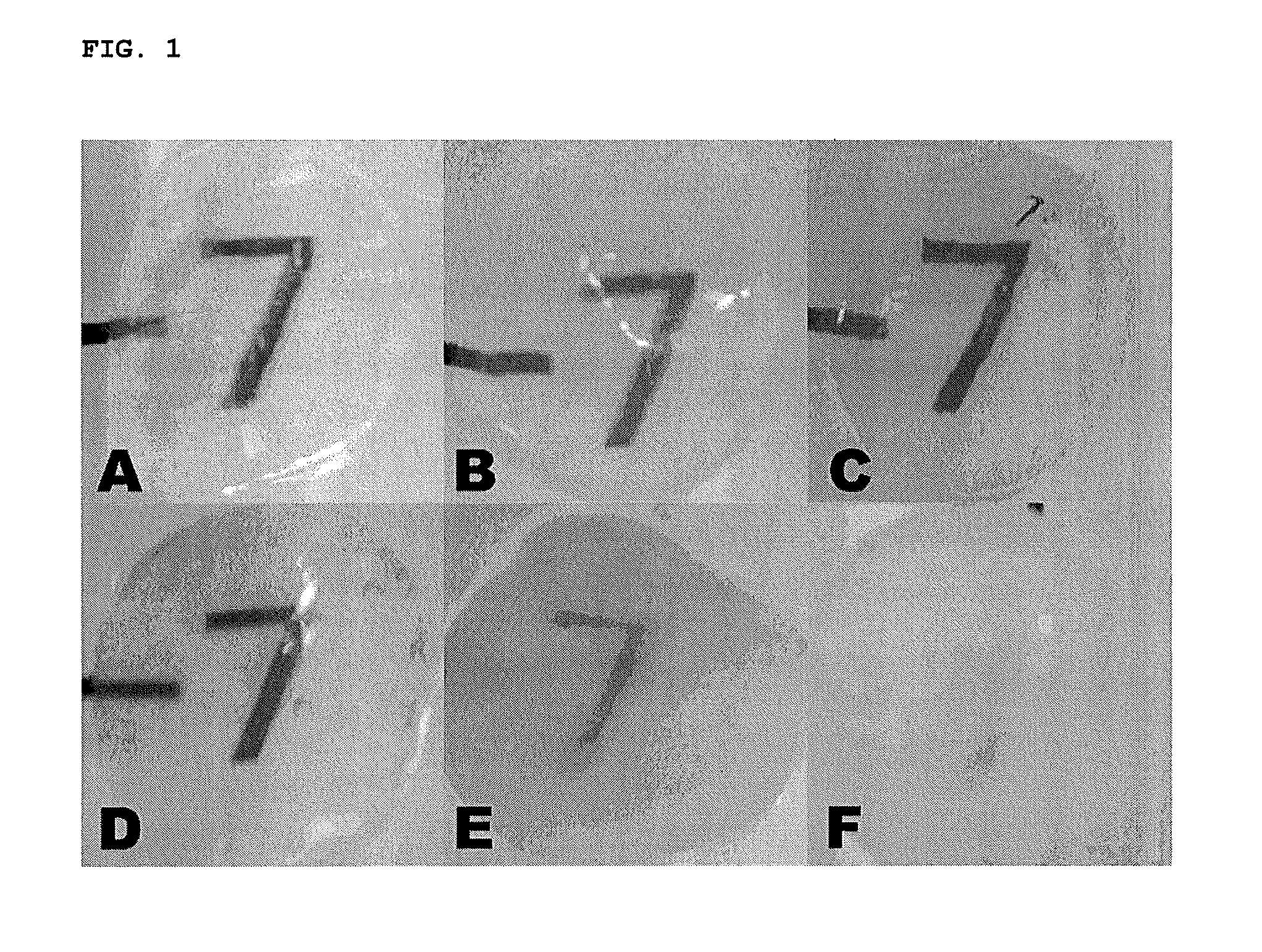
[0054]Macro-analysis revealed that the corneas for the control group, freezing group, 3-times freezing-thawing group, hypertonic saline-treated group, and hyperosmolar glycerol-treated group remained transparent, whereas the corneas for trypsin / Dispase / SDS-treated group and DNase / RNase-treated group became opaque (FIG. 1).

[0055]2. Microscopic Analysis

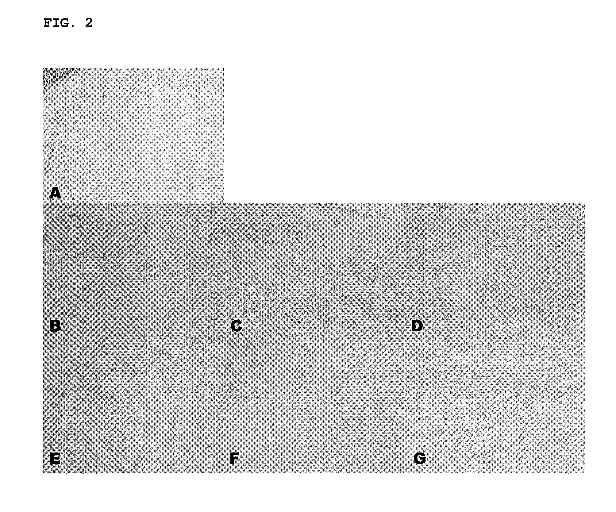
[0056]First, the porcine cornea for each group was sectioned and stained with a hematoxylin and eosin (H&E) staining method. Then, the H&E-stained slices were observed under an optical microscope (Olympus Optical Co. Ltd., Tokyo, Japan).

[0057]The H&E-stained 3-times freezing-thawing group, hypertonic saline group, hyperosmolar glycerol group and trypsin / dispase / SDS group had almost no cells. On the other hand, the control group and the freezing group had many nuclei throughout the corneal stroma (F...

experimental example 2

Identification of In Vivo Efficiency of Decellularized Corneal Stroma on Xenograft Model

[0067]1. Evaluation for Clinical Course of Corneal Xenograft

[0068]A 2-3 kg adult New Zealand white rabbit (Orient Bio Inc., Seoungnam, Korea) was used as a recipient animal for keratoplasty in the present experimental Example 2. The rabbit was anesthetized by intramuscularly administering 10 mg / kg zolazepam (Zoletil®, Yuhan Corp., Seoul, Korea) and 6.8 mg / kg xylazine hydrochloride (Rompun®, Bayer, Frankfurt, Germany).

[0069]For graft of porcine corneas, a 250 μm-thick anterior lamellar graft of the porcine cornea was marked with an 8.0 mm-diameter trephine and manually detached with a crescent knife (Alcon Surgical, Fort Worth, Tex., USA). The anterior lamellar grafts of the porcine cornea (N=7 for each group) were sutured with eight stitches into the recipient cornea of the rabbit by interrupted suture using a 10-0 nylon suture (Ethicon, Somerville, N.J., USA). At one week, all sutures were remov...


PUM


Abstract

Disclosed is a method for processing porcine cornea using an aqueous NaCl solution and an aqueous trypsin/EDTA solution to decellularize enucleated porcine cornea. The porcine cornea processed by the method causes neither inflammation nor immune rejection. The porcine corneal stroma decellularized by the method can be recellularized together with host keratocytes after transplantation.

Description

CROSS-REFERENCES TO RELATED APPLICATIONS[0001]This application is a continuation of PCT / KR2009 / 001770 filed on Apr. 6, 2009 which claims priority to Korean Application No. 10-2009-0018294 filed on Mar. 4, 2009. The entire contents of the aforementioned patent applications are incorporated herein by this reference.BACKGROUND[0002]1. Technical Field[0003]The present invention relates to a method for processing porcine cornea, and more particularly to a method for processing porcine cornea for decellularization.[0004]2. Related Art[0005]The cornea is an organ refracting light, which accounts for one-sixth of the outermost tunic of the eyeball. The cornea is comprised of a total of five layers, i.e., corneal epithelium, Bowman's membrane, corneal stroma (also substantia propria), Descemet's membrane and corneal endothelium. The corneal epithelium consists of five to six layers of cells. The basal cells of the corneal epithelium arise from stem cells at the limbus, migrate towards the ce...

Claims


Application Information

Patent Timeline
IPC IPC(8): C07G15/00A61K35/44
CPCA01N1/00A61F2/142A61K35/44A61L2430/40A61L27/3687A61L2430/16A61L27/3604A01N1/0226
Inventor WEE, WON-RYANGKIM, MEE-KUMOH, JOO-YOUN
Owner SEOUL NAT UNIV R&DB FOUND
Why Eureka
Try Eureka