System and a method for spatial estimation and visualization of multi-lead electrocardiographic st deviations

a multi-lead electrocardiographic and spatial estimation technology, applied in the field of system and a method for spatial estimation and visualization of multi-lead electrocardiographic st deviations, can solve the problems of intellectually challenging handling, no vcg-based technique has achieved widespread clinical use, and difficult to determine lead contiguity criteria, etc., to achieve fast, easy and accurate diagnosis.

Inactive Publication Date: 2011-07-28
AALBORG UNIV
View PDF5 Cites 15 Cited by
  • Summary
  • Abstract
  • Description
  • Claims
  • Application Information

AI Technical Summary

Benefits of technology

[0025]The object of the invention is on the basis of standard ECG recordings (such as the 12-lead configuration) to graphically display measured ST deviations from multiple leads as spatially ordered vectors. A further purpose of the invention is to estimate the underlying ST injury current vector that best explains the measured ST deviations and to evaluate how well the single ST injury current vector explains the observed ST deviations and to graphically display the estimated ST injury current vector as an indicator of the extent and location of myocardial ischemia allowing for fast, easy and accurate diagnosis of myocardial infarction and related conditions.

Problems solved by technology

However, recent publications in the field of electrocardiography have questioned the strict distinction between STEMI and non-STEMI on two key points.
The usual print order of the frontal leads follows a non-sequential order with respect to spatial contiguity, which makes it difficult to determine the lead contiguity criteria.
The ST deviation measurement in each lead represents a projection of the ST injury vector in a given direction in space and it is intellectually challenging to handle and combine several lead directions at once to form a spatial estimate of the ST injury current vector.
However, none of the VCG-based techniques have achieved widespread clinical use due to severe practical drawbacks in comparison with the standard 12-lead ECG.
However, to form the vectorcardiographic display, the user must use ECG recording methods that are impractical and unrecognized in the typical clinical setting.
This lead configuration requires the placement of an electrode on the back of the patient, making it unsuited for emergency care.
However, because of individual differences in body anatomy and size, the XYZ approximation is imprecise and does not offer the diagnostic accuracy required by the medical environment.
The large number of electrodes required in U.S. Pat. No. 5,419,337 makes this technique unsuited for clinical use.
Finally, the 24-lead ECG does not present an overall estimation of the ST injury current vector, still demanding the user to form this vector mentally.

Method used


Image

  • System and a method for spatial estimation and visualization of multi-lead electrocardiographic st deviations
  • System and a method for spatial estimation and visualization of multi-lead electrocardiographic st deviations
  • System and a method for spatial estimation and visualization of multi-lead electrocardiographic st deviations

Examples


application example

[0055]“ST Compass: Spatial visualization of ST segment deviation”

Introduction:

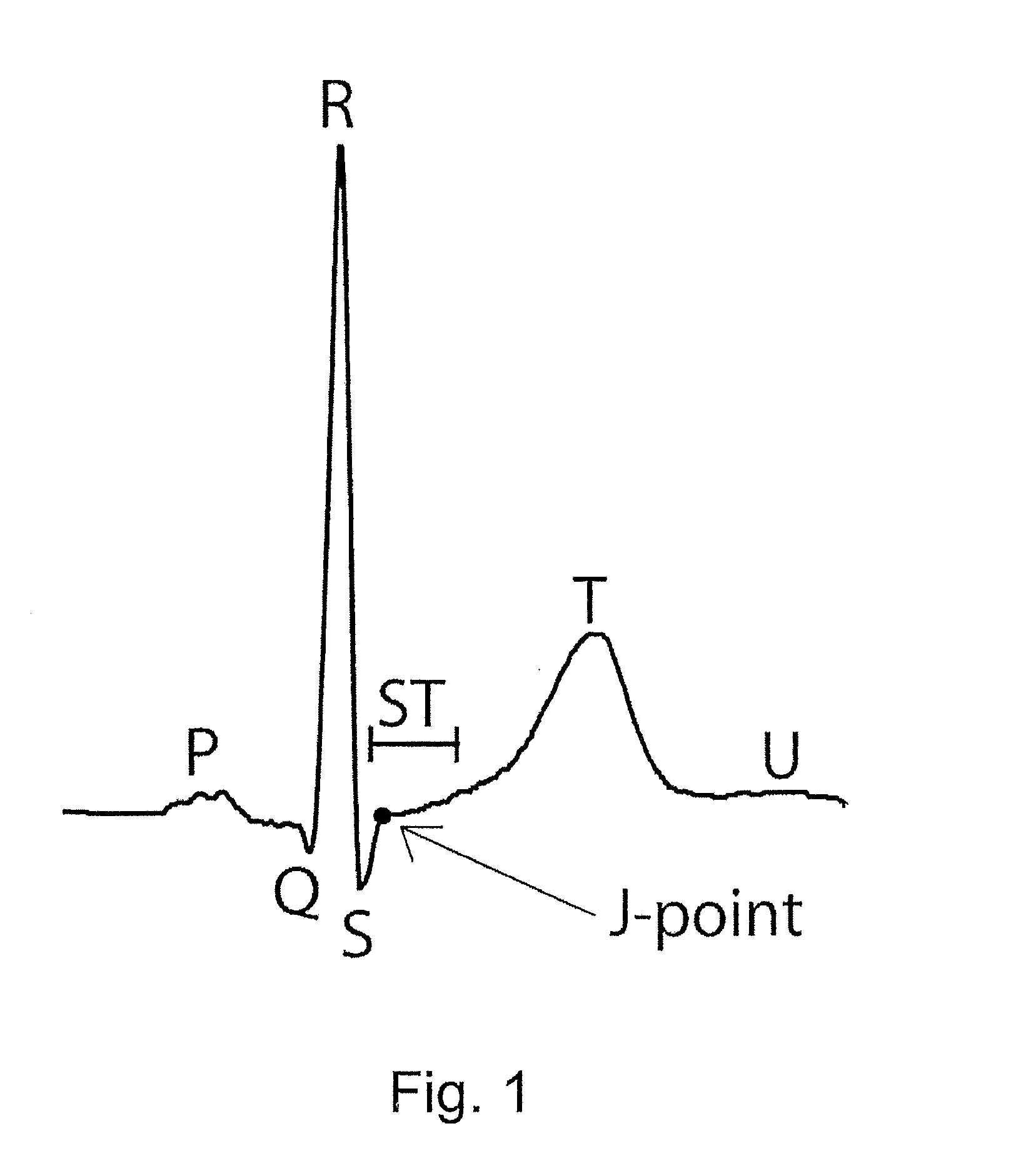
[0056]The importance of the electrocardiogram (ECG) in the diagnosis of acute myocardial infarction (AMI) is underlined by the clinical definition of the AMI subgroups based on ECG-findings: ST elevation myocardial infarction (STEMI), Bundle Branch Block myocardial infarction (BBBMI) and non-ST elevation myocardial infarction (non-STEMI). Patients with signs of STEMI or BBBMI are triaged for acute reperfusion therapy to ensure maximal myocardial salvage and optimal outcome.

[0057]In clinical practice, the fulfillment of well-defined STEMI criteria (1) determines whether the patient is eligible for reperfusion therapy. The current criteria require ST segment elevation in two contiguous leads of at least 0.1 mV (0.2 mV in leads V2-V3 for men and 0.15 mV for women). If the STEMI criteria are not fulfilled, and newly or presumably newly developed bundle branch block is not present (BBBMI), then the patient is c...

case 1

is a 57-year old male. CAG showed one-vessel disease with occlusion of the left anterior descending artery (LAD) resulting in TIMI-flow 0 immediately prior to intervention.

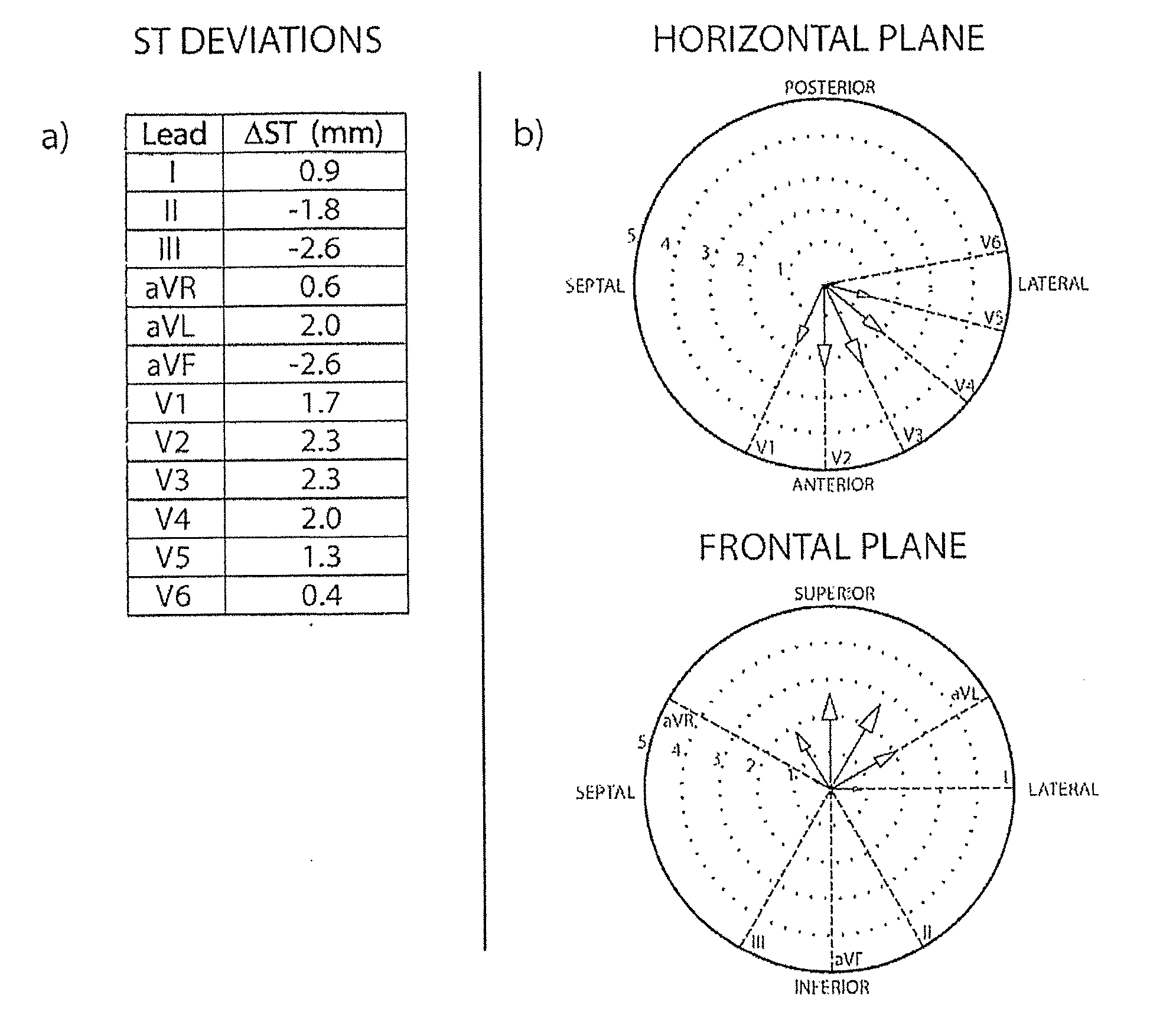
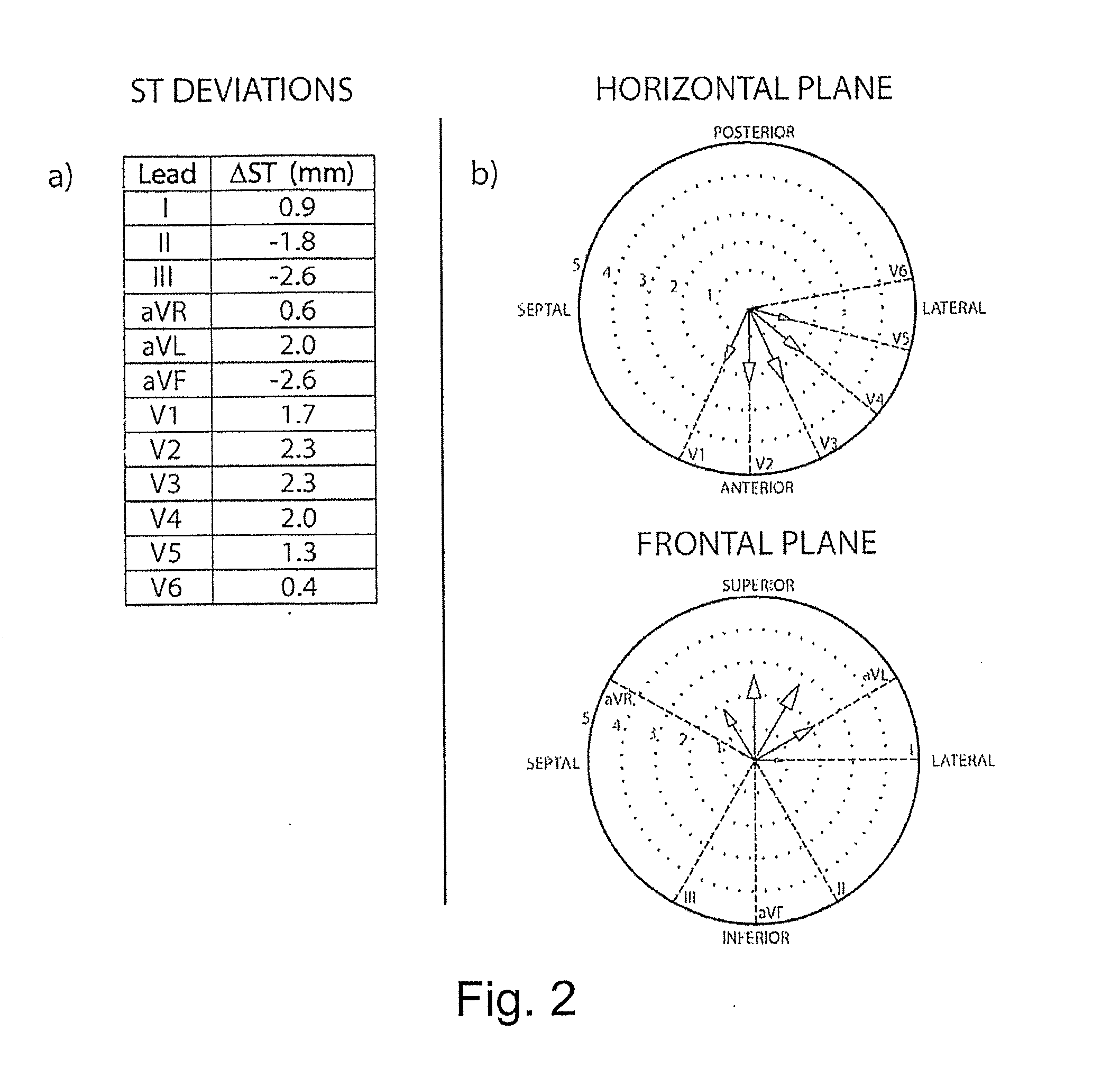
[0082]The ACC / ESC STEMI criteria are not met by the patient's ECG, since no ST elevation of >2 mm is present in V2-V3 and only one additional lead, lead V4, shows >1 mm ST elevation. Furthermore, the ECG shows signs of a single premature ventricular complex (PVC) at the end of the V1-V3 tracings shown in FIG. 4 (a).

[0083]The expert cardiologists agreed on a classification of “antero-lateral infarction” based on ST elevations in the anterior and lateral precordial leads.

[0084]SPECT-imaging showed myocardial ischemia in the apical part of the left ventricle with a slight anterior extent.

In the ST Compass the ST elevations in leads V2-V6 form a distinct pattern in the antero-lateral direction in the horizontal plane. There are no significant ST deviations in the frontal plane compass. The estimated ST injury vector p...

case 2

[0085]Results from Case 2 are presented in FIG. 5.

[0086]Case 2 is a 62-year old male. CAG showed one-vessel disease with occlusion of the right coronary artery (RCA) resulting in TIMI-flow 0 immediately prior to intervention.

[0087]The ACC / ESC STEMI criteria were met by the patient's ECG, which showed ST elevation of >1 mm in the three contiguous leads II, aVF and III.

[0088]The expert cardiologists agreed on a classification of “postero-infero-lateral infarction” based on ST elevations in the inferior limb / augmented leads (II, aVF, III), ST deviation in the anterior precordial leads (V1-V3) and ST elevation in the lateral precordial leads (V5-V6).

[0089]SPECT-imaging showed myocardial ischemia in the basal part of the left ventricle and on the inferior wall.

[0090]In the ST Compass, the ST depressions in leads V1-V4 and the ST elevations in leads V5-V6 form a distinct pattern in the posterior direction in the horizontal plane. ST elevations in leads II, aVF and III and ST depressions i...


PUM


Abstract

A system and a method for spatially ordered estimation and visualization of multi-lead electrocardiographic ST deviations induced by myocardial ischemia, in which system a plurality of ECG signals are recorded from a ECG source, which signals are stored by a processor in a memory, which processor processes the signals to obtain ST deviation, which processor performs measurement of ST deviation from each lead where the processor performs a multi-dimensional estimation of an vector representing of the spatial direction and magnitude of the underlying cardiac injury-current giving rise to the measured ST deviations, which processor hereby estimates the spatial location and severity of myocardial ischemia.

Description

BACKGROUND OF THE INVENTION[0001]1. Field of the Invention[0002]The present invention relates to a system and a method for spatial estimation and visualization of multi-lead electrocardiographic ST deviations induced by myocardial ischemia, in which system a plurality of ECG signals are recorded from a ECG source, which signals are stored by a processor in a memory, which processor processes the signals to obtain ST deviation, which processor performs measurement of ST deviation from each lead.[0003]2. Description of Related Art[0004]Electrocardiographic recordings (ECG) are made by means of electrodes on the limbs and torso of a person or animal connected to an electrocardiographic apparatus to amplify and possibly filter and store the signal in digital format. The signals can then be graphically displayed and visually interpreted as well as analyzed in a computer unit.[0005]By using several electrodes at different body positions, it is possible to obtain several different ECG reco...

Claims


Application Information

Patent Timeline
Patent Type & Authority Applications(United States)
IPC IPC(8): G06F15/00
CPCA61B5/0452A61B5/04011A61B5/341A61B5/349A61B5/358
Inventor ANDERSEN, MADS PETER
Owner AALBORG UNIV
Why Eureka
Try Eureka