Labeling composition for intraocular tissue, labeling method of intraocular tissue, and screening method

a technology of labeling composition and intraocular tissue, which is applied in the direction of anthracene dyes, biological material analysis, medical preparations, etc., can solve the problems of inability to visualize the morphology of cells forming the retina, low resolution of current oct image, and marked impairment of patient quality of life, etc., to achieve high precision, simple and easy way, and high definition

Inactive Publication Date: 2011-09-29
CANON KK
View PDF11 Cites 19 Cited by
  • Summary
  • Abstract
  • Description
  • Claims
  • Application Information

AI Technical Summary

Benefits of technology

[0022]The present invention provides a labeling composition for an intraocular tissue, whereby noninvasive labeling of the intraocular tissue, which has heretofore been impossible to conduct, becomes feasible, and so it is possible to image the condition of a layer structure of the intraocular tissue and the cell morphology thereof in a simple and easy way and with high definition. It is thereby possible to conduct evaluation and analysis of the intraocular tissue with high precision. In addition, the labeling composition for the intraocular tissue according to the present invention is combined with an observing and analyzing apparatus for intraocular tissues, whereby the condition of an intraocular tissue, or the retina in particular, of a living individual, which has heretofore been difficult to be observed, can be grasped with accuracy and high sensitivity, and so a new tool effective for researches and diagnoses in an ophthalmic field can be provided.

Problems solved by technology

The progress of amblyopia by an ophthalmic disease leads to blindness in the worst case, and the patient is markedly impaired in the quality of life.
However, a current OCT image is low in resolution, and so it has been unable to visualize the morphology of cells forming the retina.
However, the quantity of the drug transferred to the retina is extremely small.
However, an advanced technique is required, and a burden on a patient is also great, and so it is extremely difficult to use it except for the case of surgery or curing, and such administration has been unable to be used for the morphological or functional evaluation of the fundus retina.

Method used


Image

  • Labeling composition for intraocular tissue, labeling method of intraocular tissue, and screening method
  • Labeling composition for intraocular tissue, labeling method of intraocular tissue, and screening method
  • Labeling composition for intraocular tissue, labeling method of intraocular tissue, and screening method

Examples


example 1

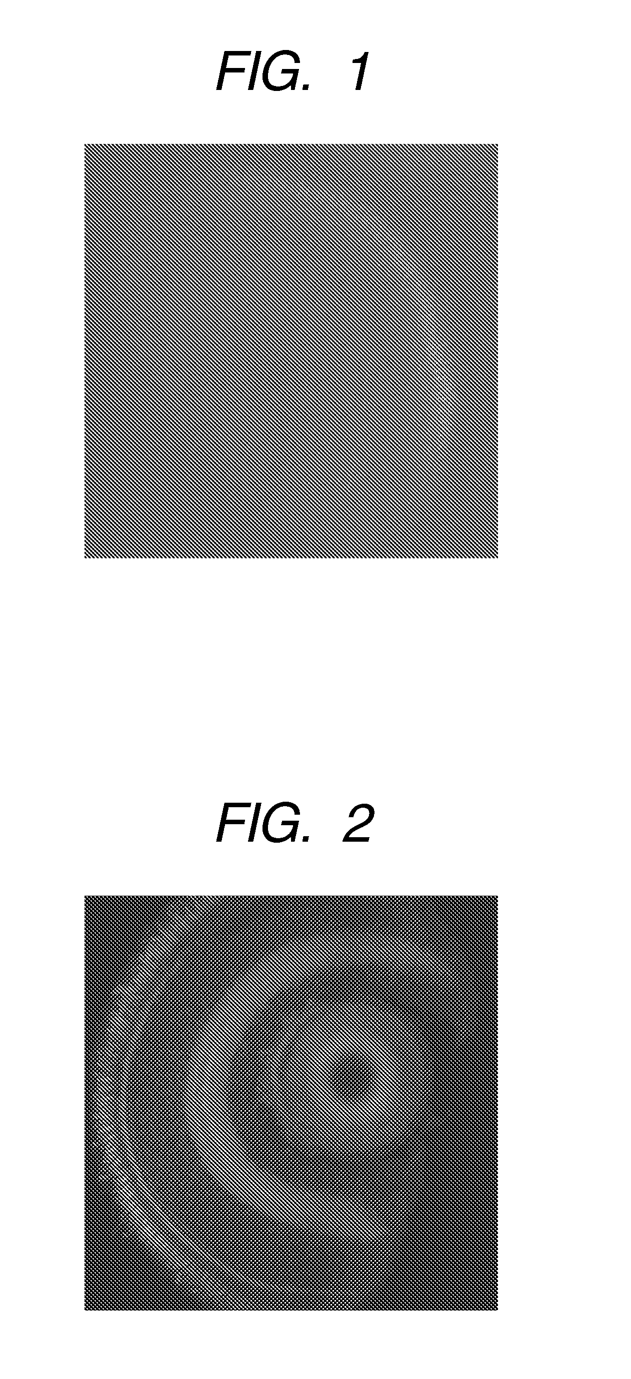
[0300]Distilled water was added to a 1 mg / mL DMSO solution of Staining compound 1 to obtain Staining Liquid 1 containing Staining compound 1 at a concentration of 1 μg / mL. In an arbitrary well of a 24-well multiplate (product of IWAKI), were put 1 mL of Staining Liquid 1 and a fry of Zebrafish 7-day-old embryo (7 dpf), and the plate was left to stand for 1 hour. A process of removing Staining Liquid 1 in the well and replacing it by 1 mL of distilled water was then performed 3 times. Further, 1 mL of a phosphate buffer solution containing 4% paraformaldehyde was added, and the plate was left to stand for 1 hour. The fry was then taken out of the well and embedded in a 5% low-temperature fused agarose gel to prepare a section of an ocular tissue using Linear Slicer PRO 7 (manufactured by Dosaka EM Co., Ltd.). The thus-prepared ocular tissue section was placed on a slide glass to observe the ocular tissue through a confocal microscope (Pascal Exciter, manufactured by Zeiss Co.).

examples 2 to 106

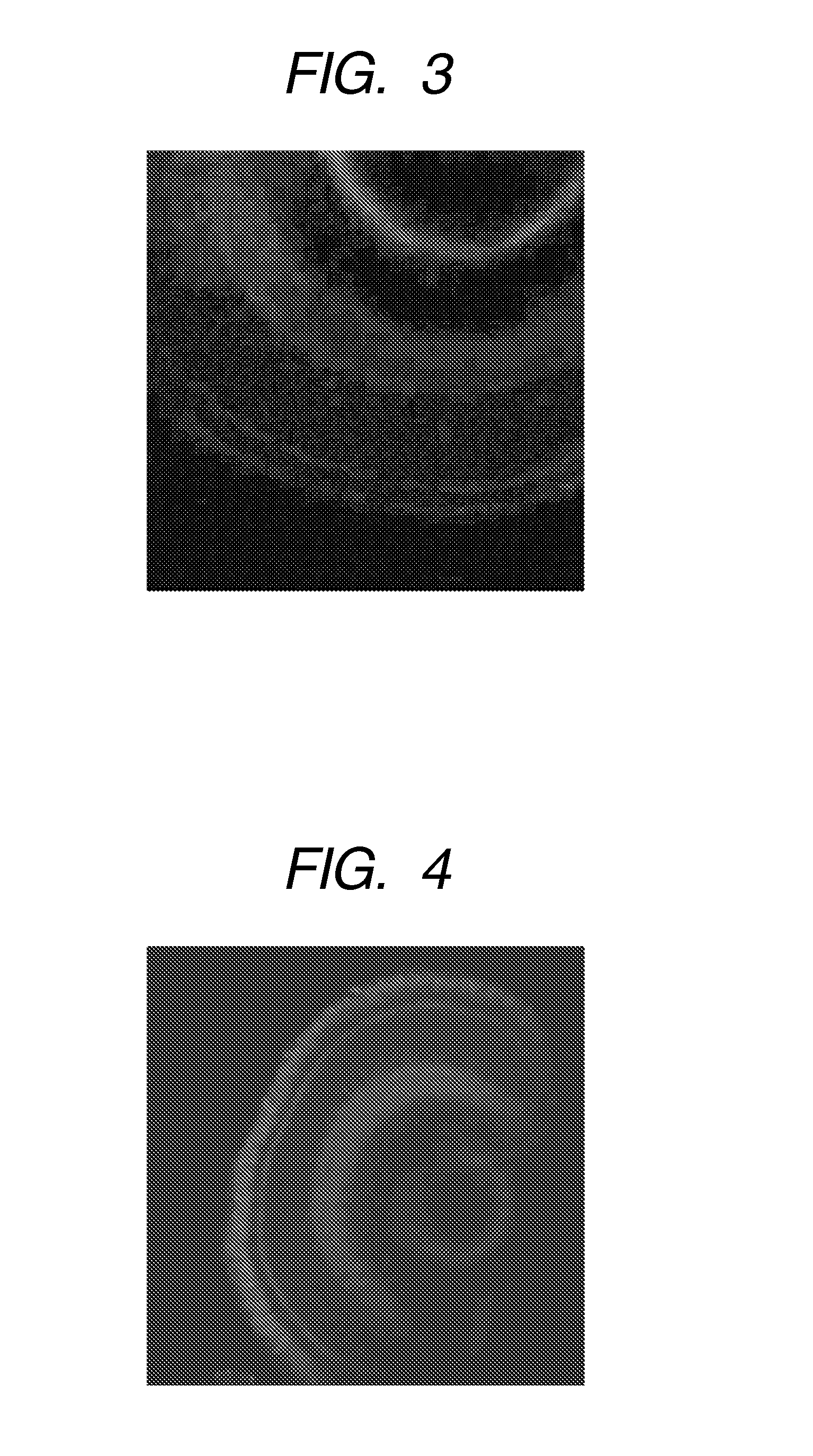
[0301]Sections were prepared by the same process as in Example 1 except that Staining compound 1 in Example 1 was changed to staining compound s described in Table 1, and observed.

example 107

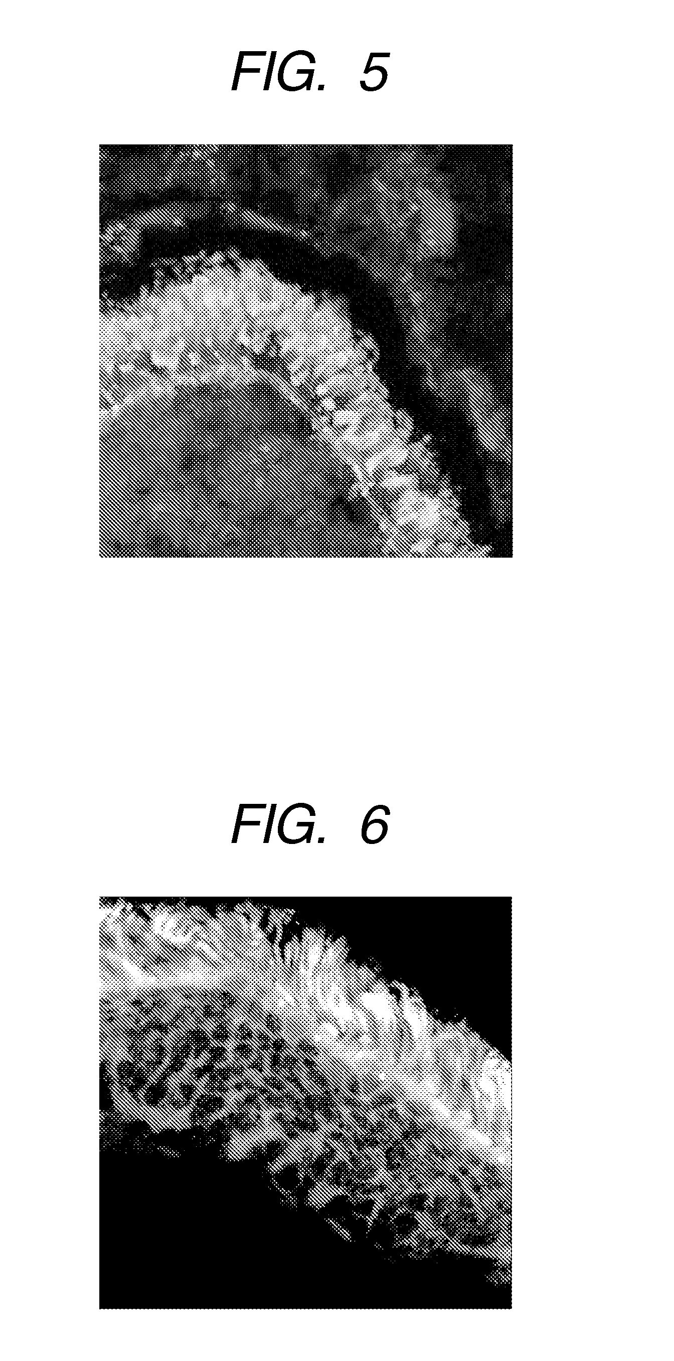
[0306]Distilled water was added to a 1 mg / mL DMSO solution of Staining compound 5 so as to give a concentration of 1 μg / mL to obtain Staining Liquid 2. In an arbitrary well of a 24-well multiplate, were put 1 mL of Staining Liquid 2 and a fry of Zebrafish, and the plate was left to stand for 1 hour. A process of removing Staining Liquid 2 in the well and replacing it by 1 mL of distilled water was then performed 3 times.

[0307]The fry was then put in a 0.7% agarose solution and placed on a slide glass to observe the fundus of the fry through a confocal microscope.


PUM

PropertyMeasurementUnit
concentrationaaaaaaaaaa
temperatureaaaaaaaaaa

Abstract

The invention provides a labeling composition for an intraocular tissue of a living individual, which specifically labels the intraocular tissue without need of an invasive operation such as exposure of an ocular tissue or injection of a staining agent into the ocular tissue or a nerve tissue linking to the ocular tissue, a method of noninvasively labeling an intraocular tissue of a living individual, and a screening method using the labeling composition for the intraocular tissues. The composition contains a compound capable of labeling at least a photoreceptor cell layer of a retina, wherein the compound is a staining compound having a particular structure as a partial structure thereof.

Description

TECHNICAL FIELD[0001]The present invention relates a labeling composition for an intraocular tissue, which is used for noninvasively labeling an intraocular tissue of a living sample, a labeling method of the intraocular tissue, and a screening method.BACKGROUND ART[0002]In recent years, there has been a tendency to increase the number of patients suffering ophthalmic diseases with the progress of population aging. The progress of amblyopia by an ophthalmic disease leads to blindness in the worst case, and the patient is markedly impaired in the quality of life. Therefore, it is required to detect the disease in its early stage and take a measure to cure the disease or retard the progress thereof. Typical diseases forming a cause of halfway blindness include diabetic retinopathy, macular degeneration diseases and glaucoma, and the fundus retina is concerned with many of them. For diagnoses of these diseases, diagnoses by evaluation of visual function by inspection of visual field, a...

Claims


Application Information

Patent Timeline
Patent Type & Authority Applications(United States)
IPC IPC(8): A61K49/00C07D221/18A61K51/04C07D491/14C09B1/54C07D311/82C07D417/06A61K51/08
CPCA61K49/0002G01N2333/4603A61K49/0039A61K49/0041A61K49/006C07D209/70C07D209/96C07D215/00C07D221/14C07D221/18C07D263/54C07D277/34C07D277/36C07D277/64C07D311/82C07D311/86C07D405/04C07D405/06C07D413/04C07D413/06C07D417/04C07D417/06C07D417/14C07D491/052C07D491/12C07D497/10G01N33/5088G01N33/582A61K49/0021
Inventor WATANABE, KOHEISHINTOU, TAICHINOMOTO, TSUYOSHIMIYAZAKI, TAKESHITANAKA, TOSHIONISHIMURA, YUHEISHIMADA, YASUHITONISHIMURA, NORIHIRO
Owner CANON KK
Why Eureka
Try Eureka