

Compression device for enhancing normal/abnormal tissue contrast in MRI including devices and methods related thereto
a compression device and contrast technology, applied in the field of magnetic resonance imaging, can solve the problems of cancer, potentially life-threatening, and difficult to detect tumors that are too deep or too small, and achieve the effects of improving contrast-to-noise ratio, prolonging scan times, and improving resolution
- Summary
- Abstract
- Description
- Claims
- Application Information
AI Technical Summary
Benefits of technology
Problems solved by technology
Method used
Image

Examples
example 1
SENC MRI
[0166]The Strain Encoded (SENC) MRI technique was introduced to measure local strain distribution of deforming tissues. In SENC MRI, the magnetization of the object under examination at point p and time t is modulated in the z-direction with a sinusoidal pattern with the spatial frequency, ω(p,t). The z-direction here is defined as the direction orthogonal to the imaging plane. Once induced, this pattern lasts for a fraction of a second, during which, if the tissue is deformed, the frequency ω(p,t) proportionally changes with the degree of deformation at the pixel p. The resulting image intensity at this pixel is given by
I(p_,t)≈12ρ(p_,t)S(ωT-ω(p_,t)),(1)
[0167]where ρ(p,t) is a term representing the proton density of the voxel including the T1 relaxation effect, S(ω) is the Fourier transform of the slice profile determined by the envelope of the applied slice selection RF pulse and ωT is called the tuning frequency, which is determined during the image acquisition by an appl...
example 2
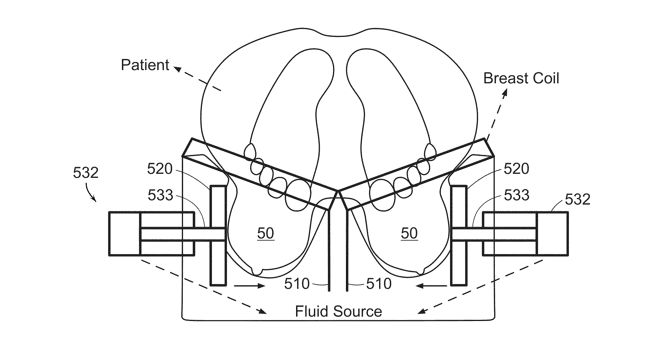
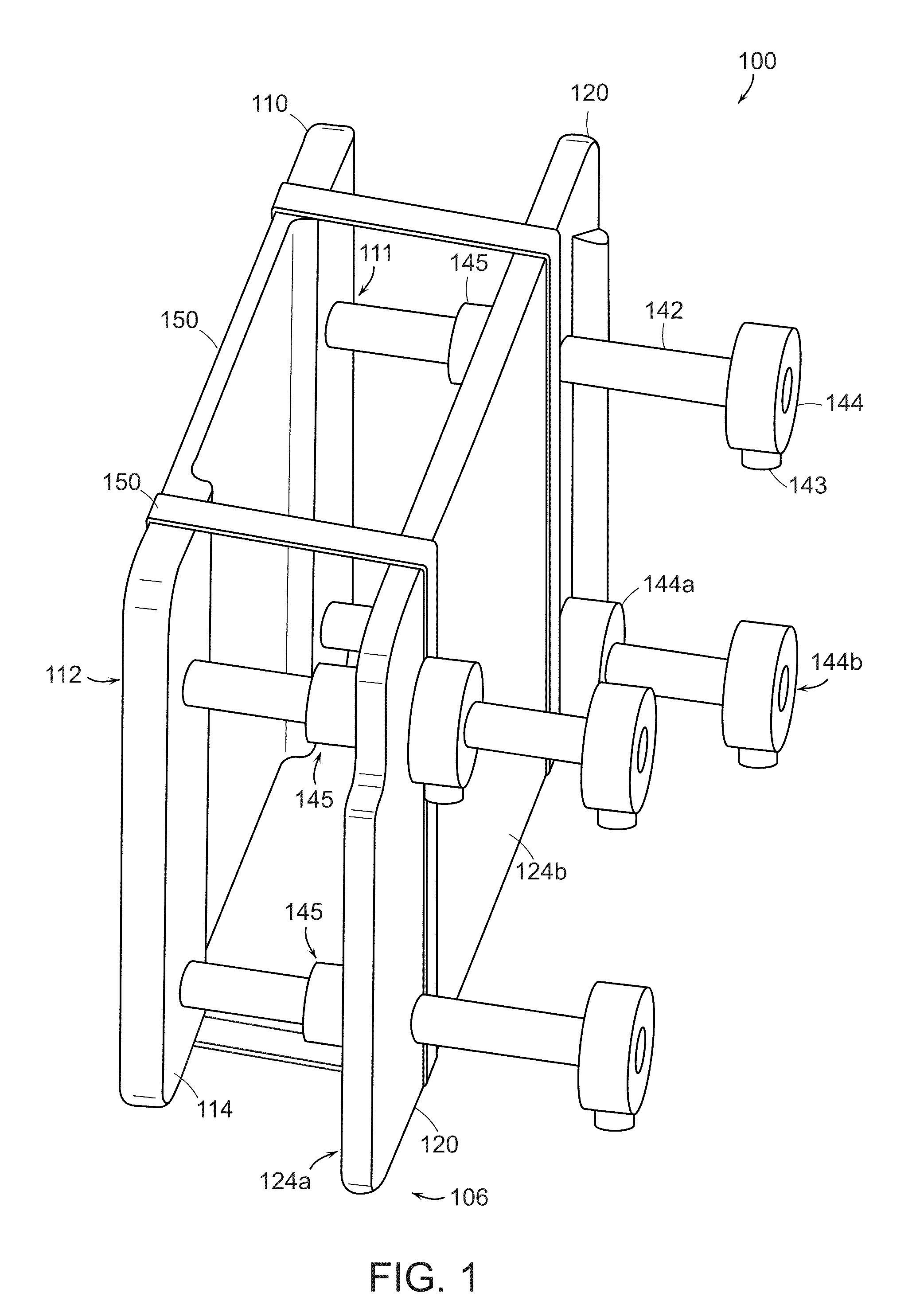
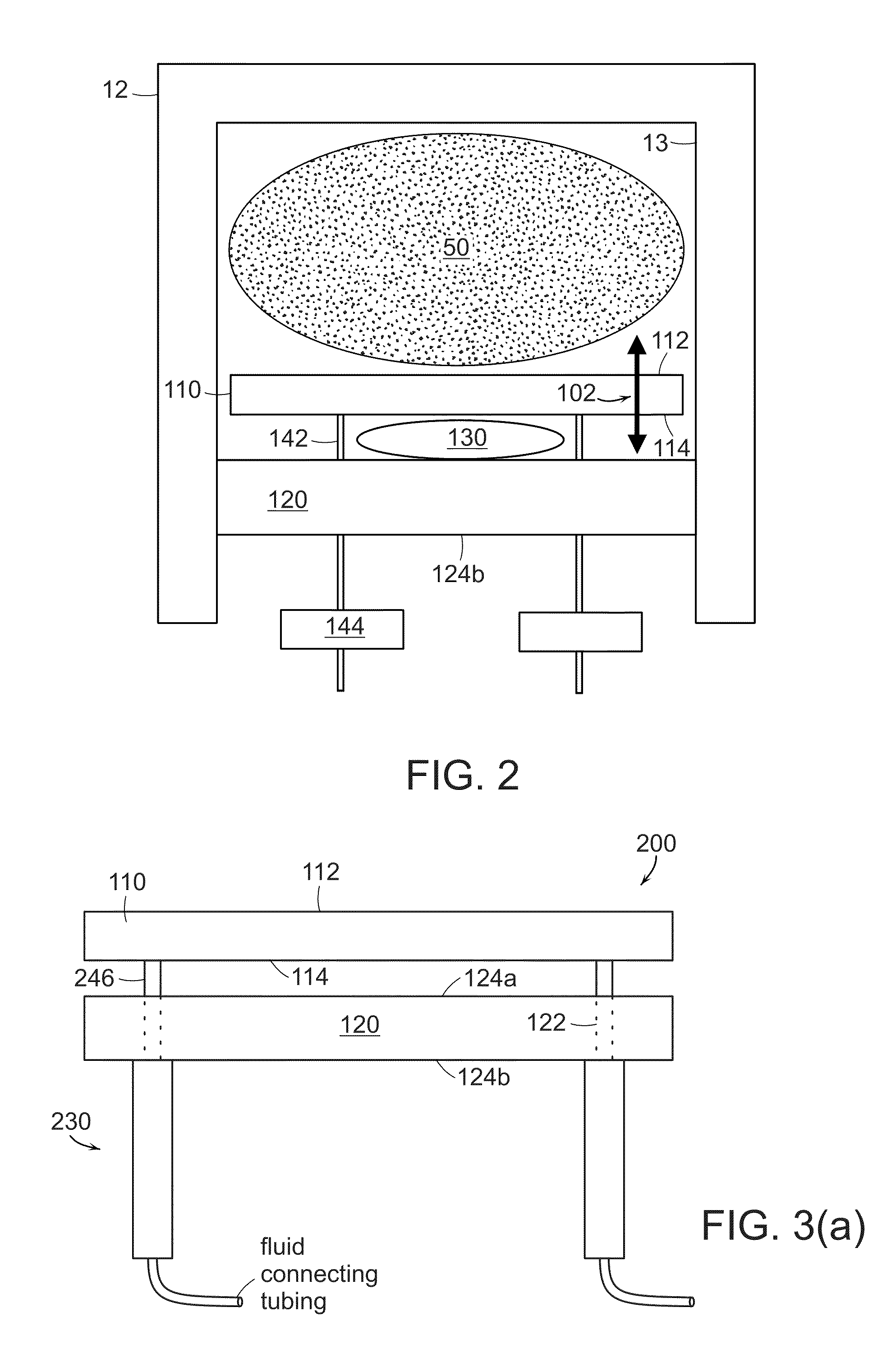
[0184]A phantom experiment was used in order to test the tissue compression device of the present invention. The phantom was made of a silicon gel material prepared by mixing two compounds, A and B (kit 3-1450, Dow Corning). The ratio in the mixture determines the stiffness of the resultant gel. The phantom was composed of a small stiff sphere 1 cm in diameter immersed in a cube made from softer gel material with dimensions 8×8×10 cm3. A top-view of the phantom is shown in FIG. 19. It is worth noting that the stiff inclusion was intentionally placed near the corner of the phantom to test the ability of the system to detect lesions at the periphery of the breast (difficult situation). The stiffness of the inclusion was made very close to that of the surrounding soft gel. In fact, manual palpation on the surface of the phantom did not detect the existence of the inclusion.
[0185]The phantom experiment was done on a 3.0 T MR scanner (Achieva, Philips Medical Systems, Be...
example 3
[0239]Breast cancer is one of the most common causes of cancer related mortality. Early detection of breast lesions using mammography and ultrasound has resulted in lower mortality-rates. However, some breast lesions are mammography occult and the use magnetic resonance imaging (MRI) is recommended. MRI has high sensitivity and moderate specificity, thus, there is a need to increase specificity so that unnecessary biopsies procedures can be reduced. Higher specificity can be achieved by incorporating the stiffness property, as cancer tumors are 3-13 times stiffer than both normal tissue and benign tumors. Previously, fast strain-encoded (FSENC) MR with simple hardware was introduced to detect different stiffness by measuring the strain directly in a simple way; however, the simple hardware limited the scanning time thus limiting the imaging resolution. In this work, a new apparatus or device is introduced that is capable of periodically compressing the breast, which allows for longe...
PUM
 Abstract
Description
Claims
Application Information