
Device and method for measuring contrast agent

a contrast agent and device technology, applied in the field of tomography devices, can solve the problems of affecting the spatial encoding of fast mr sequences, affecting the measurement of arterial signal, and the shortest echo time is still too long for measuring arterial signal, so as to achieve the effect of reducing the frequency of mr sequences

Inactive Publication Date: 2011-11-10
UNIVERSITATSKLINIKUM FREIBURG
View PDF4 Cites 0 Cited by
  • Summary
  • Abstract
  • Description
  • Claims
  • Application Information

AI Technical Summary

Benefits of technology

[0026]An embodiment of the invention is advantageous in which the further measurement unit encloses a smaller volume than the first measurement unit. This makes the further measurement unit easier to handle and it can be flexibly attached even to difficult-to-access parts of the body.
[0030]One variant of the inventive method must be seen as especially advantageous in which the excitation of the magnetization of the blood and / or contrast agent carried in the artery of the living organism under examination is performed before and / or after the imaging portion of the measurement sequence of the first measurement unit, wherein the read-out operation of the further measurement unit is performed during and / or after the imaging portion of the measurement sequence of the first measurement unit. In this way, an especially long time is available for the measurement by the at least one measurement unit. Moreover, the measurement by the at least one measurement unit can be repeated after repeated excitation of the magnetization after the imaging portion of the measurement sequence of the determination device.
[0031]A further advantageous variant of the inventive method is characterized in that the excitation slice is chosen to be sufficiently thick that the change in the signal acquired by the further measurement unit caused by the time-variable blood flow in the at least one artery is negligibly small. In this way, it is no longer necessary to determine the time-variable blood flow in the at least one artery. This permits especially simple evaluation of the signal by the at least one measurement unit.
[0033]In a further variant of the inventive method, the contrast between the artery and the tissue surrounding the artery can be set using the relevant flip angle of the excitations of the magnetization. Adapting the flip angle chosen for excitation of the magnetization advantageously enables the contrast between the artery being observed and the surrounding tissue to be set to be as large as possible so that determination of the arterial contrast agent concentration by the at least one measurement unit is simple.
[0034]In another variant of the inventive method, subsequent to the excitation and read-out operation executed before the imaging portion of the measurement sequence, one or more additional excitations with a read-out operation are appended after the imaging portion of the measurement sequence and with variable starting times. In this way, the transverse tissue magnetization surrounding the artery is additionally suppressed. Moreover, the temporal sampling rate can be increased according to the number of read-out operations.
[0035]A variant of the inventive method is advantageous in which the contrast agent concentration is determined by isolating the portion of the spectrum caused by the contrast agent out of the measured signal. By determining the contrast agent concentration using the complex spectrum measured by the at least one further measurement unit, the concentration is especially stable and can be performed precisely by isolation of the corresponding resonance function.

Problems solved by technology

There are methods by which multiple echoes of the same excitation can be acquired [4], but it has been shown that the shortest echo time is still too long for measurement of the arterial signal.
Moreover, the presence of contrast agent results in a considerable displacement of the Larmor frequency.
This disturbs the spatial encoding in fast MR sequences, such as the EPI sequence.
However, with this method, local AIFs are determined in arteries that are very small by comparison with the voxel size because of the relatively low spatial resolution.
This effect, called the partial volume effect, results in a serious loss of the arterial contribution in the measured signal.
However, such a vessel is hard to find.
In particular, blood vessels that can be used as local AIFs are not usually parallel to the magnetic field and are thus not correctable.
Because the signal is not acquired using a separate coil, the problem of the limited dynamic range remains.
A further problem in determining the perfusion parameters is the time resolution of the bolus passage.
This is particularly insufficient for arteries where the blood flow is high.
The problems of the prior art stated above can be summarized as follows:Greatly differing concentration of the contrast agent in large vessels and tissue make precise, simultaneous measurement of large vessels and tissue with the same sequence impossible.This results in a decline in the arterial signal below the noise level and in a shift in the phase of the signal.The time resolution of the DSC measurement is very low, especially for the AIF.Arterial input functions that can be determined by means of the prior art techniques are distorted by partial volume effects.

Method used


Image

  • Device and method for measuring contrast agent
  • Device and method for measuring contrast agent
  • Device and method for measuring contrast agent

Examples


Embodiment Construction

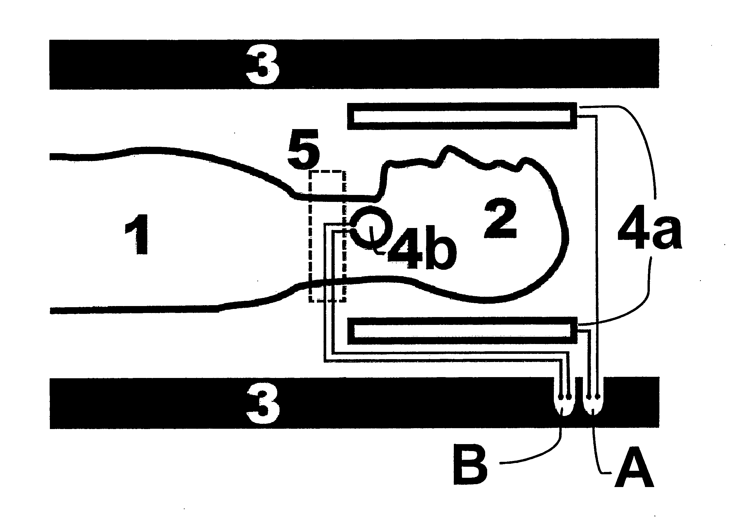
[0041]In the schematic representation FIG. 1, one part of the body 2 (in this case the head) of the living organism 1 under examination (in this case a human being) is positioned in the measurement volume defined by the first measurement unit 4a of the tomography device 3. The further measurement unit 4b constituted as a surface coil is attached to the neck of the patient. The magnetization from the excitation slice 5 is used as a signal source for the further measurement unit 4b.

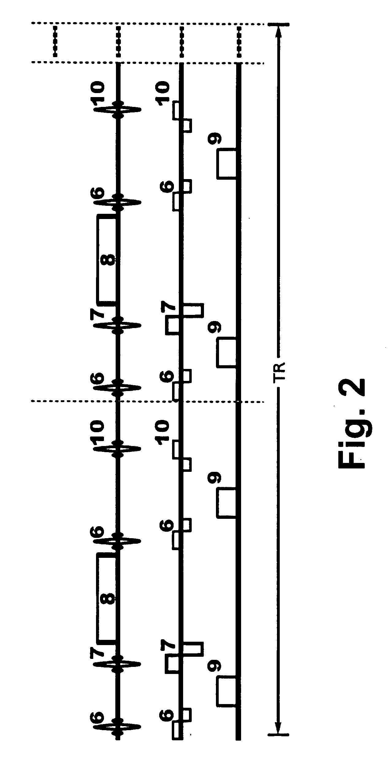
[0042]FIG. 2 shows the measurement sequence for operation of an inventive tomography device. In the sequence, after excitation of the tissue magnetization 7 in each slice, an imaging block 8 is inserted to perform the in-slice encoding. The excitation of the contrast agent magnetization 6 in the slice that results in the signal 9 in the further measurement unit 4b can be performed before and / or after the imaging block 8 and the excitation 7. In the case of excitation before the imaging block 8, the influen...


PUM


Abstract

A tomography device (3) producing magnetic resonance (MR) images of a part of the body of a living organism (1) disposed in a measurement volume of the tomography device (3), with a first measurement unit (4a) for acquiring a spatially resolved temporal series of MR images of the part of the body under examination, wherein the temporal series of MR images represents the passage of a contrast agent injected into the blood stream of the living organism through an organ located in the part of the body under examination, is characterized in that at least one further measurement unit (4b) is provided that comprises a local receiver coil that measures, close to at least one artery that supplies the part of the body (2) of the living organism disposed in the tomography device, the concentration of contrast agent with temporal resolution and concurrently with measurement of the temporal series of MR images determined by the first measurement unit. A system and an associated method are thereby provided that permit simultaneous measurement of large vessels and tissue with one and the same sequence with an adapted dynamic range.

Description

[0001]This application claims Paris Convention priority of DE 10 2010 028 749.0 filed May 7, 2010 the complete disclosure of which is hereby incorporated by reference.BACKGROUND OF THE INVENTION[0002]The invention relates to a tomography device for producing magnetic resonance (MR) images of a part of the body of a living organism disposed in a measurement volume of the tomography device, with a first measurement unit for acquiring a spatially resolved temporal series of MR images of the part of the body under examination, wherein the temporal series of MR images represents the passage of a contrast agent injected into the blood stream of the living organism through an organ located in the part of the body under examination.[0003]Such a device is known from EP 0 958 503 B1.[0004]The aim of dynamic susceptibility-compensated (DSC) measurement is to determine perfusion parameters such as cerebral blood volume (CBV), cerebral blood flow (CBF), and mean transit time (MTT) with spatial r...

Claims


Application Information

Patent Timeline
Patent Type & Authority Applications(United States)
IPC IPC(8): A61B5/055
CPCG01R33/56366G01R33/5601
Inventor KISELEV, VALERIJGALL, PETERKELLNER, ELIAS
Owner UNIVERSITATSKLINIKUM FREIBURG
Learn More