Compact Endocavity Diagnostic Probes for Nuclear Radiation Detection

a diagnostic probe and nuclear radiation technology, applied in the field of radiographic imaging, can solve the problems of unsuitable functional imaging required in cancer imaging and diagnosis, benign and cancerous tumors cannot be easily distinguished by ultrasound, and the ultrasonic technology is not an ideal tool for cancer detection and diagnosis, etc., to achieve high spatial resolution, high image contrast, and high image quality

Active Publication Date: 2011-11-24
HYBRIDYNE IMAGING TECH
View PDF17 Cites 30 Cited by
  • Summary
  • Abstract
  • Description
  • Claims
  • Application Information

AI Technical Summary

Benefits of technology

[0007]In accordance with the present invention, a novel radiation imaging probe comprising a plurality of semiconductor radiation detectors, complete signal processing circuitry, and a power supply within a cylindrical sheath is disclosed. More particularly, the semiconductor radiation detector within the radiation imaging probe of the present invention is preferably based on solid state semiconductors capable of operating as photon-charge direct conversion devices arranged as a radiation imaging camera that can localize the distribution variations of radiation sources in tissues or inanimate objects. We recognized that the novel radiation imaging probe based on the solid-state semiconductor detector of the present invention has unique features that ensure high image contrast, high spatial resolution, and high image quality, suitable for gamma ray and X-ray imaging. Moreover, with the highly integrated signal processing circuits and the readout control logic, the probe of the present invention can be designed to be very compact and easy to carry, handle, and operate. Alternatively, the probe of the present invention may have dual functionality by incorporating a radiation imaging module(s) and the ultrasound modules: one detecting radiation to produce a functional image and the other to simultaneously produce an anatomical image.

Problems solved by technology

However, ultrasound technology is not an ideal tool in cancer detection and diagnosis because ultrasound can only generate anatomical images, whereas functional images are needed, especially at the early stages of cancer.
The probe generates images at different angles to help the physician estimate the size of the prostate and detect abnormal growths; however, benign and cancerous tumors cannot easily be distinguished by ultrasound.
Hence, while the ultrasound probes can be designed to be very compact and easy to carry, handle, and operate, their inability to distinguish benign and cancerous tumors makes them unsuitable for functional imaging required in cancer imaging and diagnosis.
Although HPGe itself is small, it needs a complex cooling system to work at cryogenic temperatures (e.g., −180° C.).
Hence, all of these systems, either based on scintillator detectors or HPGe crystals, are bulky and can only be integrated into an external detection system.
However, since the detectors of such external systems are located far away from the imaged organs, they have poor detection efficiency and low spatial resolution, which limit such detector's ability to pinpoint the exact positions of cancerous tissues in a small organ.
All these drawbacks limit the usefulness of such radiation detection systems in diagnosing cancer in small organs, e.g. prostate glands, particularly for small tumors.

Method used


Image

  • Compact Endocavity Diagnostic Probes for Nuclear Radiation Detection
  • Compact Endocavity Diagnostic Probes for Nuclear Radiation Detection
  • Compact Endocavity Diagnostic Probes for Nuclear Radiation Detection

Examples


Embodiment Construction

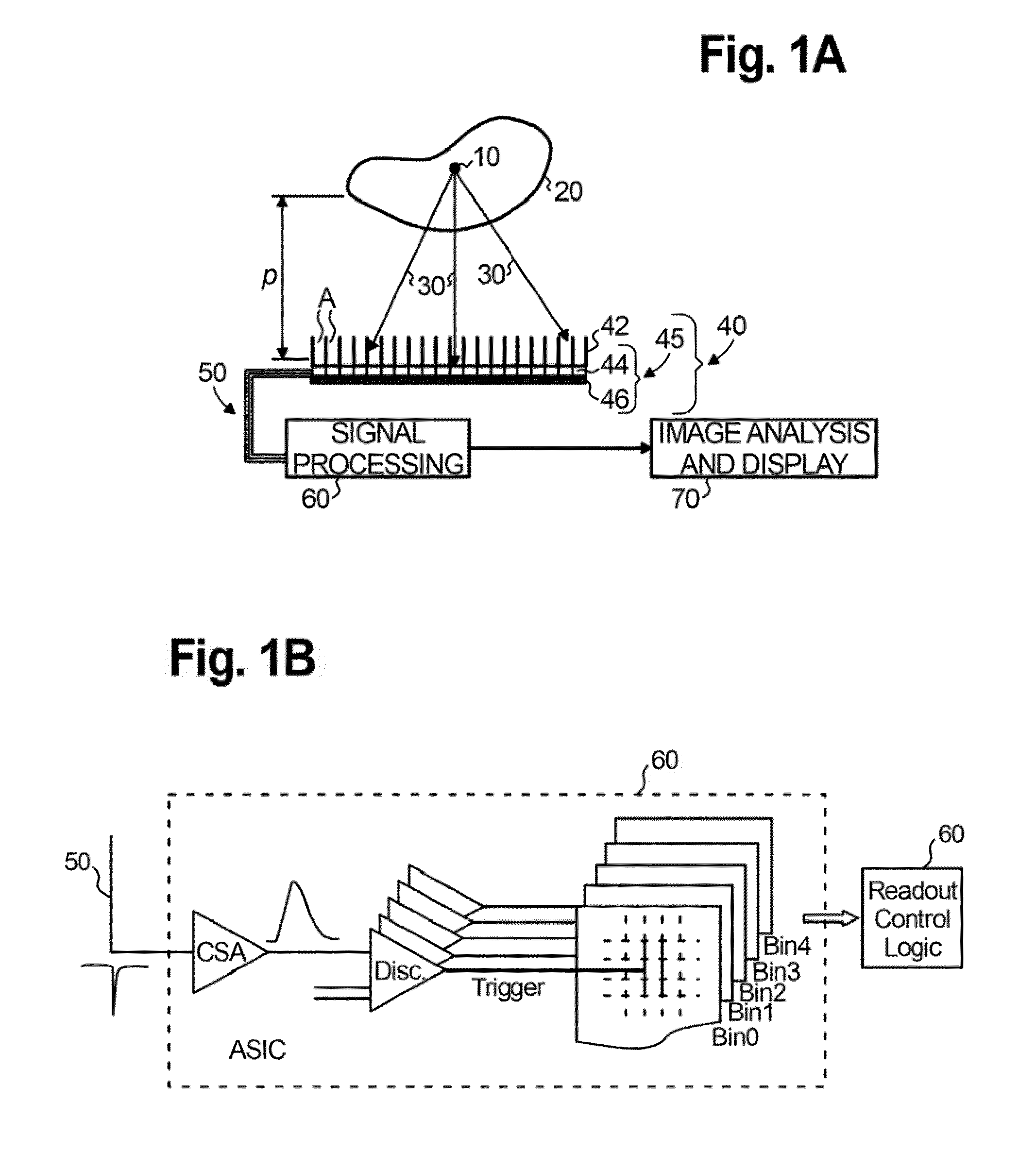
[0025]The present invention is directed to a novel radiation imaging probe that offers a compact size that is easy to carry, handle, and operate, and exhibits high energy resolution and detection efficiency. In the following detailed description of the preferred embodiments and various examples of the probe according to the present invention, reference is made to the accompanying drawings where like reference numerals refer to like parts. The drawings illustrate various embodiments in which a hybrid probe for radiation imaging applications may be practiced. It is to be understood, however, that those skilled in the art may develop other structural and functional modifications without significantly departing from the scope of the instant disclosure.

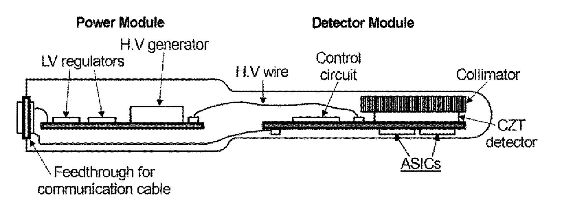
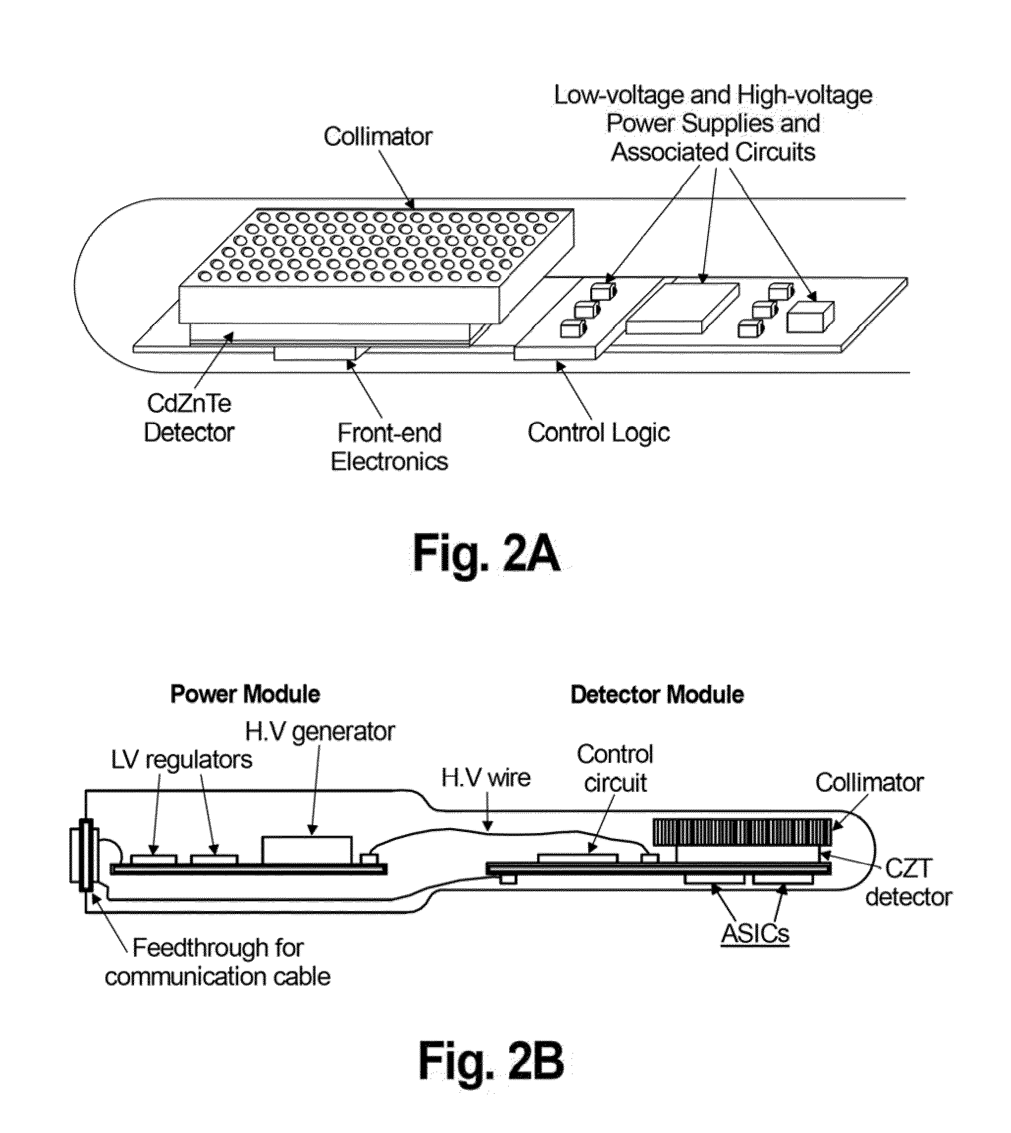
[0026]FIG. 2A and FIG. 2B illustrate two embodiments of the radiation imaging probe of the present invention. The probe is encapsulated within a tube (sheath or sleeve) and includes a detector module and a collimator. The collimator is fab...


PUM


Abstract

This invention relates to the field of radiation imaging. In particular, the invention relates to an apparatus and a method for imaging tissue or an inanimate object using a novel probe that has an integrated solid-state semiconductor detector and complete readout electronics circuitry.

Description

CROSS-REFERENCE TO RELATED APPLICATIONS[0001]This application claims the benefit of U.S. Provisional Application No. 61 / 320,157, filed Apr. 1, 2010, which is incorporated herein by reference in its entirety.STATEMENT OF GOVERNMENT RIGHTS[0002]This invention was made with Government support under contract number DE-AC02-98CH10886, awarded by the U.S. Department of Energy. The Government has certain rights in the invention.FIELD OF THE INVENTION[0003]This invention relates to the field of radiation imaging. In particular, the invention relates to an apparatus and a method for imaging tissue, such as the colon and prostate, or an inanimate object using a novel probe that has an integrated solid-state semiconductor detector and a complete readout electronics circuitry.BACKGROUND OF THE RELATED ART[0004]In medical imaging applications, two competing technologies are generally used: ultrasound and nuclear medical imaging. The benefit of the ultrasound technology is that it enables a very ...

Claims


Application Information

Patent Timeline
Patent Type & Authority Applications(United States)
IPC IPC(8): A61B6/00
CPCA61B1/051A61B6/4258A61B6/4208A61B6/425A61B6/4291A61B6/4417A61B6/48A61B6/485A61B6/50A61B8/08A61B8/0875A61B8/12A61B8/4444G01T1/161G21K1/025H01L27/14618H01L27/14661A61B6/06H01L2924/0002H01L2924/00
Inventor CUI, YONGGANGJAMES, RALPHBOLOTNIKOV, ALEKSEY
Owner HYBRIDYNE IMAGING TECH
Why Eureka
Try Eureka