Collimators and methods for manufacturing collimators for nuclear medicine imaging systems

a collimator and nuclear medicine technology, applied in nuclear engineering, instruments, and handling using diaphragms/collimeters, etc., can solve the problems of increasing restrictions and limitations, dangerous manufacturing of collimators using lead, and affecting the safety of users

Inactive Publication Date: 2012-04-12
GE MEDICAL SYST ISRAEL
View PDF5 Cites 21 Cited by
  • Summary
  • Abstract
  • Description
  • Claims
  • Application Information

AI Technical Summary

Benefits of technology

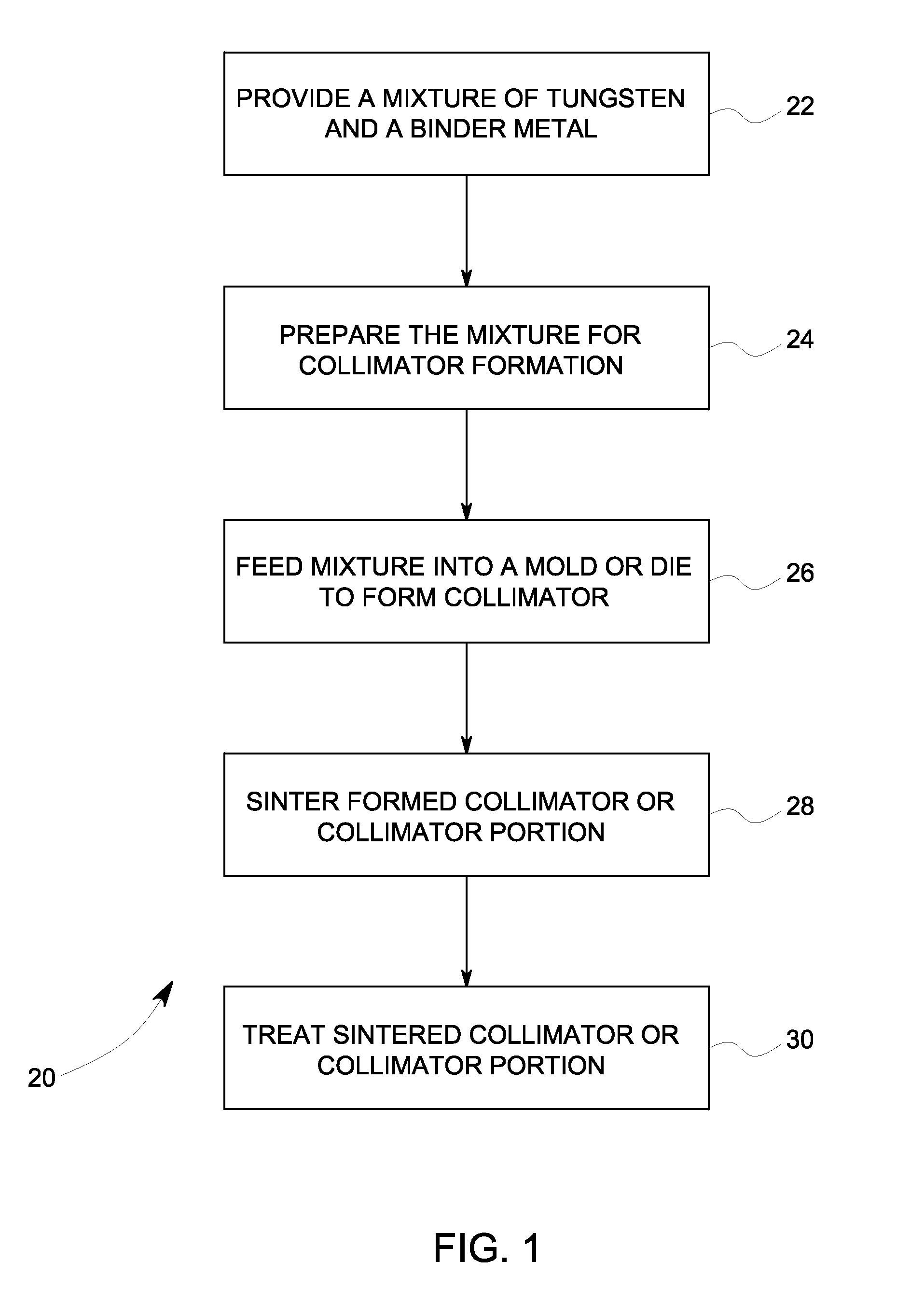
[0006]In accordance with various embodiments, a method for forming a collimator for detectors of a nuclear medicine (NM) imaging system is provided. The method includes forming a plurality of collimator segments from powdered tungsten, wherein the plurality of collimator segments have opposing faces with edges therebetween. The method also includes sintering the powdered tungsten segments and joining the plurality of sintered powdered tungsten segments at least at one or more of the edges to form the collimator for the NM imaging system.
[0007]In accordance with other embodiments, a collimator for a nuclear medicine (NM) imaging detector is provided that includes a plurality of individual powdered metal segments joined together at least at one or more of a plurality of edges between a front face and a rear face of the individual powdered metal segments to form a collimator body. The collimator also includes a plurality of bores extending through the plurality of individual powdered metal segments from the front face to the rear face of the collimator body.

Problems solved by technology

Because lead is toxic, the manufacture of collimators using lead can be dangerous, as well as harmful to the environment.
Moreover, the use of lead is only permitted in a limited number of fields, which is becoming more restrictive and limiting.
Additionally, lead collimators have a lead x-ray fluorescence that can interfere with low energy imaging.
For example, when excited with gamma rays of greater than about 80 keV, lead produces x-ray fluorescence at about 70 keV, which interferes with low energy imaging, such as imaging with Americium and Thallium.
Thus, this fluorescence can be problematic when imaging dual isotopes such as Technetium and Thallium (Tc+Tl), which results in having to perform multiple scans with a longer total scan time because of the interference.
Additionally, registration of the images for the two different scans acquired at different times can be difficult.

Method used


Image

  • Collimators and methods for manufacturing collimators for nuclear medicine imaging systems
  • Collimators and methods for manufacturing collimators for nuclear medicine imaging systems
  • Collimators and methods for manufacturing collimators for nuclear medicine imaging systems

Examples


Embodiment Construction

[0022]The foregoing summary, as well as the following detailed description of certain embodiments will be better understood when read in conjunction with the appended drawings. To the extent that the figures illustrate diagrams of the functional blocks of various embodiments, the functional blocks are not necessarily indicative of the division between hardware circuitry. Thus, for example, one or more of the functional blocks (e.g., processors or memories) may be implemented in a single piece of hardware (e.g., a general purpose signal processor or random access memory, hard disk, or the like) or multiple pieces of hardware. Similarly, the programs may be stand alone programs, may be incorporated as subroutines in an operating system, may be functions in an installed software package, and the like. It should be understood that the various embodiments are not limited to the arrangements and instrumentality shown in the drawings.

[0023]As used herein, an element or step recited in the ...


PUM

PropertyMeasurementUnit
sizeaaaaaaaaaa
sizeaaaaaaaaaa
sizeaaaaaaaaaa

Abstract

Collimators and methods for manufacturing collimators for nuclear medicine (NM) imaging systems are provided. One method includes forming a plurality of collimator segments from powdered tungsten, wherein the plurality of collimator segments have opposing faces with edges therebetween. The method also includes sintering the powdered tungsten segments and joining the plurality of sintered powdered tungsten segments at least at one or more of the edges to form the collimator for the NM imaging system.

Description

BACKGROUND OF THE INVENTION[0001]The subject matter disclosed herein relates generally to nuclear medicine (NM) imaging systems, and more particularly to methods for manufacturing a collimator for NM imaging systems.[0002]NM imaging systems, for example, Single Photon Emission Computed Tomography (SPECT) and Positron Emission Tomography (PET) imaging systems, use one or more image detectors to acquire image data, such as gamma ray or photon image data. The image detectors may be gamma cameras that acquire two-dimensional views of three-dimensional distributions of emitted radionuclides (from an injected radioisotope) from a patient being imaged.[0003]In order to acquire NM imaging information for a region of interest (ROI), the ROI, such as a heart of a patient, must be positioned within a field-of-view (FOV) of the gamma camera. The gamma cameras also may include collimators for focusing the FOV of the gamma camera. The collimators may create different sizes of FOVs for the gamma c...

Claims


Application Information

Patent Timeline
Patent Type & Authority Applications(United States)
IPC IPC(8): G21K1/02B22F3/12B22F7/00
CPCG21K1/025B22F3/225C22C1/045B22F2998/10B22F1/0059B22F3/20B22F3/10B22F1/10
Inventor BIRMAN, YOSSIHEFETZ, YARON
Owner GE MEDICAL SYST ISRAEL
Why Eureka
Try Eureka