Methods and systems for image-guided treatment of blood vessels

a technology of image-guided treatment and blood vessels, which is applied in the field of ultrasound imaging and therapy, can solve the problems of difficult to obtain an objective measure indicating the completeness of the therapeutic treatment, and difficult to evaluate the extent of the applied therapeutic treatment, so as to achieve general applicability, avoid side effects and/or drug resistance, and limit blood flow

Inactive Publication Date: 2012-05-10
THE TRUSTEES OF THE UNIV OF PENNSYLVANIA
View PDF6 Cites 28 Cited by
  • Summary
  • Abstract
  • Description
  • Claims
  • Application Information

AI Technical Summary

Benefits of technology

[0082]Another advantage of the present invention is that system 100 does not require treatment of individual cancer cells. Because the survival of several thousand cells depends on every individual blood vessel, disrupting a few blood vessels may trigger cell death in many cancer cells.
[0083]A further advantage of the present invention is that the target body structure, the endothelial cells, are in close proximity of the microbubbles. Because of the easy access to the target body structure, system 100 is not limited by drug delivery problems common with therapies which target cancer cells in the extravascular space. Furthermore, system 100 uses access to the surface of the endothelial cells, unlike other antivascular drugs that need to penetrate the cells to affect their cytoskeleton.
[0084]Another advantage of the present invention is that, unlike antivascular compounds that target molecular pathways or molecular events specific to individual tumor types, system 100 targets endothelial cells present in all tumor types and, therefore, may have a general applicability to any type of tumors. Furthermore, the present invention makes it feasible to treat tumors locally and may not cause side effects and / or drug resistance often associated with systemic treatments with chemotherapeutic and other molecular agents.
[0085]According to another embodiment of the present invention, an agent using microbubbles may be used with LIU to limit blood flow to and from the tumor, and retain a therapeutic agent within the tumor. For therapeutics to be effective, the agents are transported from the capillaries to the interstitial space. The trans-capillary flow is determined by the hydrostatic and colloid osmotic pressure difference between the intravascular pressure and the interstitial fluid pressure (IFP). In normal tissue there is net outward filtration pressure of about 1-3 mm. In tumors there is an increase in microvessel density and the individual blood vessels are not well formed and leak excess fluid to the interstitial spaces. Due to a poor or non-existent lymphatic system, within the cancer mass excess fluid is not drained and as a result fluid accumulates in the stroma, leading to local hypertension. A build up of high IFP that equals or exceeds the intravascular pressure inhibits the outflow of cancer drugs from capillaries to the extravascular space surrounding the cancer cells.
[0086]To increase the drug uptake several pharmaceutical agents are being developed to reduce the fluid pressure in the interstitium. According to an embodiment of the present invention, LIU in combination with microbubbles may disrupt tumor microvessels. This ultrasound vascular disruption may be used as a vehicle for improving drug delivery by trapping the drugs in a cancer volume.
[0087]Referring to FIG. 6, a flow chart illustrating an exemplary method of treating a tumor with a therapeutic agent (referred to herein as sonic trapping) is shown. At optional step 600, step 400 (FIG. 4) may be repeated to determine an initial dosing condition. At step 602, a therapeutic agent is introduced into the bloodstream to be directed to a tumor. The delivery of the therapeutic agent may be intravascular or oral.

Problems solved by technology

Typically, imaging and therapeutic ultrasound are performed separately, because simultaneous application may introduce artifacts in the acquired image.
Even with use of imaging ultrasound, it is typically difficult to evaluate the extent of the applied therapeutic treatment.
It may also be difficult to obtain an objective measure indicating that the therapeutic treatment is complete.

Method used


Image

  • Methods and systems for image-guided treatment of blood vessels
  • Methods and systems for image-guided treatment of blood vessels
  • Methods and systems for image-guided treatment of blood vessels

Examples


example 1

Effect of Antivascular Therapy on Survival Time

[0099]To determine whether the antivascular effects of ultrasound improve the survival rate, thirteen animals with melanoma implanted subcutaneously were studied. The animals were randomly divided into two groups: a control group and a test group. In the test group, 8 animals received one 3 minute treatment with 3 MHz ultrasound at 2.3 W / cm2. In the control group, the remaining 5 animals did not receive any treatment. The growth of tumors in all the animals was determined by measuring the tumor size with ultrasound imaging. The size was measured approximately every two days. The time to reach tumor size of 3 ml was used as the endpoint for the survival time.

[0100]The volume (mean±standard deviation) of the tumor on the treatment day for the control and test groups was 873±386 mm3, and 700±211 mm3. A two tailed Student's t-test showed the difference in volume for the two groups to not be significant (p≦0.394).

[0101]Acute change in tumor ...

example 2

Effectiveness of Antivascular Ultrasound Therapy

[0103]Longitudinal studies in mice with implanted tumors were performed to evaluate the effectiveness of antivascular ultrasound therapy. The animal studies were performed in 32 mice (6 to 8 weeks of age; C3HV / HeN strain), randomly placed into treated (n=15) or control (n=17) groups. In each mouse two million murine melanoma cells (K173522) were injected subcutaneously in the right flank. About a week later the mouse was anesthetized with isoflurane and oxygen, and the hair coat overlying the injection site was removed by clipping and applying a depilation cream. As soon as the tumor was visually detected, the mouse was re-anesthetized and a B-mode ultrasound examination was performed (7-15 MHz broad-band probe). In each of two orthogonal B-mode images, the length (L), width (W) and depth (D) of the tumor was measured and its volume (ml) was calculated by the formula V=0.5 LWD, where D was measured in the two image planes and averaged....

example 3

Numerical Simulation of Ultrasound Heating in the Presence of Microbubbles

[0113]As discussed above, microbubbles may enhance the thermal effects of ultrasound therapy and may have a dominant role in disrupting the tumor neovasculature. In this example, computer simulations are performed, to assess the role of microbubbles in enhancing tissue heating. Because blood perfusion rate, heating rate (the product of ultrasound intensity and sonication time) and sonication frequency may be related to the thermal dose delivery, their potential roles are also studied. The approach, in this example, is to vary each of the parameters systematically and evaluate the heating response.

[0114]Heat deposition by oscillating microbubbles is a function of their equilibrium radius and the incident sonication frequency. In this example, the equilibrium radii of a contrast agent present in an animal's blood pool is modeled to be distributed over a range of values described by a probability density function...


PUM


Abstract

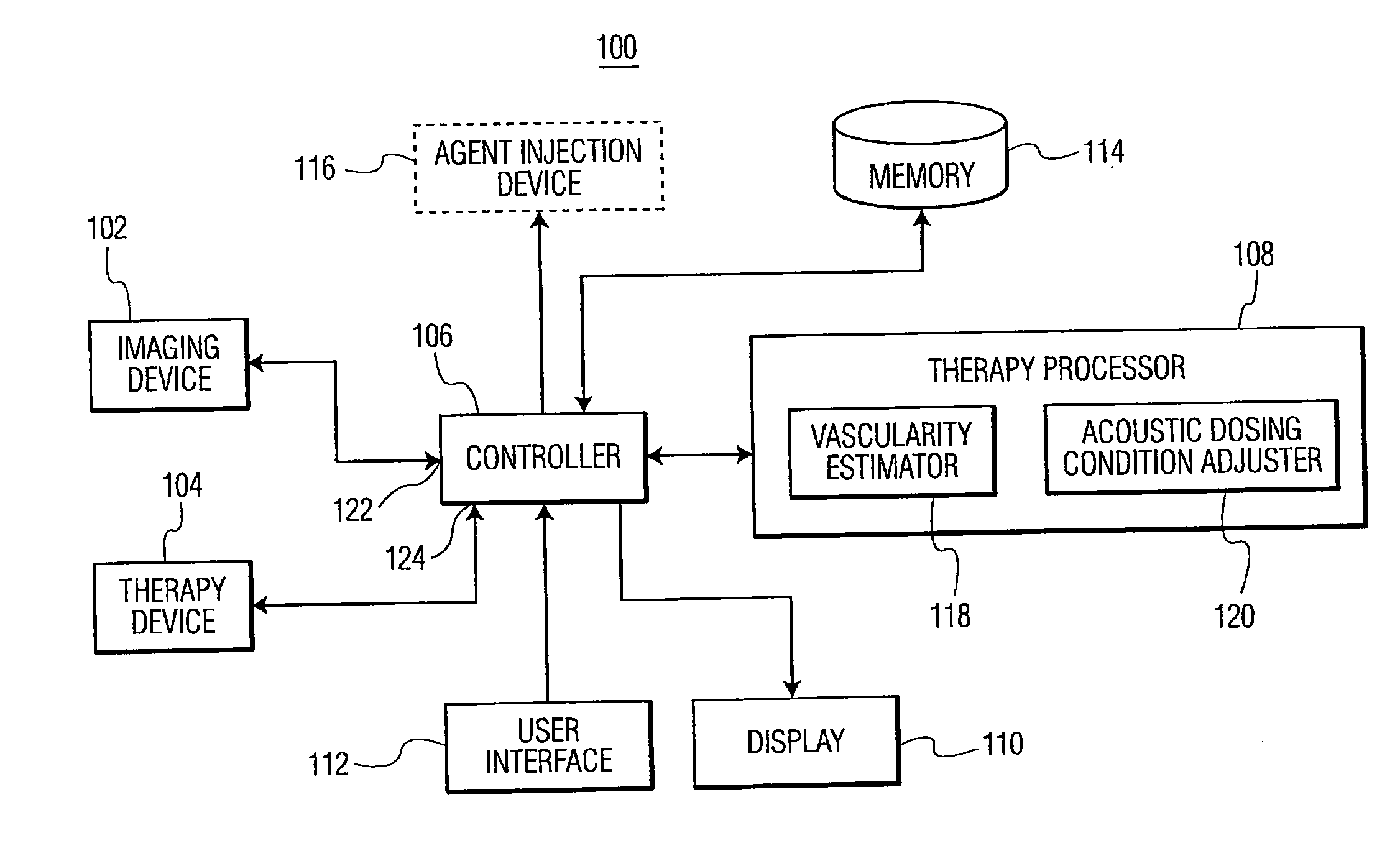
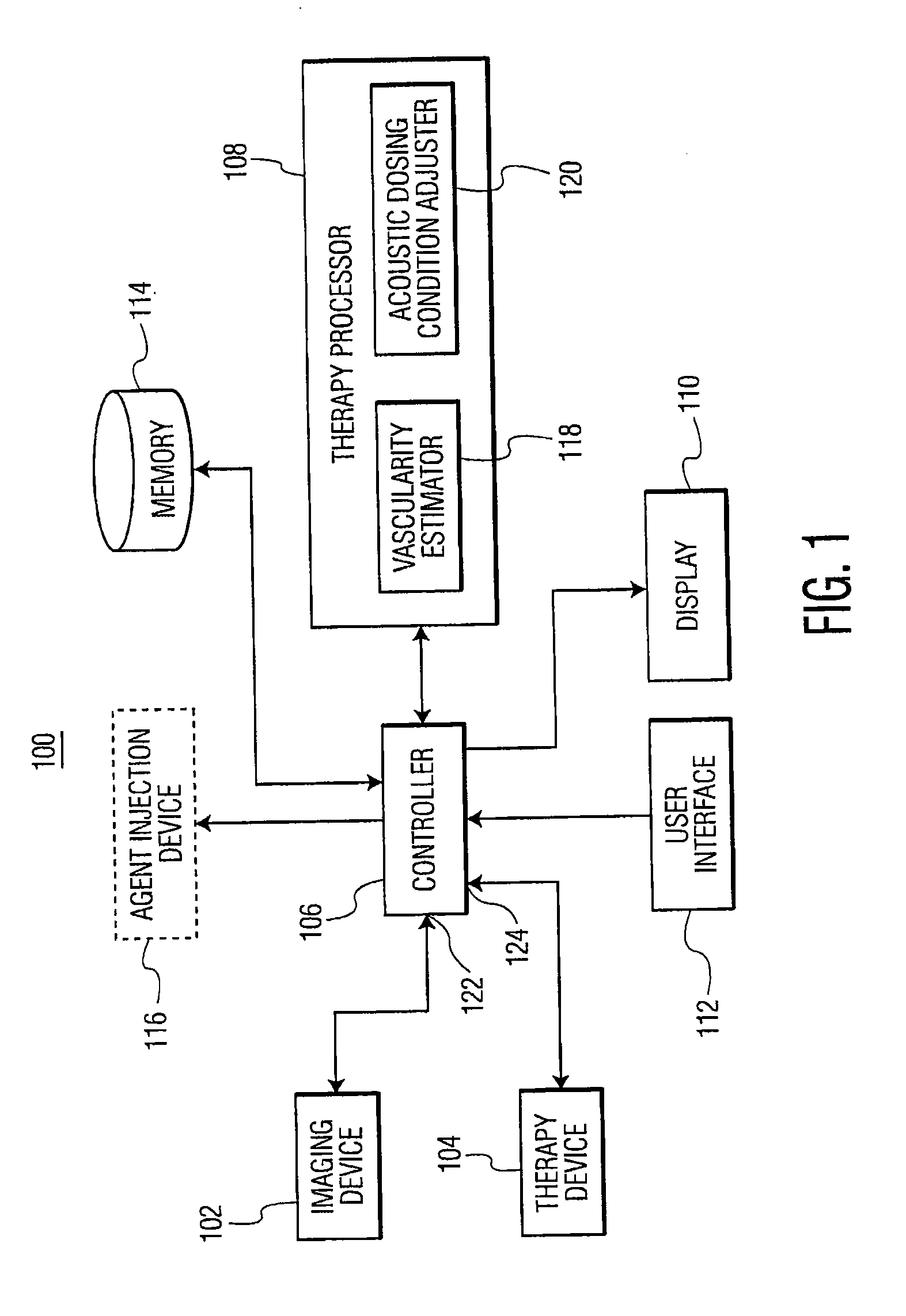
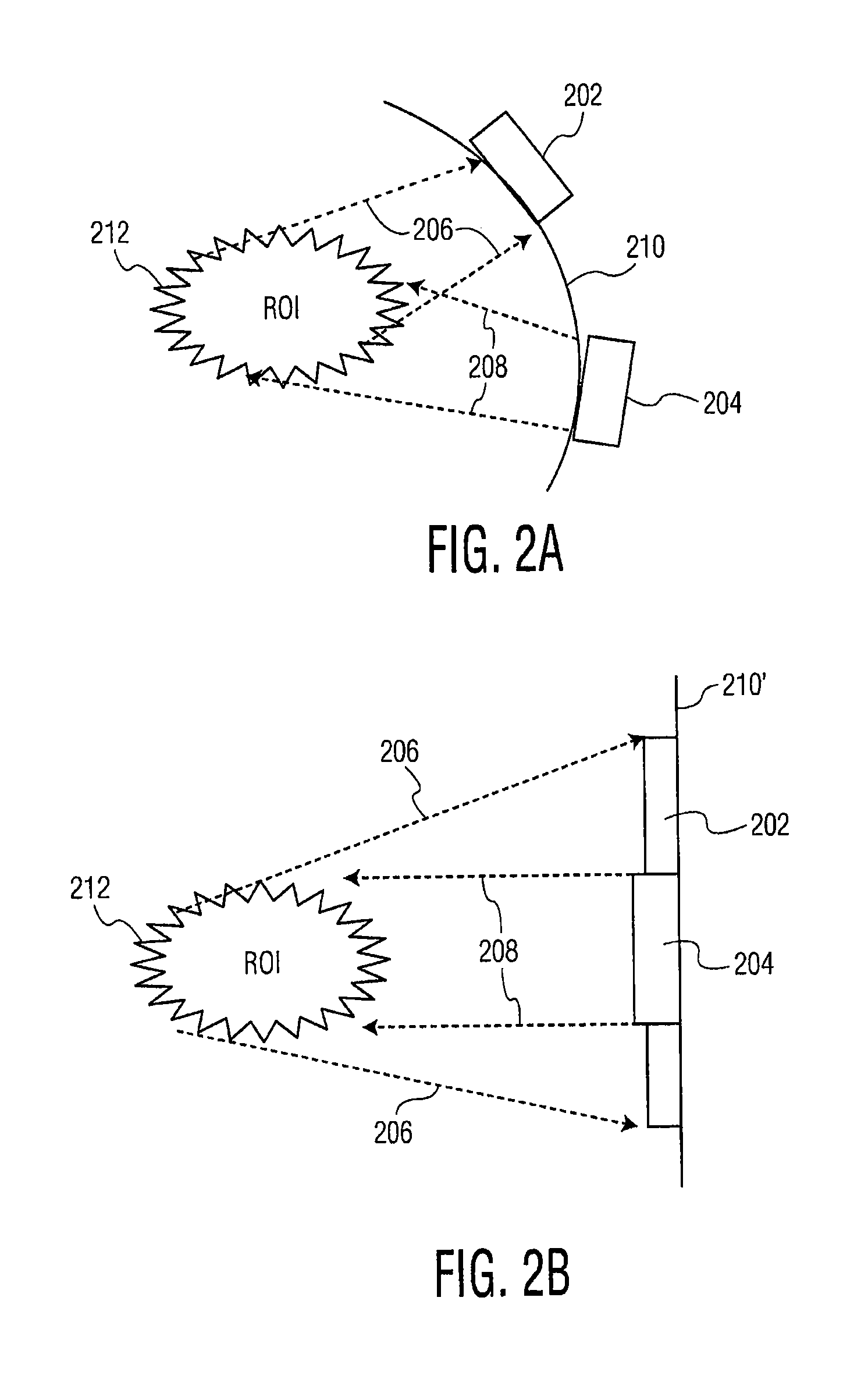
Methods and systems of treating at least one blood vessel involves the application of therapy ultrasound to the blood vessel(s) using one or more dosing conditions. An image of the region of interest is acquired responsive to the applied therapy ultrasound. A change in vascularity of the blood vessel(s) is estimated, responsive to the applied therapy ultrasound, using the acquired image to determine whether to adjust at least one of the dosing conditions. The therapy ultrasound is applied with an intensity to modify the blood vessel(s) without damaging a surrounding tissue. A method of treating a tumor comprises introducing a therapeutic agent into a bloodstream and applying therapy ultrasound to blood vessel(s). The therapy ultrasound, along with an agent, disrupts the blood vessel(s) to limit flow to and from the tumor, thereby retaining the therapeutic agent within the tumor.

Description

CROSS REFERENCE TO RELATED APPLICATIONS[0001]This application is related to and claims the benefit of U.S. Provisional Application No. 61 / 168,075 entitled METHODS AND SYSTEMS FOR IMAGE-GUIDED TREATMENT OF BLOOD VESSELS filed on Apr. 9, 2009, the contents of which are incorporated herein by reference.STATEMENT REGARDING FEDERALLY SPONSORED RESEARCH[0002]This invention was made with partial government support under the grants EB 001713 and CA 139657 awarded by the National Institutes of Health. The government has certain rights in the invention.FIELD OF THE INVENTION[0003]The present invention relates to ultrasound imaging and therapy. More particularly, the present invention relates to methods and systems of image-guided treatment of blood vessels with low intensity ultrasound.BACKGROUND OF THE INVENTION[0004]It is generally known to use ultrasound for clinical imaging of a region of a patient's anatomy. For clinical imaging, an ultrasound transducer transmits ultrasound waves to a s...

Claims


Application Information

Patent Timeline
Patent Type & Authority Applications(United States)
IPC IPC(8): A61N7/00A61B8/00
CPCA61B2019/5276A61N2007/0078A61N2007/0039A61N7/00A61B2090/378
Inventor SEHGAL, CHANDRAWOOD, ANDREW KENNETH
Owner THE TRUSTEES OF THE UNIV OF PENNSYLVANIA
Why Eureka
Try Eureka