Method of Diagnosis of Infection by Mycobacteria and Reagents Therefor

a technology of mycobacteria and reagents, applied in the direction of antibacterial agents, peptide/protein ingredients, drug compositions, etc., can solve the problems of poor compliance, serious complications and death, and worsening problems, and achieve cost-effective and improved diagnostic accuracy.

Inactive Publication Date: 2012-06-28
TYRIAN DIAGNOSTICS LTD
View PDF0 Cites 13 Cited by
  • Summary
  • Abstract
  • Description
  • Claims
  • Application Information

AI Technical Summary

Benefits of technology

[0021]There clearly remains a need for improvements in diagnostic accuracy, particularly sensitivity, of diagnostic and prognostic reagents, that provide cost-effective and rapid determination of infection by M. tuberculosis and / or disease conditions associated therewith.

Problems solved by technology

Tuberculosis (TB) is a major disease in developing countries, as well as an increasing problem in developed areas of the world, with about eight million new cases and three million deaths each year.
If left untreated, M. tuberculosis infection may progress beyond the primary infection site in the lungs to any organ in the body and generally results in serious complications and death.
As treatment for M. tuberculosis infection requires many months of therapy with multiple drugs, such an extensive course of treatment can result in poor compliance and the emergence of multidrug resistant (MDR) and extensively drug resistant (XDR) TB.
The problem is worsened by HIV infection.
Despite the enormous global burden of tuberculosis, case detection also continues to be a problem.
In the detection of active tuberculosis infection, microscopy is rapid and inexpensive, but has low sensitivity requiring the visualization of bacilli in sputum smear samples.
Culture is more sensitive, but results can take several weeks and has the incidence of 10-20% of false-positives.
Despite these tests being in use for nearly a century, they have been insufficient to control tuberculosis particularly in developing countries and where HIV is epidemic.
The development of diagnostic tests for tuberculosis have been hampered by numerous difficulties.
Furthermore, expression in different environments has complicated the identification of infectivity markers and markers that would distinguish active infection compared to latent infection.
Laboratory systems and the use of laboratory strains do not meet this deficit.
Moreover, it has been difficult to identify such a marker that would be useful for the detection of the other mycobacterium of the M. tuberculosis complex.
Even with the identification of putative markers, diagnosis of TB is further complicated by the lack of sensitivity of current tests, and reproducibility across a large cohort of clinical samples.
Molecular tests relying on the amplification of DNA and rRNA target sequences have not been suitable as tests for acute infection, or as infectivity markers.
Additionally, molecular tests based on detection of mRNA meet with the same obstacles as tests based on protein detection, with the further complication of message stability particularly during storage of clinical samples.
Whilst the sequencing of the Mycobacterium tuberculosis genome has facilitated an enormous research effort to identify M. tuberculosis proteins and mRNA that theoretically may be expressed by the organism sequence data alone are insufficient to conclude that any particular protein or mRNA is expressed in vivo by the organism, let alone during infection of a human or other animal subject.
Moreover, the known instability of mRNA during storage of clinical samples makes diagnostic testing based on mRNA detection a particularly challenging feat, in terms of actual detection, and reproducibility across large cohorts of clinical samples.
Recent evidence indicates that the protein expression profile of intracellular parasites varies markedly depending on environmental cues, such that the expression profile of the organism in vitro may not accurately reflect the expression profile of the organism in situ.
It is thought that bacilli can replicate to varying degrees in all these environments, however, little is known about the environmental conditions at each site.
Although systems for the amplification of DNA, and rRNA target sequences that are specific for members of the M. tuberculosis complex have been described, such tests have not been suitable for monitoring the response of patients to treatment due to the persistence of the nucleic acid targets.
The persistence of these nucleic acid species is thought to reflect shedding of dead or dormant bacilli from pulmonary lesions, and as a consequence are not a good indicator of acute infection.
Such nucleic acid based tests have also not been successful or recommended by the WHO as a stand-alone test, without smear or culture confirmation.
Sensitivity and specificity estimates of NAATs in respiratory specimens are highly variable, with sensitivity being lower and more inconsistent than specificity and summary measures of diagnostic accuracy are not considered clinically meaningful.
Meta-analysis studies have confirmed that NAATs that are currently available cannot be recommended to replace conventional tests for diagnosing pulmonary TB, nor have they demonstrated usefulness for monitoring treatment progress, as NAAT are still problematic for the detection of non-viable bacteria and give false-positive results.

Method used


Image

  • Method of Diagnosis of Infection by Mycobacteria and Reagents Therefor
  • Method of Diagnosis of Infection by Mycobacteria and Reagents Therefor
  • Method of Diagnosis of Infection by Mycobacteria and Reagents Therefor

Examples


example 1

Sample Collection, Processing, Nucleic Acid Extraction, Synthesis, and Quantatitation, Antibody Production and Immunoassays

[0416]Subject to the disclosure in the subsequent examples i.e., Example 2 et seq., the following general methods were employed for sample collection and nucleic acid extraction, processing, synthesis, and quantatitation. A method referred to in this example has been utilized unless an alternate method is specifically recited in a subsequent example i.e., Example 2 et seq. Methods referred to in the subsequent examples are to be construed with reference to that specific example.

1. Collection of Patient Sputum Samples

[0417]TB-negative and TB-positive sputa were used to evaluate nucleic acid based assays using primer pairs, with optional antigen based assay and with optional antibody based assay for a TB diagnostic as described in the subsequent examples. Eighty (80) patient sputa samples were recruited from Cameroon in 2007. Samples were treated with protease inh...

example 2

Detection of Active TB Complex in Clinical Sputa Using Quantitative PCR

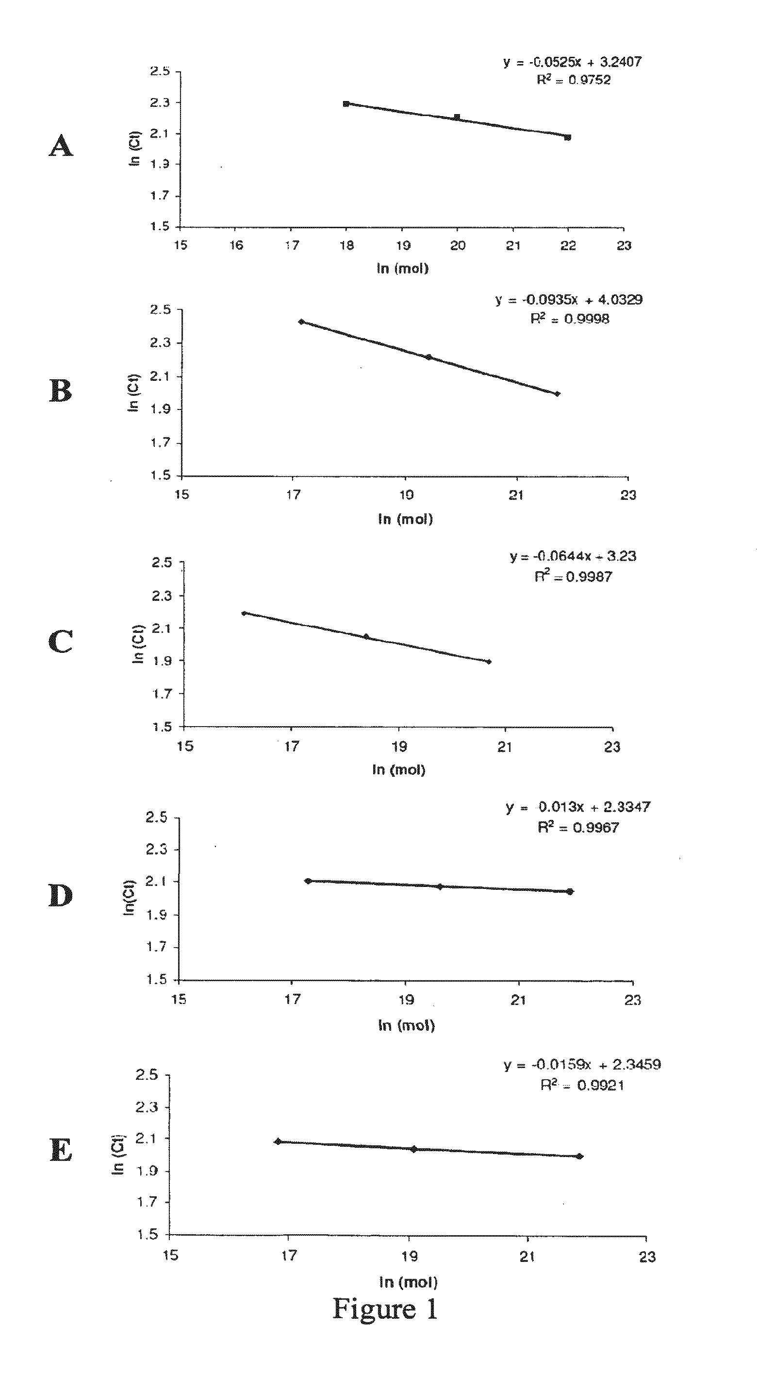
[0467]In the first set of experiments the inventors sought to develop a quantitative real-time PCR assay for the detection of ilvC biomarker (SEQ ID NO:2) encoding Mycobacterium tuberculosis complex KARI protein (SEQ ID NO:1), as a means of detection of tuberculosis or an infection by one or more organisms of the Mycobacterium tuberculosis complex. The inventors also sought to correlate the results obtained with severity of infection as demonstrated by smear status grading and to compare the sensitivity of the assay for the detection of ilvC biomarker with detection of other biomarkers of one or more organisms of the Mycobacterium tuberculosis complex. Other biomarkers assayed included nucleic acids (SEQ ID NOs: 4, 6, 8 and 28) encoding, Mycobacterium tuberculosis BSX protein (SEQ ID NO:3), Mycobacterium tuberculosis Rv1265 protein (SEQ ID NO:5) and Mycobacterium tuberculosis S9 protein (SEQ ID NO:7). The 16S rRN...

example 3

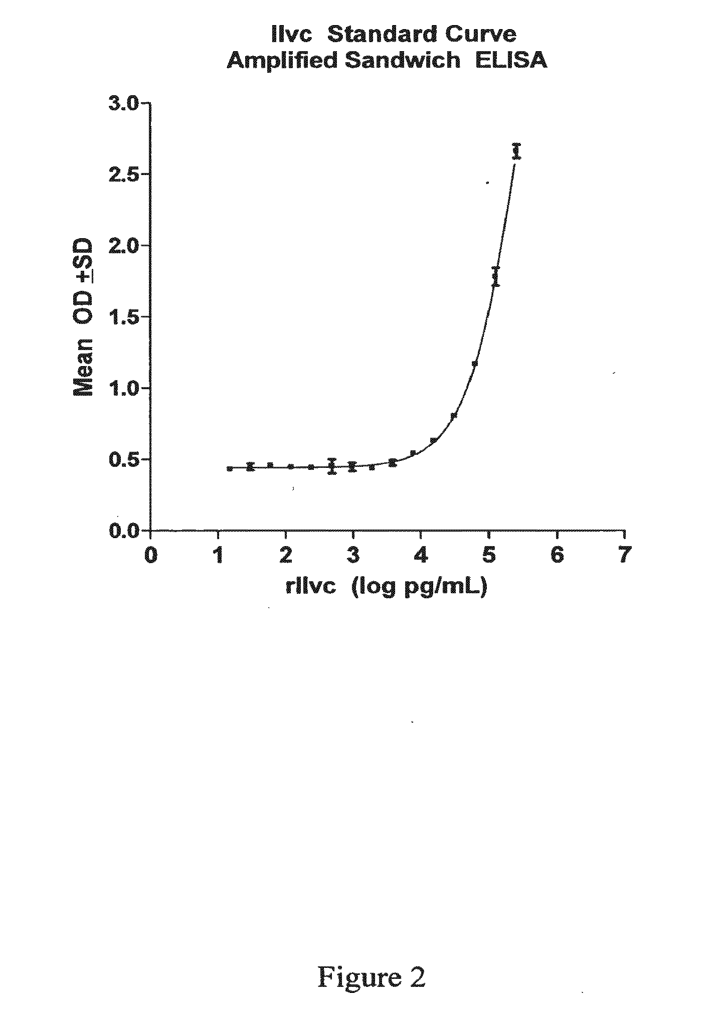
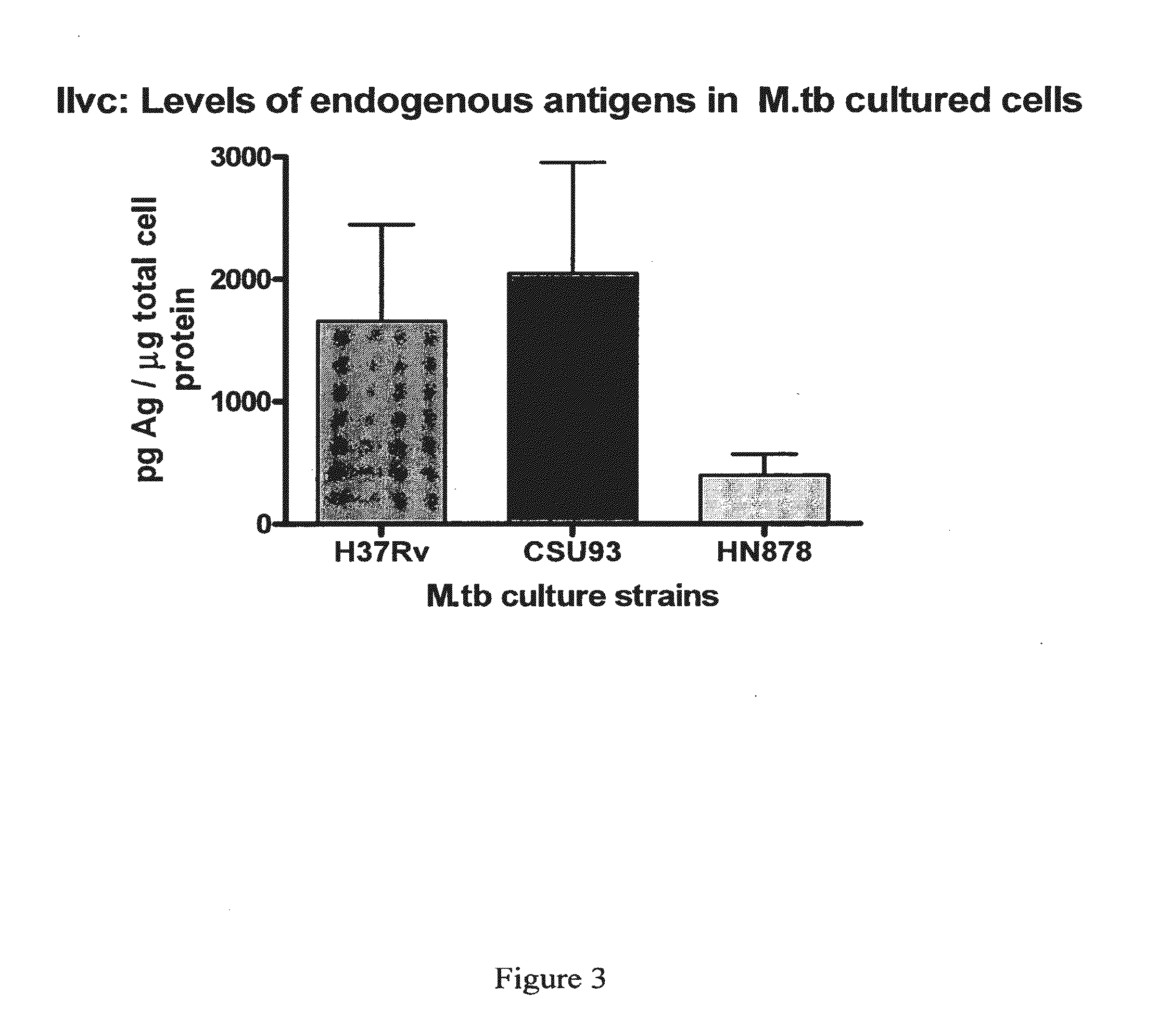
Antigen-Based Diagnosis of Tuberculosis or Infection by M. tuberculosis Using Antibodies that Bind to M. tuberculosis Ketol-Acid Reductoisomerase (KARI)

1. Identification of KARI Protein in TB-Positive Subjects

[0481]A protein having a molecular weight of about 36 kDa was recognized in TB+ samples. The sequences of ten peptides from MALDI-TOF data matched a sequence encoded by the ilvC gene of M. tuberculosis set forth in SEQ ID NO: 1. The percent coverage of SEQ ID NO: 1 by these 10 peptides was about 37%, suggesting that the peptide fragments were derived from this same protein marker.

[0482]The identified protein having the amino acid sequence set forth in SEQ ID NO: 1 is a putative Ketol-Acid Reducto Isomerase and was designated as “KARI”.

2. Antibodies

[0483]Antibodies were prepared against recombinant KARI protein encoded by the ilvC gene of M. tuberculosis (SEQ ID NO:2) using procedures described herein. Ten (10) antibodies were produced and screened for their suitability as descr...


PUM

PropertyMeasurementUnit
Tmaaaaaaaaaa
Tmaaaaaaaaaa
Tmaaaaaaaaaa

Abstract

The present invention provides a method of specifically detecting the presence of one or more Mycobacteria of the M. tuberculosis complex, said method comprising detecting ilvC nucleic acid of one or more Mycobacteria of the M. tuberculosis complex in a sample under conditions that do not detect ilvC nucleic acid of the M. avium complex. The invention also provides methods of diagnosis and treatment of tuberculosis in a subject employing the specific detection ilvC nucleic acid of one or more Mycobacteria of the M. tuberculosis complex.

Description

CROSS-REFERENCE TO RELATED APPLICATIONS[0001]This application claims priority from Australian Patent Application No. 2009900876 filed Feb. 26, 2009 the contents of which are incorporated herein in their entirety.BACKGROUND OF THE INVENTION[0002]1. Field of the Invention[0003]The invention provides relates to the detection of one or more Mycobacteria of the M. tuberculosis complex, and to the diagnosis and prognosis of infection of an animal subject such as a human by Mycobacteria of the M. tuberculosis complex, and the diagnosis of conditions associated with such infections, especially tuberculosis. More particularly, the present invention relates to the expression in an infected subject of a Ketol-acid reductoisomerase (KARI) mRNA of M. tuberculosis and encoded protein (SEQ ID NO:1) thereof, and reagents for novel diagnostic and prognostic methods and for monitoring efficacy of therapy of infection and / or treatment of tuberculosis.[0004]2. Description of the Related Art[0005]Tuberc...

Claims


Application Information

Patent Timeline
Patent Type & Authority Applications(United States)
IPC IPC(8): A61K38/16C12Q1/70A61P31/06C12Q1/68
CPCC12Q1/689A61P31/06C12Q2600/158
Inventor LINDNER, ROBYNGARTHWAITE, IAN
Owner TYRIAN DIAGNOSTICS LTD
Why Eureka
Try Eureka