Erythrocyte-binding therapeutics

a technology of erythrocytes and therapeutics, applied in the field of medical compositions, can solve problems such as blockage of tumor blood supply, and achieve the effect of blocking tumor blood supply

Active Publication Date: 2012-07-12
ECOLE POLYTECHNIQUE FEDERALE DE LAUSANNE (EPFL)
View PDF3 Cites 53 Cited by
  • Summary
  • Abstract
  • Description
  • Claims
  • Application Information

AI Technical Summary

Benefits of technology

[0007]Embodiments are also directed to treating cancer by embolizing tumors. Many antigens for tumors and/or tumor microvasculature are known. Antibodies may readily be made that specifically bind these antigens. Such tumor-binding ligands are molecularly fuse

Problems solved by technology

These fusions bind at the tumor site and also bind eryt

Method used


Image

  • Erythrocyte-binding therapeutics
  • Erythrocyte-binding therapeutics
  • Erythrocyte-binding therapeutics

Examples


example 1

Screening for Erythrocyte-binding Peptides with Mouse Erythrocytes

[0123]The PhD naïve 12 amino acid peptide phage library commercially available from New England Biolabs (NEB) was used in the selection. In each round of screening, 1011 input phage were incubated with mouse erythrocytes in PBS with 50 mg / mL BSA (PBSA-50). After 1 h at 37° C., unbound phage were removed by centrifugation in PERCOLL (GE Life Sciences) at 1500 g for 15 min. A subsequent dissociation step was carried out in PBSA-50 in order to remove low-affinity binding phage. Dissociation duration and temperature were increased in later rounds of screening to increase stringency of the selection process. In round 1, phage binding was followed by a 2 min dissociation step at room temperature prior to washing and elution. In round 2, phage binding was followed by a 10 min dissociation at 37° C. In rounds 3 and 4, two separate and sequential dissociation steps were conducted at 37° C.: 10 min followed by 15 min in round 3...

example 2

Characterizing of Binding to Mouse Erythrocytes

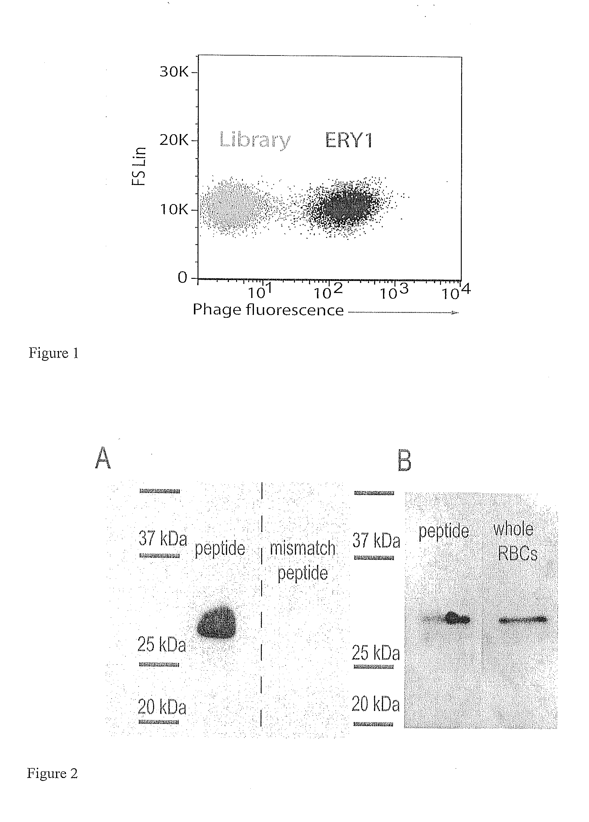
[0124]Result: Microscopy confirmed that the ERY1 phage binds the erythrocyte cell surface without altering cell morphology and without cytoplasmic translocation. Fluorescence and phase contrast images reiterated the erythrocyte-binding capacity of ERY1 phage relative to the non-selected library. High-resolution confocal imaging revealed that ERY1 phage are distributed across the cell surface (as opposed to being clustered at a single site) and bind preferentially to the equatorial periphery of the cell surface, and that binding was homogeneous among erythrocytes (FIG. 1).

[0125]Method: For all samples sample, 1011 input phage were incubated with mouse erythrocytes in PBS-50. After 1 h at 37° C., unbound phage were removed by centrifugation at 200 g for 3 min. For regular fluorescence microscopy samples, cells were incubated with anti-M13 coat protein-PE antibody (Santa Cruz Biotechnology) at a 1:20 dilution in PBSA-5 for 1 h at room temp...

example 3

Characterizing the Molecular Target of Binding to Mouse Erythrocytes

[0126]Result: To search for the molecular target for the ERY1 peptide, affinity pull-down techniques using a biotinylated soluble peptide were employed; this method revealed glycophorin-A (GYPA) as the ERY1 ligand on the erythrocyte membrane. When whole erythrocytes were incubated with ERY1 peptide functionalized with biotin and a photoactivatable crosslinker, a single 28 kDa protein was conjugated with the peptide-biotin complex, as detected by a streptavidin Western blot (FIG. 2A). The reaction lysate was extensively washed and purified using streptavidin magnetic beads to ensure no unlabeled proteins from the erythrocyte lysate remained. As expected, the mismatch peptide failed to conjugate to any erythrocyte proteins. The mismatch peptide, PLLTVGMDLWPW (SEQ ID NO:2), was designed to contain the same amino acid residues as ERY1, and to match its hydrophobicity topography. Evidence of the apparent size of the inte...


PUM


Abstract

Peptides that specifically bind erythrocytes are described. These are provided as peptidic ligands having sequences that specifically bind, or as antibodies or fragments thereof that provide specific binding, to erythrocytes. The peptides may be prepared as molecular fusions with therapeutic agents, tolerizing antigens, or targeting peptides. Immunotolerance may be created by use of the fusions and choice of an antigen on a substance for which tolerance is desired.

Description

CROSS REFERENCE TO RELATED APPLICATIONS[0001]This application is a continuation-in-part of U.S. Ser. No. 13 / 206,034 filed Aug. 9, 2011 that claims priority to U.S. provisional patent application Ser. No. 61 / 372,181 filed Aug. 10, 2010, which are hereby incorporated by reference herein.TECHNICAL FIELD OF THE INVENTION[0002]The Technical Field relates to medical compositions and uses for ligands or antibodies that bind erythrocytes. Specific uses include immunotolerization, drug delivery, and cancer therapies.BACKGROUND[0003]The rejection of transplanted tissue and autoimmune diseases are pathological conditions that involve an immunorejection of a foreign biomolecule because of its antigenic nature. Many drugs and clinical processes are involved in suppressing or treating immunorejection. Vaccines take advantage of this process by stimulating an immune response to antigens on pathogenic biomolecules to build up the immune system response against proteins or other biomolecules that ca...

Claims


Application Information

Patent Timeline
IPC IPC(8): C07K19/00C12N9/96
CPCA61K9/1075C12N9/82A61K38/00A61K39/0008A61K39/001A61K2039/6031A61K2039/6056C07K7/08C07K16/18C07K2317/56C07K2317/622C07K2317/92C07K2319/00C07K2319/21C07K2319/22C07K2319/23C07K2319/33C07K2319/41C07K2319/43C07K2319/74C07K14/62C07K14/70539C12Y305/01001A61K9/513A61K47/64A61K47/6843C12N9/96C07K16/28C07K2319/30A61K2039/605A61K47/6815A61K47/6803A61K47/6811A61K47/6849
Inventor HUBBELL, JEFFREY A.KONTOS, STEPHANEDANE, KAREN Y.
Owner ECOLE POLYTECHNIQUE FEDERALE DE LAUSANNE (EPFL)
Why Eureka
Try Eureka