Efficient proliferation method for a kupffer cell and use thereof

Inactive Publication Date: 2012-08-09
NAT INST OF AGROBIOLOGICAL SCI
View PDF0 Cites 1 Cited by
  • Summary
  • Abstract
  • Description
  • Claims
  • Application Information

AI Technical Summary

Benefits of technology

[0049]According to the present invention, an efficient proliferation method has been completed for a Kupffer cell using a mixed culture system of cells derived from a liver. The use of the proliferation method for a Kupffer cell of the present invention makes it possible to proliferate a Kupffer cell in the mixed culture system of cells derived from a liver, and to collect the Kupffer cells. Besides, the Kupffer cells can be collected from the mixed culture system repeatedly multiple times during the primary culture. Moreover, the Kupffer cells can be collected from the mixed culture system through separation by shaking a culturing vessel for the cultured cell mixture. Further, by the selection and collection based on the strong adherence to a plastic container, the purity of the Kupffer cells thus collected can be further increased.
[0050]Conventionally, specific cells derived from a liver have been produced from a mixture population of dispersed liver cells by making use of various fractionation•purification processes from the viewpoint of how high-purity target cells are collected. This approach is not suitable for mass production of Kupffer cells. Meanwhile, the method of the present invention makes it possible to produce a large amount of high-purity Kupffer cells conveniently using only conventional experimental equipment without requiring skilled techniques.
[0051]Specifically, the method of the present invention makes it possible to continuously select and collect Kupffer cells from a single tissue culture flask every 2 to 3 days for approximately 2 weeks on the basis of the adherence to a plastic container. As a result, when a tissue culture flask having a bottom area of 75 cm2 is used, approximately 1×10⁷ Kupffer cells can be collected per tissue culture flask (approximately 1×10⁶ to approximately 3×10⁶ by

Problems solved by technology

However, the isolation of Kupffer cells has problems to be solved because it requires special equipment, and furthermore complicated processes dependent on skilled techniques.
This makes it difficult to obtain a large amount of Kupffer cells conveniently.
However, this method also has difficulty obtaining a large amount of Kupffer cells conveniently because the amount of Kupffer cells obtained depends on the amount of liver that is the starting material.
In addition, the proli

Method used


Image

  • Efficient proliferation method for a kupffer cell and use thereof
  • Efficient proliferation method for a kupffer cell and use thereof
  • Efficient proliferation method for a kupffer cell and use thereof

Examples


example 1

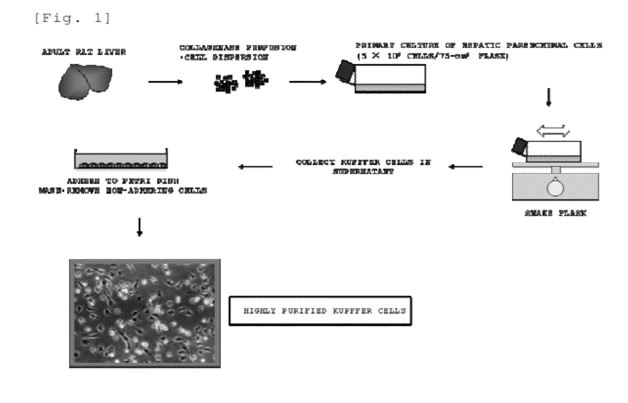
Production of Kupffer Cells from Rat Live r

[0124]In the same manner as in the method described in the known document (Non Patent Literature 4), the livers of adult male SD rats (10 to 15 weeks old) were perfused with a collagenase solution, and digested liver tissues were removed.

[0125]Each of the removed liver tissues was minced. To this, a collagenase solution was added, and a liver cell mixture suspension was obtained by pipetting. A low-speed centrifugation operation (50×g for 1 minute) at 4° C. and a washing operation with a medium (which is a Dulbecco's modified Eagle's medium (high glucose type) supplemented with fetal bovine serum (10%), bovine insulin (10 μg / ml), and 2-mercaptoethanol (0.1 mM)) (hereinbelow, unless otherwise specifically stated, a medium of the same kind was used) were repeated three times on the obtained mixture suspension. A hepatic parenchymal cell fraction that was a precipitate obtained there from was suspended in a medium, and 5×10⁶ cells were seeded ...

example 2

Production of Kupffer Cells from Cattle Liver

[0141]By the same production method as for the above-described rat Kupffer cells, hepatic parenchymal cells were separated from the livers of calves, and 5×10⁶ of the cells were seeded into each tissue culture flask (bottom area: 75 cm2) for primary culture. In this primary culture, the hepatic parenchymal cells became absent substantially at week 1 to day 10. A fibroblast-like cell layer was formed at day 8 to week 2. Spherical macrophage-like cells actively proliferated on the layer (FIG. 8). In other words, at day 8 to week 2 after the primary culture was started, a mixed culture system including the fibroblast-like cells and the macrophage-like cells was formed in the tissue culture flask.

[0142]In the same manner as for the rat, cells were selected and collected on the basis for the adherence to the plastic dish (FIG. 9). The collected cells showed quite a similar morphology to that of a macrophage. Almost all the cells were strongly ...

example 3

Primary Culture of Mouse liver Cells, and Immortalization of Kupffer Cells

[0149]In the same manner as for the rat, hepatic parenchymal cell fractions were obtained from C57BL / 6 mice (male, 7 weeks old). Each of the fractions was seeded into a tissue culture flask (bottom area: 25 cm2). The medium used was the same as that for the rat. In this primary culture, the hepatic parenchymal cells became absent substantially at day 5 to day 7. A fibroblast-like cell layer was formed at day 7 to day 14. Spherical macrophage-like cells actively proliferated on the layer (FIG. 14).

[0150]In the same manner as in the cell immortalization method described in the known documents (Non Patent Literatures 7, 8), the cells were infected with a retrovirus vector containing a human oncogene c-myc and a neomycin resistance gene from day 11 after the culturing consecutively for 3 days (FIG. 15A). After the infection, the cells were returned to a normal medium, and the culturing was continued using a tissue...


PUM

PropertyMeasurementUnit
Timeaaaaaaaaaa
Fractionaaaaaaaaaa
Concentrationaaaaaaaaaa

Abstract

The present invention has successfully established a mixed culture system capable of actively proliferating a Kupffer cell in a primary culture of a cell population derived from a liver. Additionally, the present invention has successfully established a novel production method for efficiently producing a large amount of highly purified Kupffer cells using this mixed culture system.

Description

TECHNICAL FIELD[0001]The present invention relates to an efficient proliferation method for a Kupffer cell and use thereof. More specifically, the present invention relates to an efficient proliferation method for a Kupffer cell in a mixed culture system, a method for producing a Kupffer cell using the proliferation method, a screening method for a compound influencing proliferation of a Kupffer cell and a screening method for a compound influencing a biological activity of a Kupffer cell, as well as the use of a Kupffer cell produced by the production method.BACKGROUND ART[0002]Kupffer cells are one type of liver-specific resident tissue macrophages, and are cells playing important biological defense functions such as recognition of foreign materials and induction of immune responses. Moreover, Kupffer cells are closely associated with induction and onset of liver dysfunction, hepatitis, cirrhosis, and the like. Thus, Kupffer cells are considered to be a very important target for e...

Claims


Application Information

Patent Timeline
IPC IPC(8): C12N5/071G01N21/64G01N33/566C12Q1/02C12N15/85A61K47/46A61P1/16C12N15/09
CPCC12N5/0645G01N33/5055C12N2500/84A61P1/16A61K35/407
Inventor KITANI, HIROSHITAKENOUCHI, TAKATOYAMANAKA, NORIKOYOSHIOKA, MIYAKO
Owner NAT INST OF AGROBIOLOGICAL SCI
Why Eureka
Try Eureka