Animal stroke model

a stroke model and animal technology, applied in the field of animal models of stroke, can solve the problems of tissue death, irreversible injury, brain area ceases to function and dies,

Active Publication Date: 2012-12-20
UNIVERSITY OF PRINCE EDWARD ISLAND
View PDF0 Cites 4 Cited by
  • Summary
  • Abstract
  • Description
  • Claims
  • Application Information

AI Technical Summary

Problems solved by technology

During ischemic stroke, blood flow to an area of the brain is blocked and, consequently, that area of the brain begins to cease function and will ultimately die.
Over time, irreversible injury will occur, often leading to death of the tissue, i.e. “infarction”.
However, in a detrimental and paradoxical response, the return of blood flow can increase cerebral edema and cause further brain damage, for example, through the introduction of free radicals (Dietrich, W. D. (1994) Morphological manifestations of reperfusion injury in brain.
However, tPA therapy is limited to administration within four hours of the onset of clinical signs of stroke, due to the detrimental effects of reperfusion injury (Kuroda, S. and B. K. Siesjo (1997) Reperfusion damage following focal ischemia pathophysiology and therapeutic windows.
It has been found that, after 4 hours, the damage due to reperfusion injury can exceed the damage caused by the prolonged ischemia.
The available models of ischemic stroke have limitations.
One significant limitation of prior art models is high mortality rate during the experimental procedure.
Another significant limitation of prior art models is a lack of reproducibility, and thus high variability, with respect to infarct size and / or location.
The three-point approach resulted in focal infarct areas restricted to the cerebral cortex with an associated intra-operative mortality rate of less than 1%.
While permanent occlusion of the MCA can provide a means of studying ischemic events involved in a stroke, such models do not permit examination of the effects of subsequent reperfusion.
However, due to variations in anatomy (for instance, differences in the MCA of each animal), it is difficult to place the end of the string in the same place in each animal.
It is impossible to visualize and confirm the site of occlusion, the occurrence and extent of occlusion, and the extent of reperfusion.
However, this method suffers from many of the same disadvantages of the Longa et al. method in that the placement of the suture and extent of occlusion and reperfusion cannot be visually confirmed or assessed.
Advancement of a monofilament (suture) through a vessel, as described by Longa et al. and Reddy et al. can damage the endothelium (see below).
However, there was greater variability in infarct size in the healthy Wistar rats (see Buchan et al. at page 277, paragraph 1) and studies of SHR rats may not be representative.
However, this yielded a variable infarct size in male SHRs.
Microclips are expensive and are known to damage the vascular endothelium.
In some cases, adhesion of the clipped vessel occurs which results in incomplete and / or inconsistent reperfusion.
Damaged endothelium (using clips or a monofilament advanced through CCA) can lead to release of endothelial factors which can increase brain damage or trauma on their own.
Therefore, using such methods, it can be difficult to assess stroke damage induced by occlusion and / or reperfusion versus damage due to release of endothelial factors.
However, the clinical relevance of the animal stroke model of Manabe et al. is questionable, as human stroke patients do not experienced occlusions of both carotids (leading to a global decrease in blood flow) in addition to occlusion of a vessel, such as the MCA.
One major hinderance of stroke research is the lack of good animal models.
Questionable clinical relevance, surgery-induced damage, and / or mortality during the experimental procedure are problems in existing animal models (Gerriets T. et al.
A high level of mortality during the procedure brings into question both the validity of the model and the ability of the data gleaned from the surviving animals to be representative of the group (Broderick and Hacke (2002).
Lack of reproducibility in the size and location of the infarct produced is another problem.

Method used


Image

  • Animal stroke model
  • Animal stroke model
  • Animal stroke model

Examples


example 1

Suture-Induced Middle Cerebral Artery Occlusion (MCAO)

[0081]Methods:

[0082]All experiments were carried out in accordance with the guidelines of the Canadian Council on Animal Care and were approved by the University of Prince Edward Island Animal Care Committee. All experiments were conducted on male Sprague-Dawley rats (n=4 to 6 animals per group); 200-300 g; Charles Rivers; Montreal, PQ, CAN). For all animals, food and tap water were available ad libitum.

[0083]Rats were anaesthetized with sodium thiobutabarbital (Inactin; RBI, Natick, Mass., USA; 100 mg / kg; ip). A polyethylene catheter (PE-10; Clay Adams, Parsippany, N.J., USA) was inserted into the right femoral vein to permit intravenous administration of drugs. An endotracheal tube was inserted to facilitate breathing. Body temperature was monitored by a digital rectal thermometer and maintained at 37±1° C.

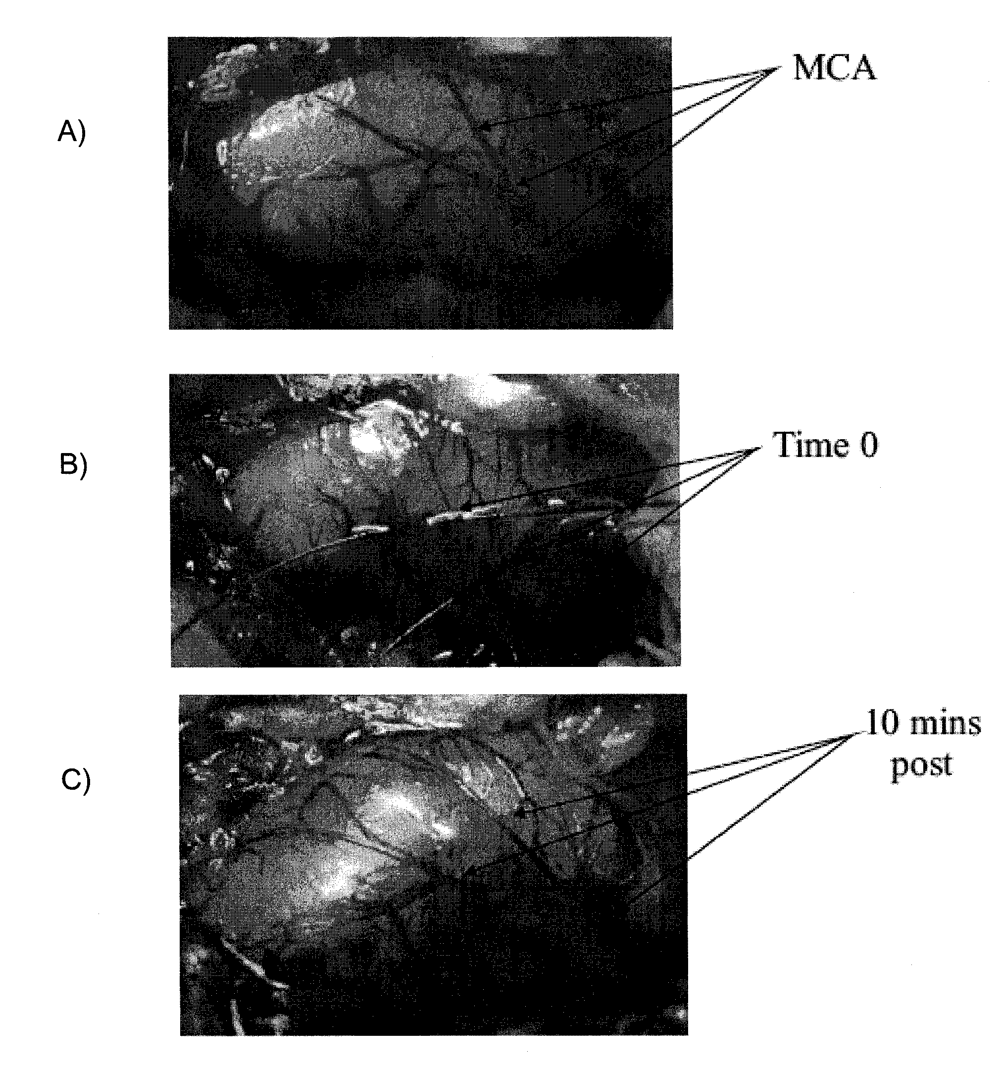
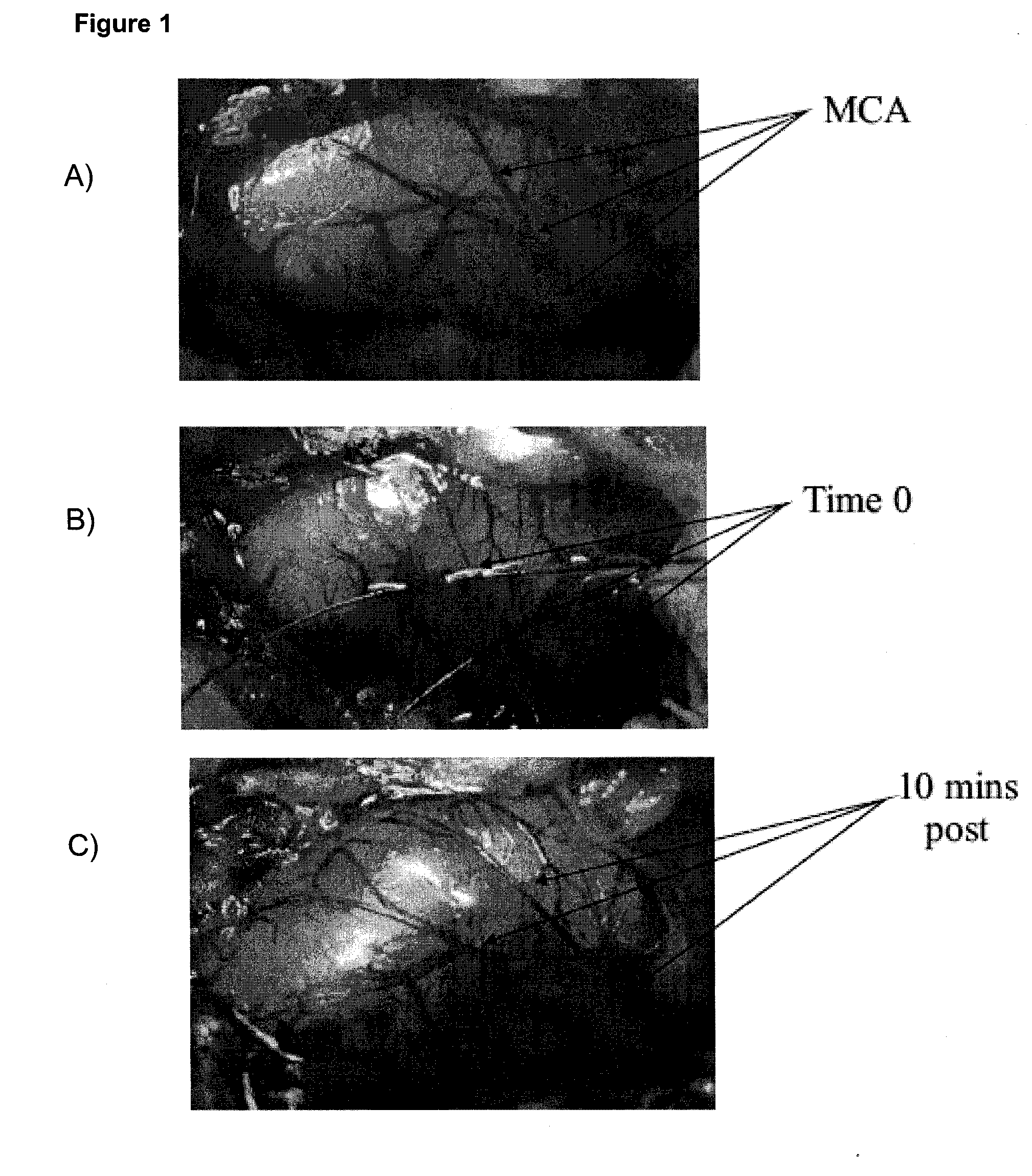
[0084]Animals were placed in a David Kopf stereotaxic frame (Tujunga, Calif., USA) and the right MCA approached through a r...

example 2

Effect of Occlusion Time on Infarct Volume

[0090]The following experiment was designed to determine if a time-dependent relationship existed between MCAO and infarct volume.

[0091]Method:

[0092]MCAO was carried out as described in Example 1. The three sutures were left in place for 0.5, 2, 4, or 6 hours. Following the removal of the sutures, all animals were perfused transcardially with phosphate buffered saline (PBS; 0.1 M; 200 mls), the brains removed and sliced into 1 mm coronal sections using a rat brain matrix (Harvard Apparatus; Holliston, Mass., USA). Sections were incubated in a 2% solution of 2,3,5-triphenol tetrazolium chloride (TTC; Sigma-Aldrich; St. Louis; MO, USA) for 5 minutes.

[0093]Infarct volumes were calculated with the use of scanned digital images of each brain section. The infarct area for both sides of each brain section was calculated using a computer-assisted imaging system (Scion Corporation; Frederick, Md., USA). The infarct areas for each side for each indivi...

example 3

Effect of Reperfusion on Infarct Volumes

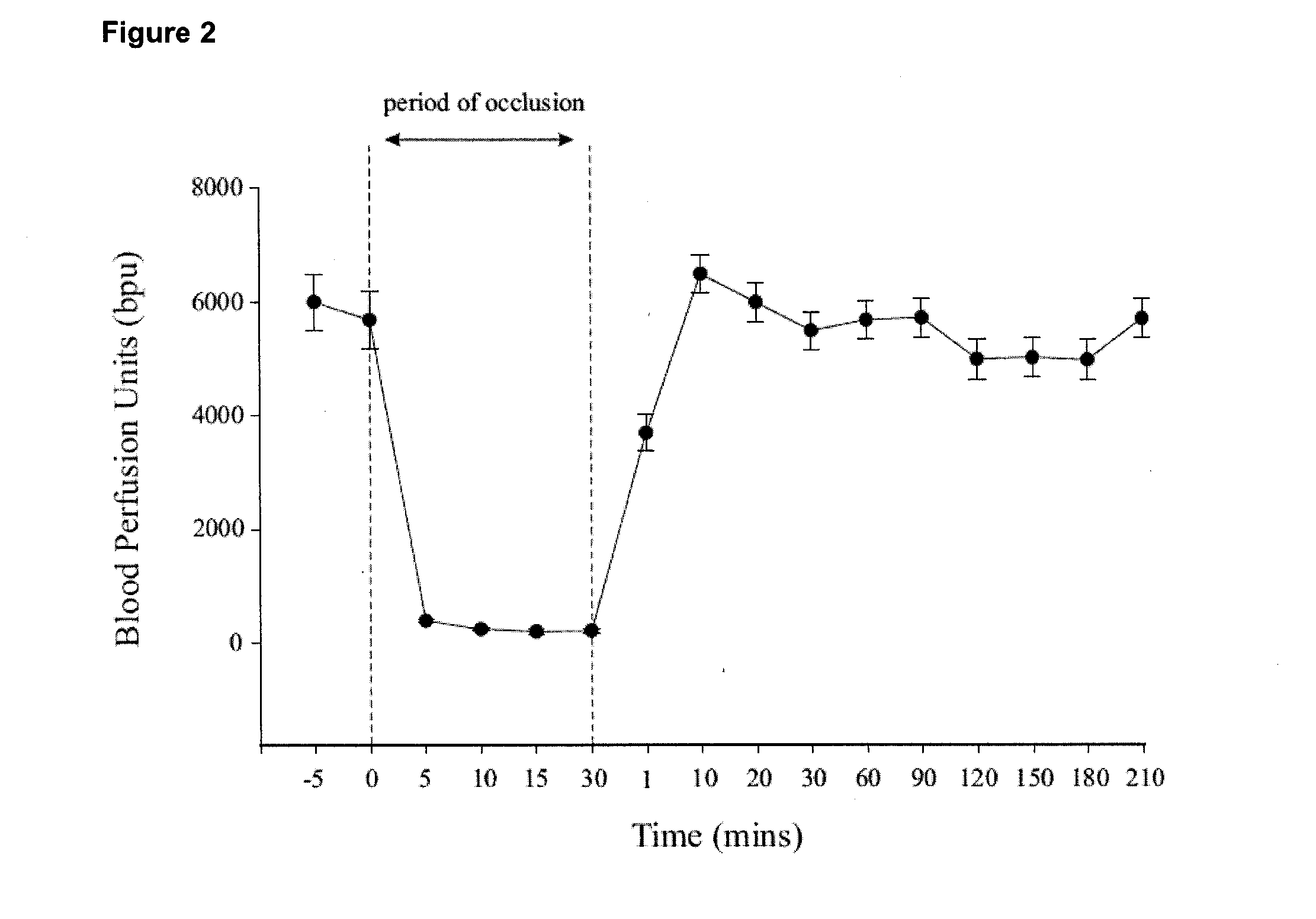
[0097]The following experiment was designed to investigate the effect of reperfusion on infarct volume following MCAO.

[0098]Method: MCAO was carried out as described in Example 1. The sutures were left in place, thus impeding blood flow through the MCA, for 0.5, 2 and 4 hrs. For the 0.5 hour group (n=9), blood flow was returned for 5.5 hours; for the 2 hour group (n=6), blood flow was returned for 4 hours; and for the 4 hour group (n=6), blood flow was returned for 2 hours. In an additional validation group (n=6), the sutures were put in place and immediately removed, and blood flow was allowed to return for 6 hours. In all cases, the return of blood flood was visually confirmed with the use of a dissecting microscope. In all groups, the experiment was terminated at the 6 hour time point.

[0099]At the end of six hours all animals were treated and infarct volumes determined as set out in Example 2.

[0100]Results:

[0101]There was a progressive incr...


PUM

PropertyMeasurementUnit
occlusion timeaaaaaaaaaa

Abstract

An animal stroke model is provided. The model is useful in the study of brain ischemia and / or reperfusron injury and in identification and testing of compounds and interventions useful in the treatment of stroke. The method is carried out in a non-human mammal, such as rat. The method of inducing ischemia and / or reperfusion injury involves exposing a portion of the middle cerebral artery (MCA) and temporarily occluding it at one or more distinct locations, preferably three distinct locations. The model results in highly reproducible and focal infarct sizes with low rate of mortality during the experimental procedure.

Description

CROSS REFERENCE TO RELATED APPLICATIONS[0001]This application claims the benefit of priority of U.S. Provisional Patent Application No. 61 / 305,016, filed Feb. 16, 2010, which is incorporated herein by reference in its entirety.FIELD OF THE INVENTION[0002]The present invention relates generally to an animal model of stroke. More particularly, the invention relates to a method of inducing reproducible brain ischemia and / or reperfusion injury in a non-human mammal, and uses thereof.BACKGROUND OF THE INVENTION[0003]A stroke is the rapidly developing loss of brain function(s) due to disturbance in the blood supply to the brain. This can occur due to a hemorrhage or due to ischemia (lack of oxygen and / or glucose supply) which may be caused, for example, by thrombosis or embolism. During ischemic stroke, blood flow to an area of the brain is blocked and, consequently, that area of the brain begins to cease function and will ultimately die. The initial area of neuronal death is called the i...

Claims


Application Information

Patent Timeline
Patent Type & Authority Applications(United States)
IPC IPC(8): A01K67/027G01N21/78
CPCA01K67/027A01K2207/30A01K2267/0356G01N33/5088
Inventor CONNELL, BARRY JAMESSALEH, TAREK
Owner UNIVERSITY OF PRINCE EDWARD ISLAND
Why Eureka
Try Eureka