Microfluidic devices and methods for separating and detecting constituents in a fluid sample
a microfluidic device and fluid sample technology, applied in the field of microfluidic devices and methods for separating and detecting constituents in fluid samples, can solve the problems of inability to meet the performance needs of applications that require high-throughput sample analysis or are impractical for clinical us
- Summary
- Abstract
- Description
- Claims
- Application Information
AI Technical Summary
Benefits of technology
Problems solved by technology
Method used
Image

Examples
example 1
1. Multi-Stage, Single-Channel Assay Under Programmable Electrophoretic Control
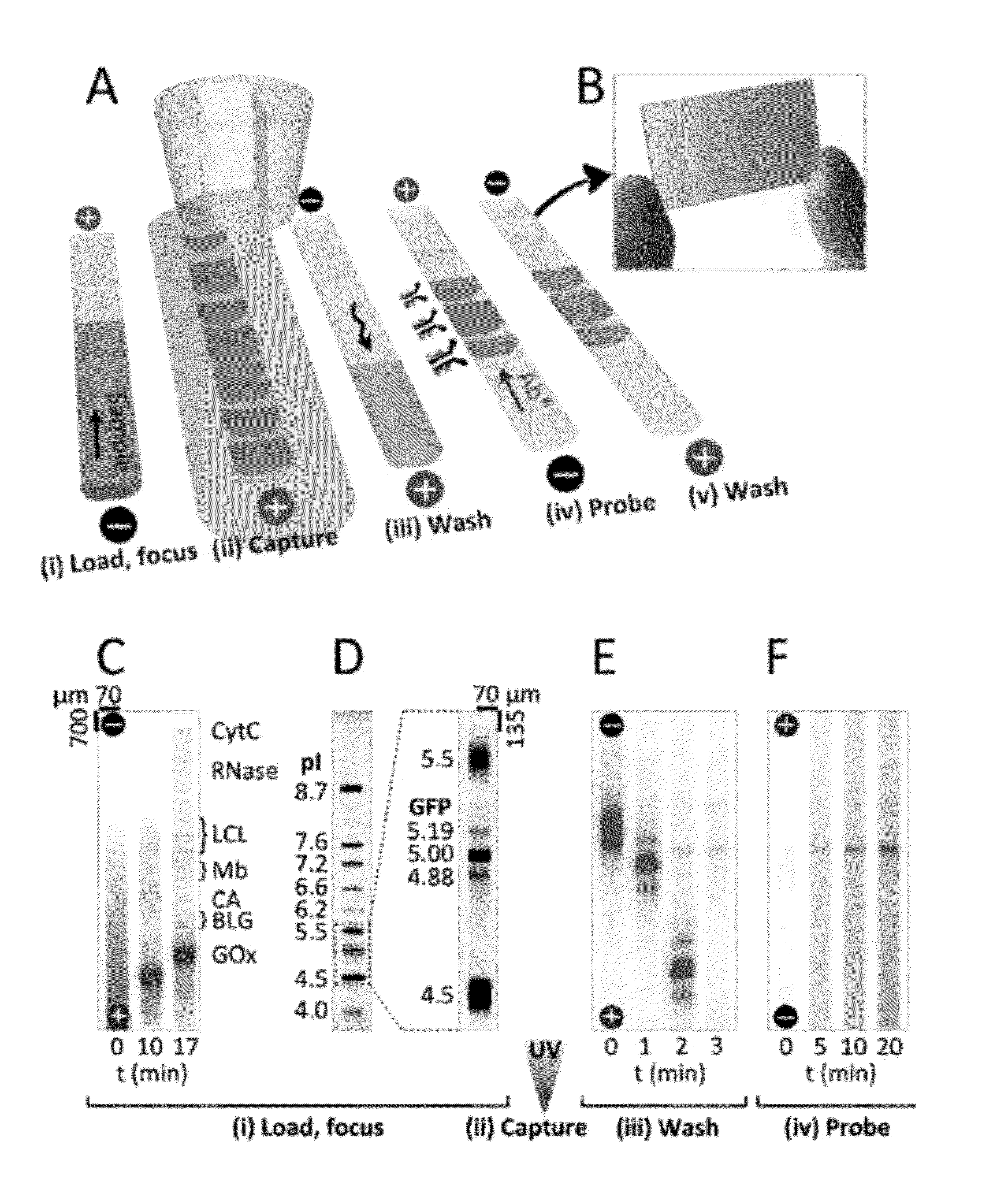
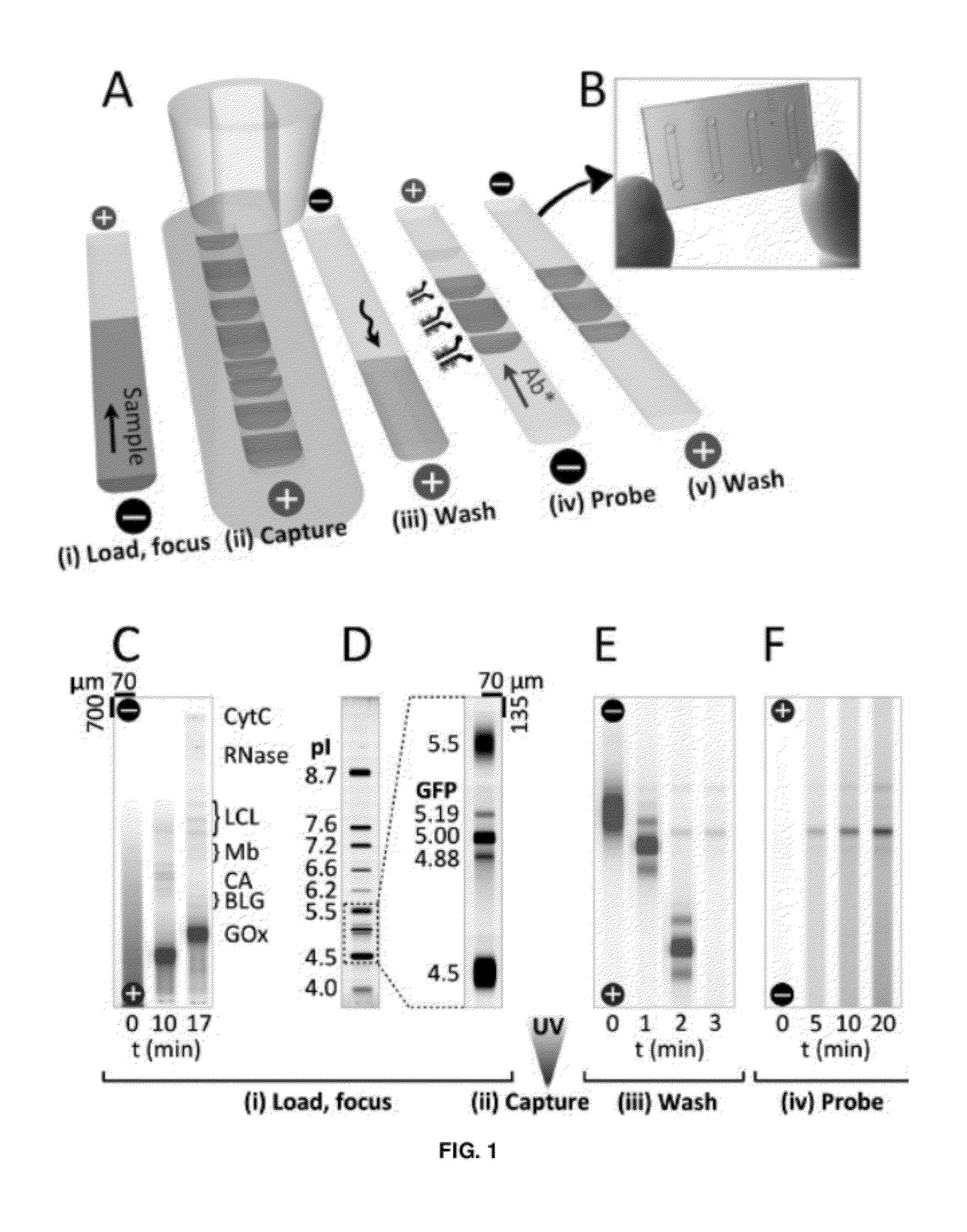
[0123]The microfluidic device included a single microchannel housing a photoactive polyacrylamide gel matrix to integrate three assay stages (FIG. 1A): Separation—(i) sample loading and isoelectric focusing; Photocapture—(ii) UV exposure to covalently attach IEF-resolved protein isoforms to the separation medium followed by (iii) electrophoretic mobilization and washout of uncaptured species; and Probing—(iv) electrophoretic-introduction of antibody to the immobilized protein bands and (v) electrophoretic washout of unbound detection antibodies. Two or more microfluidic devices can be run in parallel on a single microfluidic chip (FIG. 1B).
[0124]In the first stage of the assay, proteinaceous samples were prepared and loaded in an ampholyte buffer titrated to the alkaline limit of the buffering range (˜pH 10 for Pharmalyte 3-10) to minimize electrophoretic loading bias. After addition of anolyte and cathol...
example 2
[0191]The subject microfluidic device can be used to perform size based separations linked to immunoprobing in both native and SDS-PAGE variants. Embodiments that include size based separation (SDS-PAGE) and immunoprobing may facilitate a microfluidic “western blot”. Experiments were performed that show native (FIG. 13) and SDS-PAGE (FIG. 14) separations of Alexa Fluor 488-labeled fluorescent protein ladder species with subsequent photocapture onto the separation medium and in-situ probing for ovalbumin (OVA*) with a specific antibody labeled with Alexa Fluor 568. The assay was performed with unlabeled target analytes, and was directly analogous to Western blotting, with significantly reduced assay time, reagent and sample requirements as compared to typical Western blotting.
[0192]The first separation step was performed across a discontinuous polyacrylamide interface built using two-step chemical polymerization of a high percentage (6% T) separation gel precursor (BPMA+) and a low p...
example 3
[0196]Experiments were performed using a microfluidic device according to embodiments of the present disclosure for western blotting. In certain embodiments, the assay duration was reduced from 3-18 hours to 10-60 min as compared to typical western blotting. In some instances, 5-plex simultaneous analyte detection and quantitative readout was performed. In a single microchannel, the subject microfluidic device performed stacking sodium dodecyl sulfate-polyacrylamide gel electrophoresis (SDS-PAGE), protein immobilization with SDS removal (blotting), and subsequent antibody probing. The scalable, high-throughput nature of microfluidic design allowed 54-plex parallelization, and 10 min assay times. A photopatternable (blue light) and photoreactive (UV light) polyacrylamide gel forms the separation medium for the assay. The polymer included both an SDS-PAGE separation matrix with a defined stacking interface and, after brief UV-switching, a protein immobilization matrix offering high ca...
PUM
| Property | Measurement | Unit |
|---|---|---|
| time constant | aaaaa | aaaaa |
| concentration | aaaaa | aaaaa |
| pH | aaaaa | aaaaa |
Abstract
Description
Claims
Application Information
 - R&D
- Intellectual Property
- Life Sciences
- Materials
- Tech Scout
- Unparalleled Data Quality
- Higher Quality Content
- 60% Fewer Hallucinations
Browse by: Latest US Patents, China's latest patents, Technical Efficacy Thesaurus, Application Domain, Technology Topic, Popular Technical Reports.
© 2025 PatSnap. All rights reserved.Legal|Privacy policy|Modern Slavery Act Transparency Statement|Sitemap|About US| Contact US: help@patsnap.com