Mr compatible compression based nuclear imaging system for breast cancer

a nuclear imaging and compression-based technology, applied in the field of multi-modal medical imaging, can solve the problems of inability to achieve the effect of reducing the risk of breast cancer, affecting the diagnostic accuracy of the combined system, etc., and achieves the effect of improving the diagnostic specificity, high sensitivity, and high specificity

Active Publication Date: 2012-12-27
RGT UNIV OF CALIFORNIA
View PDF4 Cites 12 Cited by
  • Summary
  • Abstract
  • Description
  • Claims
  • Application Information

AI Technical Summary

Benefits of technology

[0020]The illustrated embodiments of the invention are also defined as a method use in diagnostic imaging of a human breast for cancer diagnosis including the steps of performing scintimammography (SMM) or positron emission mammography (PEM), and performing MRI to improve t...

Problems solved by technology

Although mammography is very sensitive in detecting early breast cancer, it does not work well in women who have dense breasts, breast implants, or scar tissues.
However, the variable specificity of MRI can lead to unnecessary biopsies and over-treatment.
However, interference between the MRI and SMM components presents a significant challenge to the design of any comb...

Method used


Image

  • Mr compatible compression based nuclear imaging system for breast cancer
  • Mr compatible compression based nuclear imaging system for breast cancer
  • Mr compatible compression based nuclear imaging system for breast cancer

Examples


Embodiment Construction

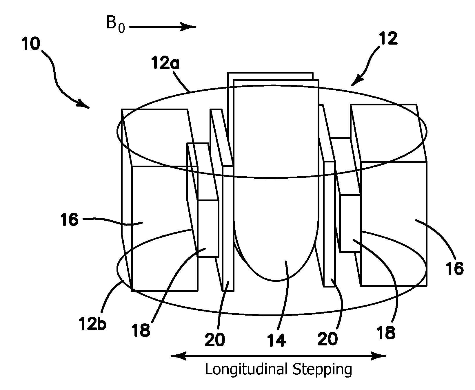
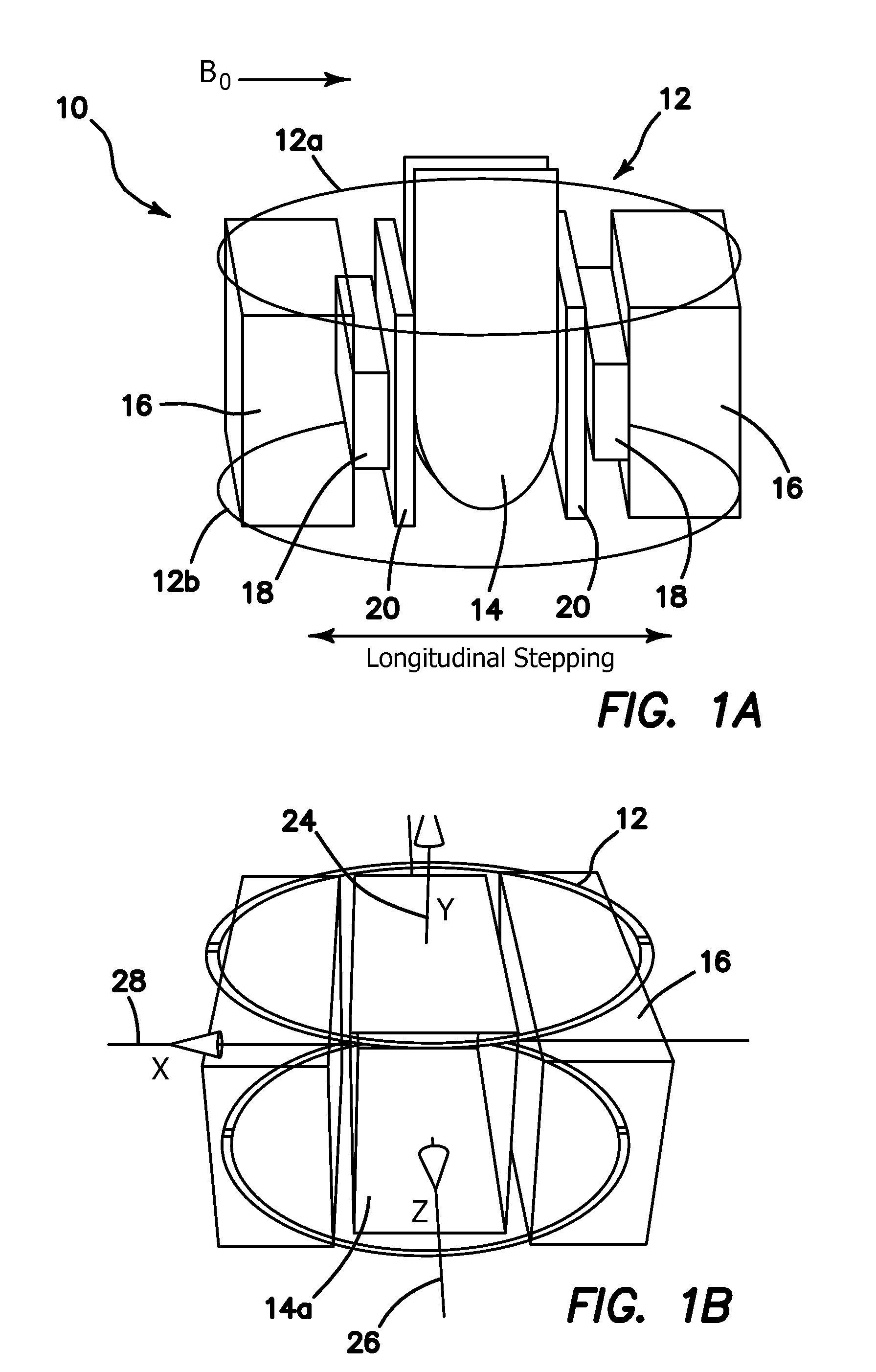
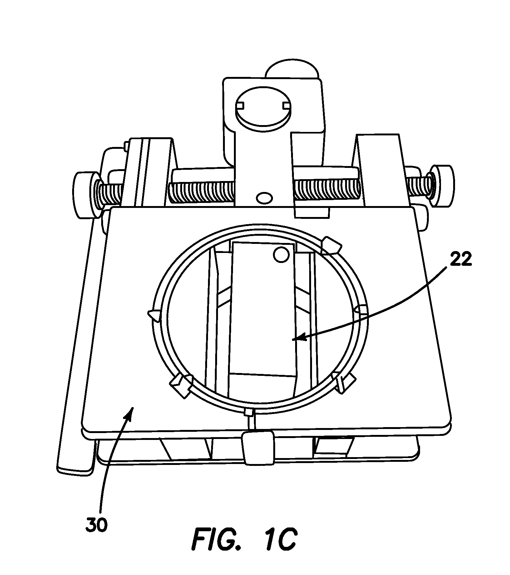
[0037]One embodiment of the present invention is the MRSSM apparatus 10. One preferable embodiment of apparatus 10 is optimized for a 3T magnetic resonance imaging (MRI). However, it is to be understood that any conventional MRI field specification or MRI imaging system may be employed, when modified as disclosed below. The geometry of the RF array coil 12 of the MRI system includes two parallel circular loops 12a and 12b each 15 cm in diameter and separated by 6.5 cm as diagrammatically shown in FIG. 1a. In the illustrated embodiment as an example only, each loop 12a and 12b is etched on FR4 laminate board using copper strips of 0.0341 mm thickness, and mounted on an acrylic plate with the material within the loops removed to allow for insertion of the breast 14. The separation between the end plates of the coils 12a and 12b allows for insertion of CZT detector modules 16 through the side of the RF coil 12. MR compatible CZT detector modules 16 have been developed with the consulta...


PUM


Abstract

An apparatus combines magnetic resonance (MR) and nuclear imaging of human breast for cancer diagnosis. An MRI system including an MR breast RF coil is combined with a nuclear imaging system having a detector disposed or disposable within the MR breast RF coil arranged and configured for the performance of simultaneous or sequential coregistered breast MRI and nuclear imaging. A selectively controlled compression mechanism for lightly compresses the breast being imaged. The remotely controlled compression mechanism is integrated with the MR breast RF coil.

Description

RELATED APPLICATIONS[0001]The present application is related to U.S. Provisional Patent Application Ser. No. 61 / 540,473, filed an Sep. 28, 2011 pursuant to 35 USC 119 and is a continuation in part of U.S. patent application Ser. No. 12 / 763,863 filed on Apr. 20, 2011 pursuant to 35 USC 120, both of which are incorporated herein by reference.FIELD OF THE INVENTION[0002]This invention relates generally to multi-modal medical imaging, and more specifically to the use of combined magnetic resonance-single photon or positron emission tomography of the human breast for diagnosis of cancer and evaluation of treatment.BACKGROUND[0003]Although mammography is very sensitive in detecting early breast cancer, it does not work well in women who have dense breasts, breast implants, or scar tissues. Alternatively, magnetic resonance imaging (MRI) has proven to the most sensitive imaging modality in delineating tumor extent and detecting multifocal or multicentric diseases. Many studies indicate tha...

Claims


Application Information

Patent Timeline
IPC IPC(8): A61B6/00A61B5/055
CPCG01R33/481A61B6/583A61B6/037G01R33/34061
Inventor NALCIOGLU, ORHANROECK, WERNER W.
Owner RGT UNIV OF CALIFORNIA
Why Eureka
Try Eureka