Compounds for the treatment of inflammation and neutropenia
- Summary
- Abstract
- Description
- Claims
- Application Information
AI Technical Summary
Benefits of technology
Problems solved by technology
Method used
Image
Examples
example 1
Materials and Methods
[0121]Neutrophil isolation, granulocytic differentiation of CD34+ precursors and cell culture. Human neutrophils from healthy (Etablissement Francais du Sang, Paris) or G-CSF-treated healthy donors (10 mg / kg for 5 days to induce hematopoietic stem cells mobilization) (Biotherapy Department, Necker Hospital, France) were isolated from EDTA-anticoagulated blood, using density-gradient centrifugation through polymorphoprep (Nycomed), as described in Witko-Sarsat et al. (1999, Blood 94:2487-2496). Blood donors gave their written informed consent to participate in this study, which was approved by the Inserm Institutional Review Board and Ethics Committee of Necker-Enfants Malades Hospital (Paris, France). Differentiation of CD34+ cells into granulocytes was induced as described in Hino et al. (2000, Br. J. Haematol. 109:314-321), with some minor modifications. Briefly, CD34+ cells were isolated from cord blood and then cultured with stem cell factor (SCF; 10 ng / ml),...
example 2
Mature Human Neutrophils Express PCNA Exclusively in their Cytoplasm
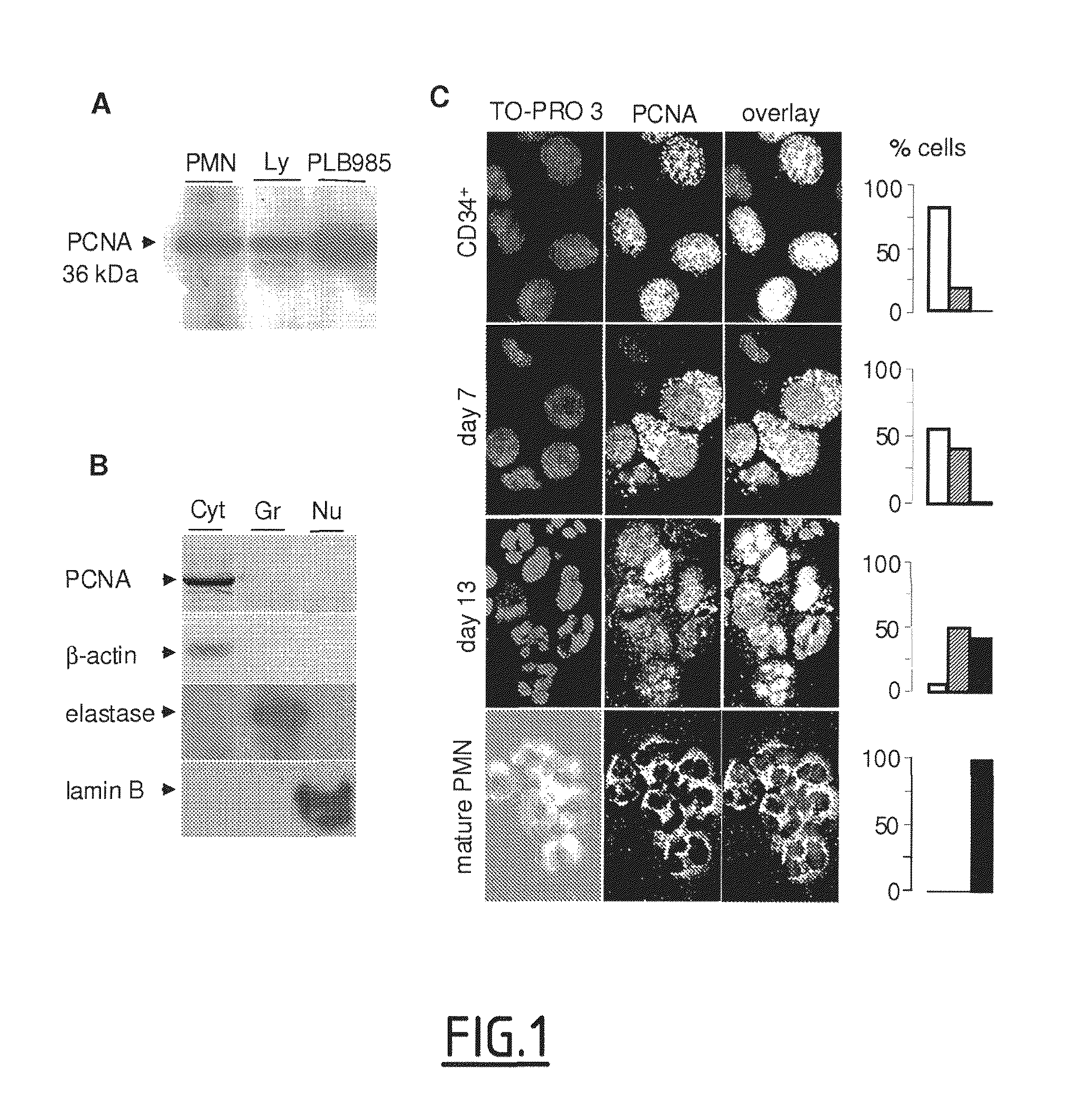
[0136]Western-blot analysis of neutrophil lysates readily detected PCNA in amounts comparable to those in lymphocytes but less than those in the PLB985 promyelocytic cell line (FIG. 1A).
[0137]Surprisingly, subcellular fractionation of neutrophils showed high PCNA contents only in the cytosol, and its absence in the nucleus and granules (FIG. 1B). The quality of the fractionation procedure was validated by the detection of specific markers: α-actin, elastase and lamin-B for cytosol, granules and nuclei, respectively.
example 3
Nuclear-to-Cytoplasmic PCNA Relocalization Occurs During Granulocyte Differentiation
[0138]PCNA subcellular localization was also studied by confocal microscopy after PCNA immunolabeling during the course of in vitro granulocyte differentiation of human CD34+ cells, isolated from umbilical cord blood and cultured with IL-3 and G-CSF. Complete granulocyte maturation was evaluated by morphological analysis after May-Grünwald-Giemsa (MGG) staining. Before inducing differentiation, PCNA was detectable almost exclusively in the nucleus of CD34+ cells, whereas 7 days post IL3-G-CSF-treatment, the protein exhibited a mixed cytoplasmic and nuclear distribution (FIG. 1C). On day 13, most cells had multilobular nuclei with PCNA located in the cytoplasm, similar to what is observed in mature peripheral neutrophils.
[0139]Quantitative analysis (see histograms, FIG. 1C), consisting of counting the cells having a nuclear, cytoplasmic or mixed nuclear-cytoplasmic localization, confirmed this PCNA re...
PUM
| Property | Measurement | Unit |
|---|---|---|
| Fraction | aaaaa | aaaaa |
| Temperature | aaaaa | aaaaa |
| Temperature | aaaaa | aaaaa |
Abstract
Description
Claims
Application Information
 - R&D
- Intellectual Property
- Life Sciences
- Materials
- Tech Scout
- Unparalleled Data Quality
- Higher Quality Content
- 60% Fewer Hallucinations
Browse by: Latest US Patents, China's latest patents, Technical Efficacy Thesaurus, Application Domain, Technology Topic, Popular Technical Reports.
© 2025 PatSnap. All rights reserved.Legal|Privacy policy|Modern Slavery Act Transparency Statement|Sitemap|About US| Contact US: help@patsnap.com


