Device and method for in vivo flow cytometry using the detection of photoacoustic waves

a flow cytometry and photoacoustic wave technology, applied in the field of in vivo flow cytometry using the detection of photoacoustic waves, can solve the problems of ineffective detection of rare metastatic cells or other antigens, unpredictable artifacts, and traditional flow cytometry techniques that are not suitable for applications such as early diagnosis, prevention and treatment,

Active Publication Date: 2013-03-07
BIOVENTURES LLC
View PDF1 Cites 29 Cited by
  • Summary
  • Abstract
  • Description
  • Claims
  • Application Information

AI Technical Summary

Problems solved by technology

Flow cytometry ordinarily requires invasive extraction of cells from the living organism, fluorescent cell labeling, and cell sorting procedures, which may lead to unpredictable artifacts such as cytotoxicity.
Traditional flow cytometry techniques are not suitable for applications such as the early diagnosis, prevention, and treatment of metastasis, inflammations, sepsis, immunodeficiency disorders, strokes, or heart attacks.
In these small volume samples, the detection of rare metastatic cells or other antigens is ineffective until the disease has progressed to a stage in which the rare antigens are numerous enough to be detected in small blood samples.
The powerful fluorescent labeling used in most in vitro and in vivo FC is prone to photobleaching, blinking, or cytotoxicity.
These technical shortcomings limit the extension of traditional FC techniques to the long-term monitoring of blood or lymph flow on humans in vivo.
The fluorescent labeling of cells may seriously compromise cell function and physiology.
Fluorescent dyes or tags used in FC may significantly distort the measured occurrence and elimination of cells in circulation, such as apoptotic or cancer cells.
Although this technique overcomes the shortcomings associated with fluorescent labeling, only a limited subset of circulating cells are sensitive to light scattering, and there is extensive background noise due to scattered light from red blood cells, which make up the majority of cells in circulation.
Further, intervening cells and tissue attenuate the scattered light from the circulating cells, thereby limiting this technique to cells circulating in vessels near the skin's surface.
Like all of the other techniques described above that utilize the detection of light to gather information about circulating cells this technique is limited to cells circulating in relatively thin tissues.
As such, the effectiveness of all of the flow cytometry techniques described above has been limited to the measurement of cells in relatively superficial blood vessels, since light may travel for only a short distance through the cells and tissues surrounding these vessels before becoming too scattered to be detected.
However, due to the challenge of coordinating the timing and characteristics of the laser illumination, limited sensitivity of the detection of the PA waves, time-consuming signal-acquisition algorithms, and poor spatial resolution, applications of PA imaging methods have been limited to the visualization of large groups of stationary cells, making this technique inappropriate for the requirements of in vivo flow cytometry.
In vivo flow cytometry techniques to date are limited to the detection of circulating cells in blood vessels only, due to intrinsic limitations in sensitivity and resolution.

Method used


Image

  • Device and method for in vivo flow cytometry using the detection of photoacoustic waves
  • Device and method for in vivo flow cytometry using the detection of photoacoustic waves
  • Device and method for in vivo flow cytometry using the detection of photoacoustic waves

Examples


example 1

In Vitro Photothermal (PT) Imaging was Used to Determine the Effect of Laser Energy Levels on Laser-Induced Cell Damage to Blood Cells and Subsequent Cell Viability

[0101]To determine whether the laser pulses using in in vivo flow cytometry caused any significant damage to cells or tissues of the organism, the following experiment was conducted. The laser-induced damage threshold of single cells was evaluated as a function of the pumped-laser energy levels at a range of wavelengths using established methods (Zharov and Lapotko 2005, Lapotko and Zharov 2005). In vitro measurements of specific changes in photothermal (PT) images and PT responses from individual cells were used to determine cell damage. During the PT imaging, individual cells were illuminated with a pulse of laser light at a specified energy level and wavelength. After absorbing the energy of the laser pulse, the short-term temperature of the cell increased by as much as 5° C. The laser-induced temperature-dependent ref...

example 2

Prototype In Vivo Photoacoustic Flow Cytometry System Used to Detect Contrast Dye Circulating in Mice

[0106]The following experiment was conducted to demonstrate the feasibility of in vivo photoacoustic flow cytometry (PAFC) for real-time, quantitative monitoring in the blood circulation of a conventional contrast agent, Lymphazurin. In this experiment, a prototype PAFC system was used to detect Lymphazurin circulating in the blood vessels of a mouse ear.

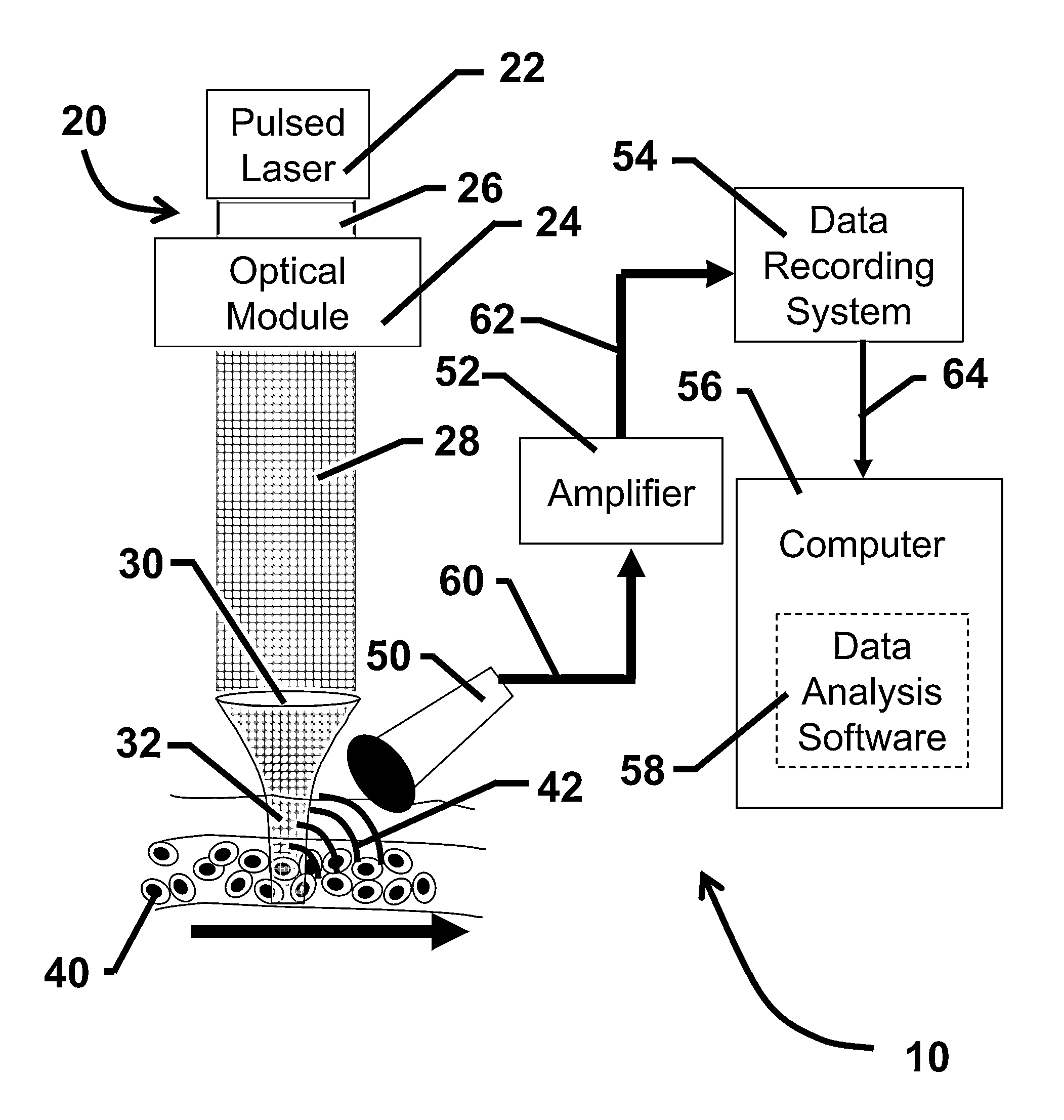
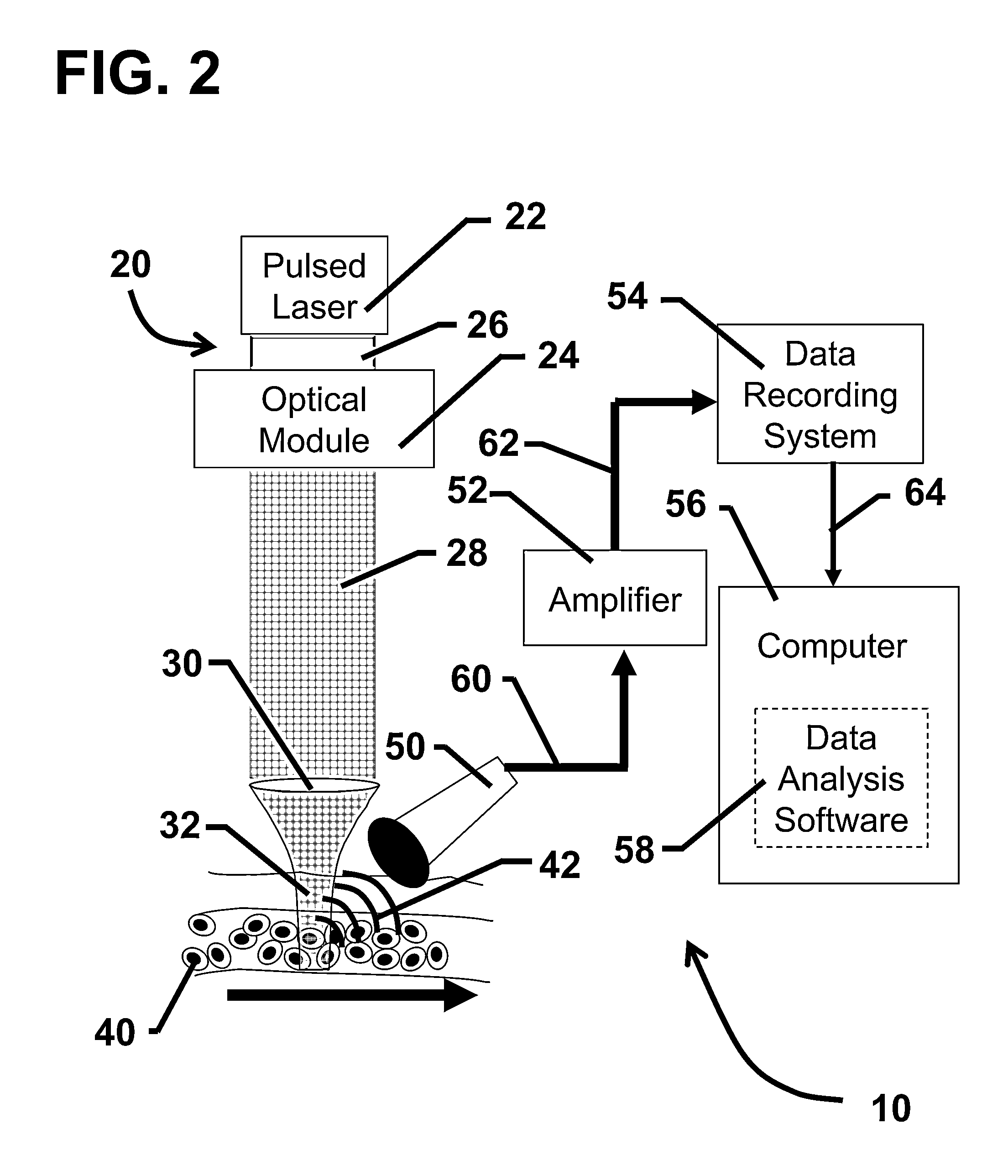
[0107]The prototype PAFC system was built on the platform of an Olympus BX51 microscope (Olympus America, Inc.) and a tunable optical parametric oscillator (OPO) pumped by a Nd:YAG laser (Lotis Ltd., Minsk, Belarus). The general layout of the PAFC system is shown in FIG. 2. Laser pulses had an 8 ns pulse width, a regular repetition rate of 10 Hz with the ability to provide short-term pulses at 50 Hz, and a wavelength in the range of 420-2,300 nm. Laser energy was directed to the blood vessels using a conventional lens, or an optical ...

example 3

Prototype In Vivo Photoacoustic Flow Cytometry System Used to Detect Nanoparticles Circulating in Rats

[0116]To demonstrate the sensitivity of a prototype in vivo photoacoustic flow cytometry (PAFC) system described in Example 2 an experiment was conducted using the prototype PAFC system to detect nanoparticles intravenously injected into the tail veins of rats.

[0117]The in vivo measurements in this experiment were performed using the rat mesentery model. The rat (White Fisher, F344) was anesthetized using ketamine / xylazine at a dosage of 60 / 15 mg / kg, and the mesentery was exposed and placed on a heated microscope stage, and bathed in Ringer's solution at a temperature of 37° C. and a pH of 7.4. The mesentery consisted of transparent connective tissue of 7-15 μm thickness, and a single layer of blood and lymph microvessels.

[0118]The nanoparticles used in this experiment were gold nanorods (GNR), obtained from the Laboratory of Nanoscale Biosensors at the Institute of Biochemistry and...


PUM


Abstract

A photoacoustic flow cytometry (PAFC) device for the in vivo detection of cells circulating in blood or lymphatic vessels is described. Ultrasound transducers attached to the skin of an organism detect the photoacoustic ultrasound waves emitted by target objects in response to their illumination by at least one pulse of laser energy delivered using at least one wavelength. The wavelengths of the laser light pulse may be varied to optimize the absorption of the laser energy by the target object. Target objects detected by the device may be unlabelled biological cells or cell products, contrast agents, or biological cells labeled with one or more contrast agents.

Description

CROSS REFERENCE TO RELATED APPLICATIONS[0001]This application is a divisional application of U.S. non-provisional application Ser. No. 12 / 334,217, entitled “Device and Method for In Vivo Flow Cytometry Using the Detection of Photoacoustic Waves” filed on Dec. 12, 2008, which claims priority from U.S. provisional patent application Ser. No. 61 / 013,543, entitled “Device and Method for In Vivo Flow Cytometry Using the Detection of Photoacoustic Waves” filed on Dec. 13, 2007, both of which are hereby incorporated by reference herein in their entirety.GOVERNMENTAL RIGHTS IN THE INVENTION[0002]This work was supported in part by the National Institutes of Health grant numbers R01EB000873 and R21EB0005123. The U.S. Government may have certain rights in this invention.FIELD OF THE INVENTION[0003]This application relates to a device and methods of using the device to non-invasively detect laser-induced photoacoustic waves emitted by target objects such as cells, pathogens, microparticles, and...

Claims


Application Information

Patent Timeline
Patent Type & Authority Applications(United States)
IPC IPC(8): A61B6/00
CPCA61B5/0059G01N2201/0697A61B5/412A61B5/415A61B5/416A61B5/418A61B8/08A61K49/22G01N15/1425G01N15/1434G01N15/147G01N21/1702G01N21/39G01N2015/1477A61B5/0095A61B2018/20351A61B2018/20357A61B2018/20361A61B2018/207A61B5/02007A61B5/7278A61B18/20A61B2018/00642
Inventor ZHAROV, VLADIMIR PAVLOVICH
Owner BIOVENTURES LLC
Why Eureka
Try Eureka