

Identification of invasive and slow-growing tumorigenic cell subsets in tumors
a tumorigenic cell and tumor technology, applied in the field of tumor heterogeneity and metastasis, can solve the problems of increasing the risk of peripheral disease and slow-growing subsets escaping treatmen
- Summary
- Abstract
- Description
- Claims
- Application Information
AI Technical Summary
Benefits of technology
Problems solved by technology
Method used
Image

Examples
example 1
Detecting Cells Actively Metabolizing Hyaluronan
[0089]We have developed a method for detecting cells that are actively metabolizing HA and have shown that this property is shared by both normal but injured cells and aggressive BCA cell lines (FIG. 10, and M. Veiseh, J. Zhang, R. C. Savani, R. E. Harrison, D. Mikilus, L. Collis, J. Koropatnick, L. Luyt, M. J. Bissell and E. A. Turley, “Imaging of homeostatic, neoplastic, and injured tissues by HA-based probes,”Biomacromolecules. 2012 Jan. 9; 13(1):12-22. Epub 2011 Dec. 12). HA metabolism was detected and quantified as the amount of fluorescent, high molecular weight (HMW) HA—referred to as Texas Red-HA that can bind to and be taken up by these cultured cells. Quiescent fibroblast monolayers showed little uptake while scratch-wounding these monolayers resulted in high TR-HA uptake at the wound edge (FIG. 10). We verified that the TR-HA probe accurately detected areas of high HA metabolism by demonstrating its rapid and HA-receptor dep...
example 2
Detecting and Imaging Tumor Heterogeneity
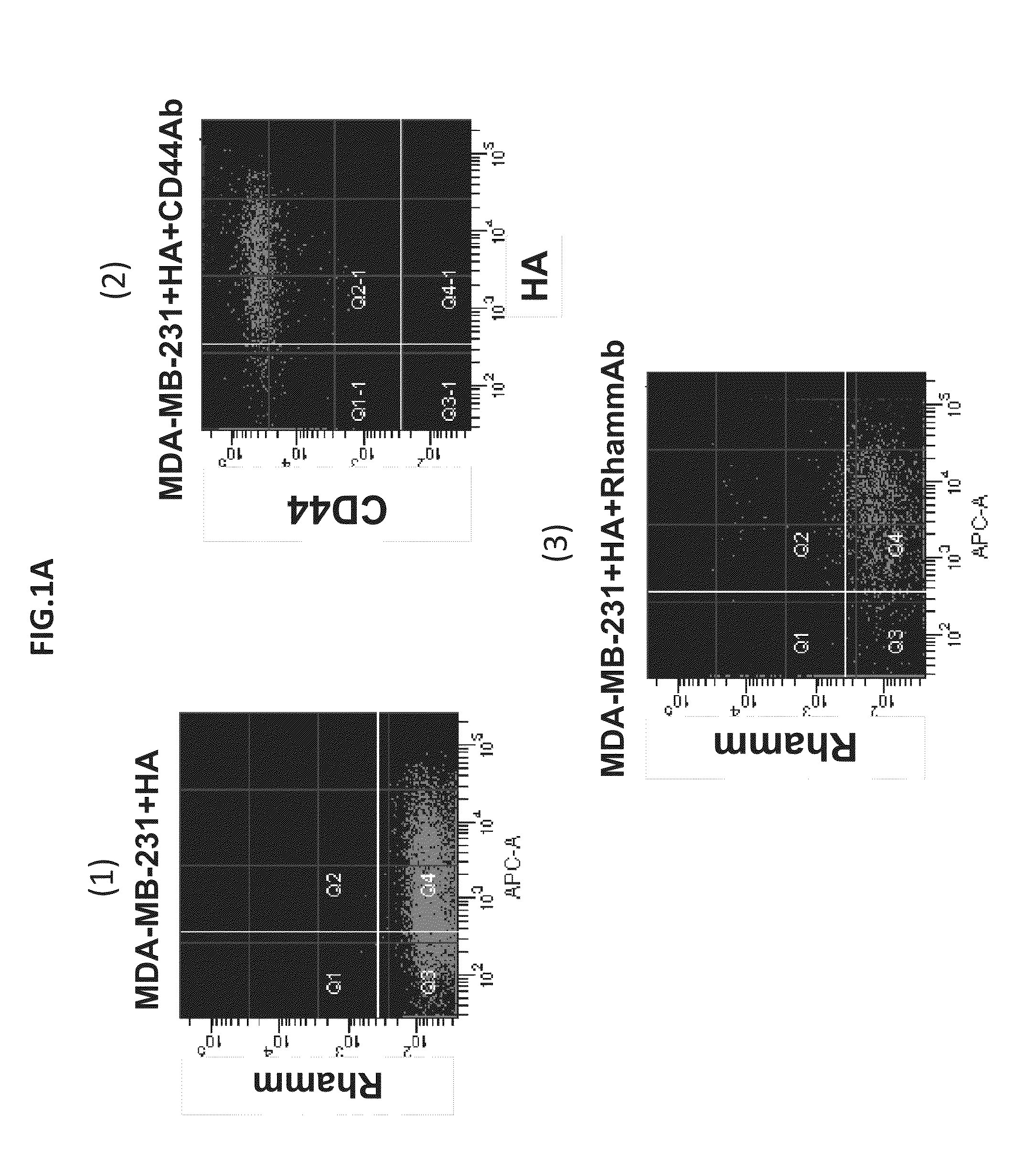
[0091]Tumor heterogeneity is a characteristic of breast cancer (BCA) at both the molecular and histological level. The presence of heterogeneous tumor cell subpopulations, which exhibit distinct cell surface markers, biological functions and tumorigenicity, has recently been implicated in both BCA progression and response to treatment. In particular, increasing evidence implicates stem / progenitor-like subpopulations in BCA initiation, metastasis, and resistance to therapy. These particular subpopulations are characterized by high surface display of CD44, what is now considered to be the minimal surface phenotype amongst additional surface markers. CD44 is an integral receptor for the tissue polysaccharide, hyaluronan (HA), which activates the signaling functions of CD44 in BCA. Another HA receptor, RHAMM / HMMR, is a co-receptor for CD44 and promotes its ability to activate MAPK pathways through HA and HA fragments (See Tolg C, Hamilton S, Zali...
example 3
Flow Cytometry Using Ha Metabolism
[0111]A biopsy of tissue can be obtained from a patient. Using flow cytometry, the cells can be sorted based on their ability to actively metabolize HA.
[0112]For example, a sample protocol using the Alexa Fluor 647 HA assay, then sorting using a flow cytometer, is described below:
[0113](1) Grow cells to 50% confluence on culture flask; (2) Rinse in Ca2+-free Hank's Buffered Saline Solution (HBSS / 20 mM HEPES, pH 7.3); (3) Harvest cells with non-enzymatic HBSS-based cell dissociation solution; (4) Resuspend in cold and sterile PBS solution that contains Antibiotic, HEPES pH 7.4, Insulin, and Transferrin (PBS / additive solution); (5) Centrifuge and eliminate supernatant; (6) Wash in cold PBS / additive solution; (7) Count cells using cell counter instrument and measure viability; (8) Treat cells (in suspension) with Alexa Flour 647-HA; (9) Incubate for 45 min on ice, in dark.
[0114]For single-color HA-binding flow cytometry: (a) wash the cells, eliminate s...
PUM
| Property | Measurement | Unit |
|---|---|---|
| Atomic weight | aaaaa | aaaaa |
| Composition | aaaaa | aaaaa |
| Molecular weight | aaaaa | aaaaa |
Abstract
Description
Claims
Application Information