Special-illumination surgical stereomicroscope

a stereomicroscope and surgical technology, applied in the field of special-illumination surgical stereomicroscopes, can solve the problems of affecting the efficiency of the white light from the surgical illumination device, affecting the patient's health, and having to undergo long approval procedures, so as to improve the efficiency of laser light, reduce the space requirements of lasers, and save spa

Inactive Publication Date: 2013-08-08
LEICA MICROSYSTEMS (SCHWEIZ) AG
View PDF6 Cites 10 Cited by
  • Summary
  • Abstract
  • Description
  • Claims
  • Application Information

AI Technical Summary

Benefits of technology

[0042]The inventive idea is based on the fact that in surgical microscopy, in particular in surgical stereomicroscopy, it is now possible to cause light emission also in endogenous tissue substances that do not exhibit visible fluorescence. In the present invention, the visualization of stimulated light emission is integrated with the conventional surgical microscopy technique, in which the object was heretofore visualized only by the reflection of white surgical illumination light or by fluorescence. In accordance with the present invention, no filters or drugs are needed, which avoids the disadvantages described above.
[0043]Excitation is accomplished using a completely different light source which is not known for use in surgical microscopes and which is cable of causing normally non-luminescent endogenous substances to emit visible light, such as, for example, hemoglobin (e.g., using 830 nm excitation light at 20 mW, for example, and a stimulation wavelength of 600 nm at 3 mW, for example). This is important, among other things, to distinguish well-perfused tumor tissue from healthy, more poorly perfused tissue. To this end, in accordance with the invention, stimulated emission is used in a novel way under the surgical microscope. This technique is clearly different from the fluorescence technique heretofore used under surgical fluorescence microscopes. Only the effect observed by the surgeon appears to be similar. However, if the light sources and the operating modes are suitably designed, the effect appears much better, especially when also taking into account the possibility of amplification by video image data processing.
[0045]The laser pulses may be coupled into the illumination beam path of the surgical illumination light source by input coupling optics, using either a physical or a geometric beam splitter. Optionally, in order to avoid interference with the illumination beam path or the observation beam path, the laser light may also be irradiated directly onto the object from the side of the microscope, thereby possibly increasing the efficiency of the laser light.
[0046]The space requirements of lasers are continuously reduced, so that, in one embodiment of the present invention, the lasers can also be fitted directly into the illumination devices used heretofore, which eliminates the need for a special input coupling device. This not only saves space, but reduces the number of additional assemblies outside of the microscope.
[0048]In one embodiment of the present invention, a controller controls the laser light sources temporally in such a way that, through modulation of the pulse duration and spacing, an optimized stimulated emission can be observed. Thus, in this configuration of the present invention, there is no need to use expensive special spectral filters, which would attenuate the visible light.

Problems solved by technology

In conventional surgical fluorescence microscopy, fluorescent drugs (e.g., 5-ALA (aminolevulinic acid)) are used, which may potentially harm the patient and have to undergo lengthy approval procedures.
The use of filters to separate the excitation light from the emission light in surgical fluorescence microscopy negatively affects the efficiency of the white light from the surgical illumination device.
The emission light is generally very weak and, in contrast to the visible white light (and, for example, also to the ambient light), is very hard to see.
Therefore, great effort is required of microscope manufacturers in terms of illumination and filter technology.
This increases equipment complexity on the one hand, and on the other hand may additionally reduce the illumination efficiency with respect to the visible light from the surgical field.
In surgical microscopy, however, the technological requirements are completely different from those in the field of high-magnification scanning microscopy or transmission microscopy.
The use of fluorescent contrast agents is associated with significant medical side effects.
Nevertheless, the contrast often leaves a lot to be desired.
With regard to the filter combinations used, compromises must be made (reduced efficiency of the illumination and excitation light).
The large number of required filters and their controls make a microscope complicated to manufacture and difficult to use.

Method used


Image

  • Special-illumination surgical stereomicroscope
  • Special-illumination surgical stereomicroscope
  • Special-illumination surgical stereomicroscope

Examples


Embodiment Construction

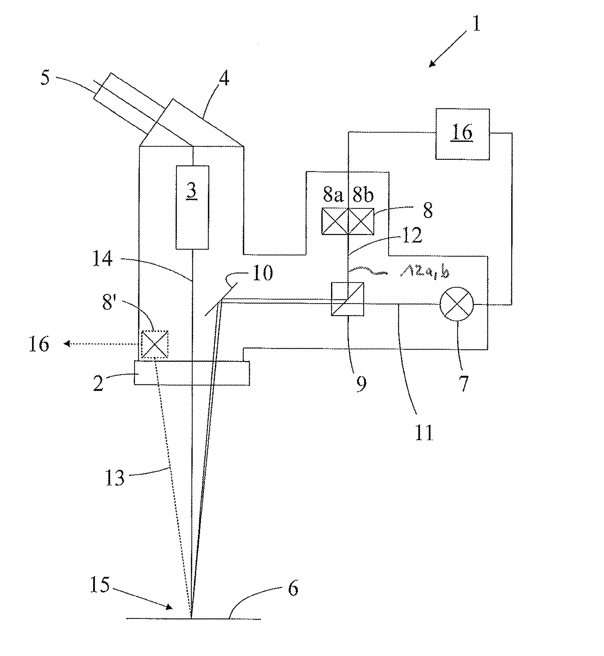
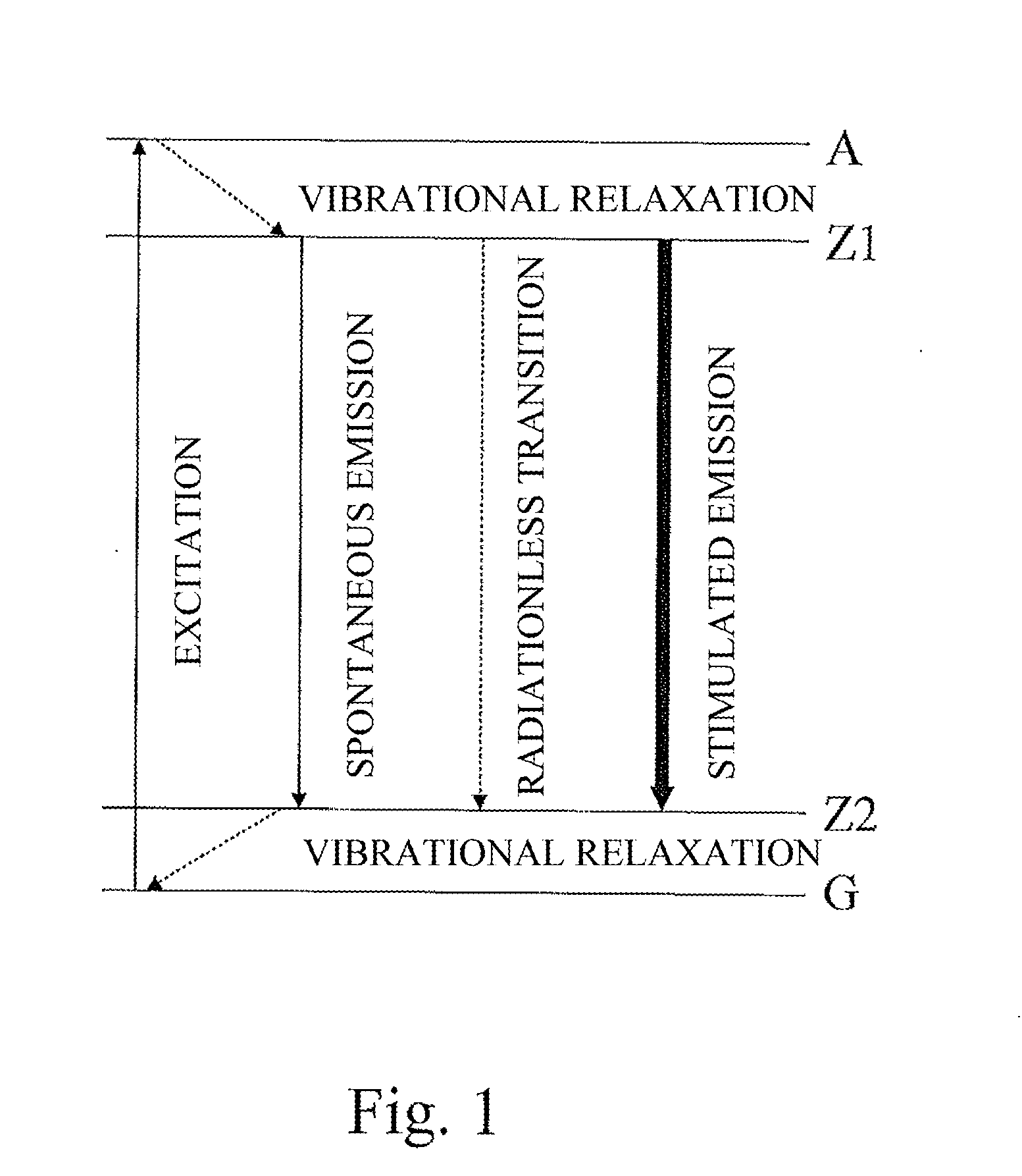
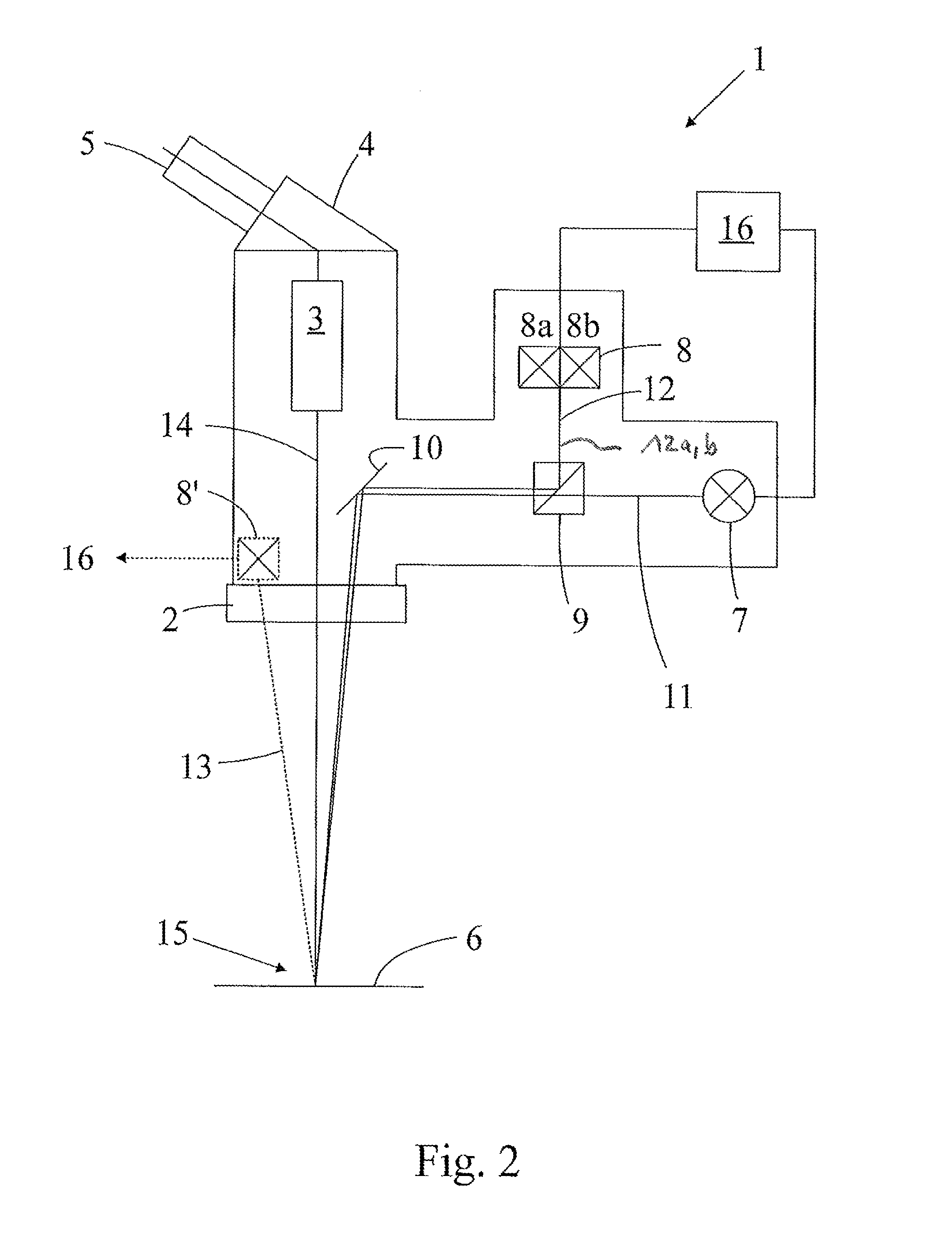
[0067]FIG. 1 shows an energy-level diagram illustrating the differences between stimulated emission, radiationless transition, and spontaneous emission (fluorescence). Radiation from an excitation light source causes an electron to go from ground state G to an excited state A. From there, the electron goes to an intermediate state Z1. In the case of non-fluorescent substances, this is followed by a radiationless transition to a lower energy state Z2, from where ground state G is reached again by vibrational relaxation.

[0068]Spontaneous emission (autofluorescence), which is also shown in FIG. 1, occurs only in fluorescent substances and without the need for separate excitation. However, since endogenous substances do not exhibit fluorescence, except for the rather low autofluorescence, and therefore foreign fluorescent substances are used, the present invention, in contrast, uses the principle of stimulated emission, which in many interesting endogenous substances in the body (e.g., ...


PUM


Abstract

The present invention relates to a special-illumination surgical stereomicroscope (1) for observing an object (6) in an object field (15) under special illumination, the special-illumination surgical stereomicroscope including a surgical illumination light source (7; 7a) for illuminating the object field (15) via an illumination beam path (11) and further including a special-illumination light source. The special-illumination light source is adapted for observation of stimulated emission and includes an excitation light source (8a) for specific excitation of a substance contained in the tissue of the object (6) via an excitation beam path (12a), and a stimulation light source (8b) for stimulating the emission of light from the previously excited substance via a stimulation beam path (12b). A common observation beam path (14) is provided for guiding the light generated by stimulated emission and the reflected surgical illumination light.

Description

CROSS REFERENCE TO RELATED APPLICATIONS[0001]This application claims priority of German patent application number 10 2012 001 854.1 filed Feb. 1, 2012, the entire disclosure of which is incorporated by reference herein.FIELD OF THE INVENTION[0002]The present invention relates to a special-illumination surgical stereomicroscope, which term includes a conventional surgical stereomicroscope, a video surgical microscope, an endoscope, or a pair of surgical loupes, and to a method for operating such a special-illumination surgical stereomicroscope.DEFINITIONS OF TERMS[0003]The definitions of certain important terms and functions are explained below.[0004]A white light surgical microscope illumination device is known to those skilled in the art because it is used in a wide variety of surgical microscopes. It covers the entire spectrum of white light, since it is used principally to illuminate the surgical field and is generally intended to do so during surgery in as color-neutral a manner...

Claims


Application Information

Patent Timeline
Patent Type & Authority Applications(United States)
IPC IPC(8): G02B21/00G02B21/06
CPCG02B21/0076G02B21/06A61B3/13A61B3/14A61B19/5223A61B5/0077G02B21/16A61B5/0071G02B21/0012A61B90/20
Inventor SANDER, ULRICH
Owner LEICA MICROSYSTEMS (SCHWEIZ) AG
Why Eureka
Try Eureka