Methods and compositions for non-invasive, dynamic imaging of intestinal motility

a dynamic imaging and intestinal motility technology, applied in the field of biomedical imaging, can solve the problems of inability to enable longitudinal monitoring, no utility in the clinic, less useful in veterinary clinical practice, etc., and achieve the effect of sufficient temporal resolution and sensitivity

Inactive Publication Date: 2013-08-22
BOARD OF RGT THE UNIV OF TEXAS SYST
View PDF3 Cites 15 Cited by
  • Summary
  • Abstract
  • Description
  • Claims
  • Application Information

AI Technical Summary

Benefits of technology

[0012]The problem of non-invasively imaging gastrointestinal (GI) motility without the use of radioactive contrast agents, but with sufficient temporal resolution and sensitivity to assess dynamic motion of the intestine can be solved using administration of a highly sensitive NIR fluorescent dye and imaging system in combination with auto fluorescent compounds present in foodstuffs. Such compositions can be used to identify abnormal patterns and physiology, provide correct diagnosis of GI motility disorders or dysfunction, and guide evaluation of pharmacologic and surgical therapy for GI motility diseases, disorders, and related symptoms in both veterinary and human patients.

Problems solved by technology

However, these ex vivo and in situ measurements while informative are invasive and do not enable longitudinal monitoring of GI motility in disease and response to pharmacological intervention but certainly have no utility in the clinic.
However there were difficulties associated with repeated oral contrast agent administration and art factual intestinal motions were observed due to the distension of the intestinal walls by the significant quantities of contrast required.
However, several of these require the contrast agents many of which are radioactive.
For example, scintigraphic evaluation of gastric emptying with oral administration of Tc99m labeled compound is widely used in the human clinical practice but it is less useful in veterinary clinical practice due mainly to long camera integration times and low temporal resolution.

Method used


Image

  • Methods and compositions for non-invasive, dynamic imaging of intestinal motility
  • Methods and compositions for non-invasive, dynamic imaging of intestinal motility
  • Methods and compositions for non-invasive, dynamic imaging of intestinal motility

Examples


example 1

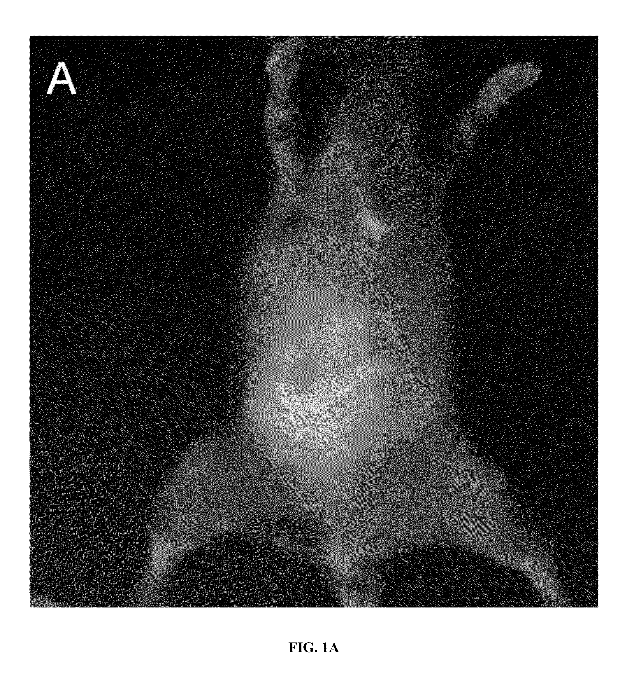
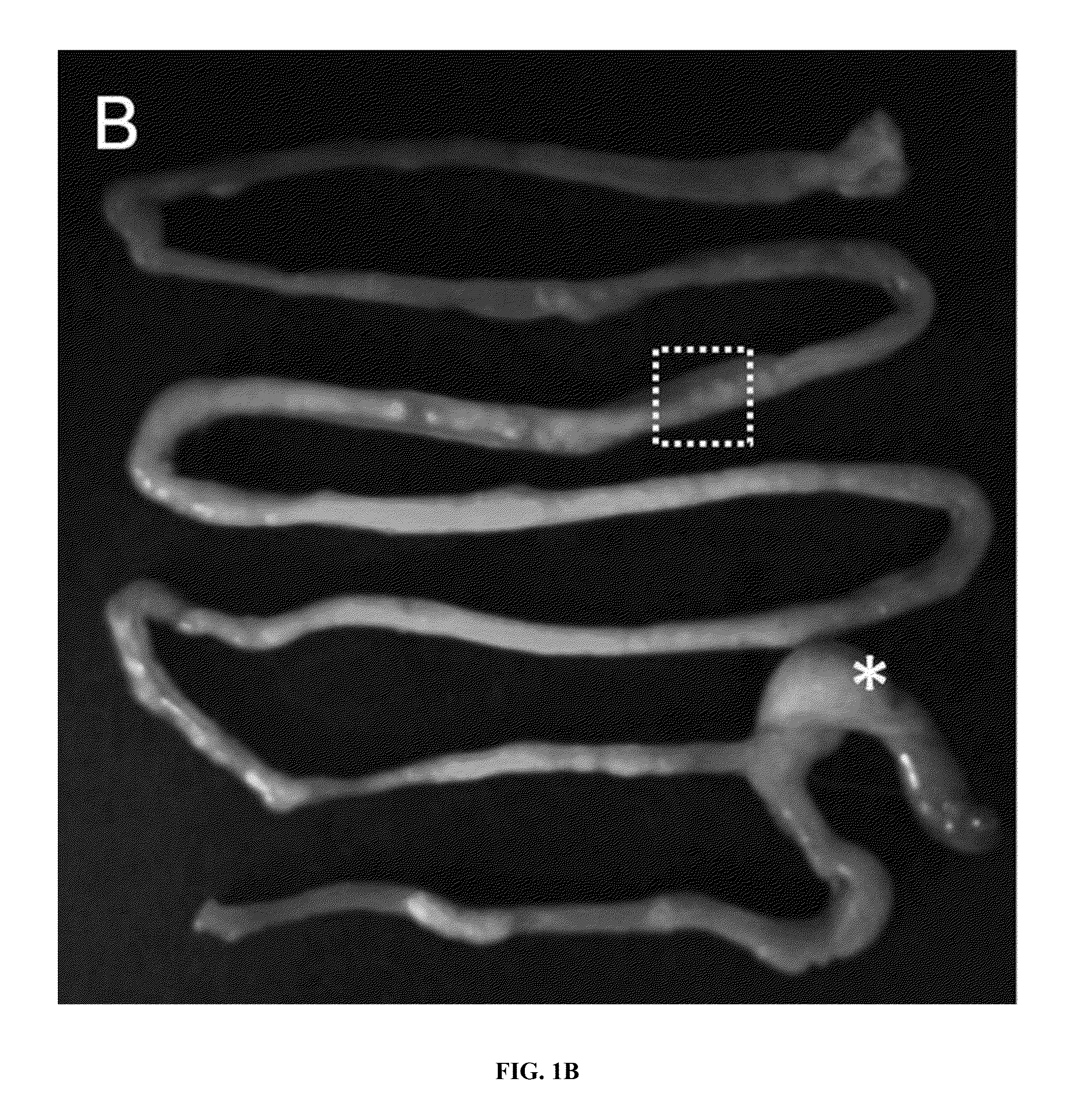
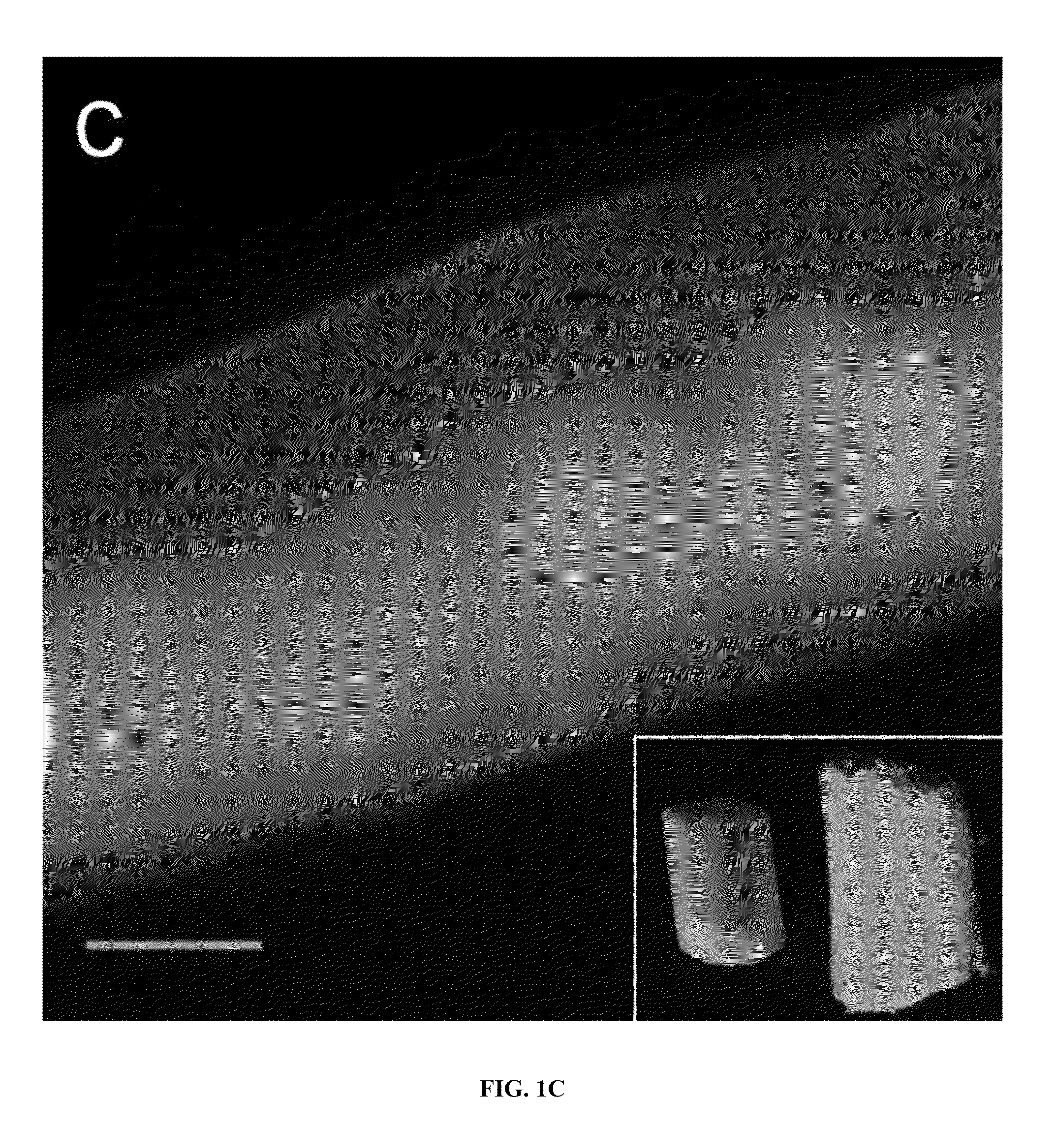
Using Auto Fluorescence to Non-Invasively Monitor Intestinal Motions

[0109]Six to eight week old mice that happened to carry a Rasa 1 floxed allele (rasa 1 fl / fl), i.e. the Rasa 1 allele is flanked by a Lox P sites to facilitate removal). Rasa I fl / fl mice are completely normal (until exposed to Cre-recombinase) with regards to GI function and were maintained in standard cages with free access to water and pellets (Purina 5053, Labdiet, PMI Nutritional International, St. Louis, Mo., USA) prior to imaging. Once anesthetized with isofluorane, the hair of the mice was clipped and depilatory agents were used to remove residual hair 24 hr prior to fluorescence imaging. Mice were housed and maintained in a pathogen-free mouse facility accredited by the American Association for Laboratory Animal Care. All experiments were performed in accordance with the guidelines of the Institutional Animal Care and Use Committee. Animal experiments were approved by University of Texas Health Science Cent...

example 2

Using Exogenous Fluorescence to Monitor Intestinal Motions

[0116]In order to clearly characterize and demonstrate the abilities of the methods disclosed, studies were carried out on mice as a model for all animals, including but not limited to those animals that can suffer a gastrointestinal motility disease or disorder. Such animals include, but are not limited to, laboratory animals, domesticated and livestock animals, wild and captured animals such as those that are in zoos, companion animals and humans.

[0117]Mice were chosen because part of the study involved opening the peritoneal cavity and removing organs to provide detailed information about the method and its abilities. Therefore, the subjects of the study were Female C57BL6 mice (4-6 wk old; Charles River, Wilmington, Mass., USA) that were housed and maintained in a pathogen-free mouse facility accredited by the American Association for Laboratory Animal Care. All studies were performed in accordance with the guidelines of ...


PUM


Abstract

Described are methods and composition for use in non-invasive imaging of intestinal structure and function. These methods can be used to identify, diagnose, assess, monitor and direct therapies for gastrointestinal diseases and disorders. Embodiments of the methods utilize highly sensitive optical imaging and fluorescent spectroscopy techniques to track or monitor packets of organic dye excreted in bile into the intestinal tract to provide quantitative information regarding intestinal propulsion and function.

Description

[0001]This application claims the benefit of U.S. Provisional Patent Application Nos. 61 / 594,880, filed Feb. 3, 2012, and 61 / 650,805, filed May 23, 2012, the entirety of which are incorporated herein by reference.[0002]The invention was made with U.S. government support under Grant Nos. DK056338, HL092923, CA128919, and CA136404 awarded by the National Institutes of Health. The government has certain rights in the invention.BACKGROUND OF THE INVENTION[0003]1. Field of the Invention[0004]The present invention relates generally to the field of biomedical imaging. More particularly, it concerns methods and compositions for functionally imaging and measuring intestinal motility in an individual using a highly sensitive near infra-red (NIR) imaging system. This disclosure also relates to the use of such methods and compositions in the diagnosis, assessment, and monitoring of diseases and disorders of the gastrointestinal tract using administration of a near infra-red (NIR) fluorescent dy...

Claims


Application Information

Patent Timeline
Patent Type & Authority Applications(United States)
IPC IPC(8): A61B5/00
CPCA61B5/0071A61B5/4255A61B5/4848G06T7/0012G06T2207/30028A61B2503/42A61B2576/00G06T2207/10048A61B2503/40G16H30/40
Inventor KWON, SUNKUKSEVICK-MURACA, EVA M.DAVIES-VENN, CYNTHIA
Owner BOARD OF RGT THE UNIV OF TEXAS SYST
Why Eureka
Try Eureka